![]()
![]()
![]()
Use LEFT and RIGHT arrow keys to navigate between flashcards;
Use UP and DOWN arrow keys to flip the card;
H to show hint;
A reads text to speech;
894 Cards in this Set
- Front
- Back
|
Approach to altered mental status |
IS IT MEATS |
|
|
Intubation preparation |
STOP I C BARS |
|
|
Diagnosing the cause of an alarming ventilator |
D - Dislodged tube |
|
|
CHF treatment |
POND |
|
|
Ingestions that are not absorbed by activated charcoal |
PHAILS-O - Oil of wintergreen |
|
|
Causes of anion-gap metabolic acidosis |
KULT |
|
|
Hydrocarbon additives that require GI decontamination |
CHAMP |
|
|
Mass casualty triage protocol |
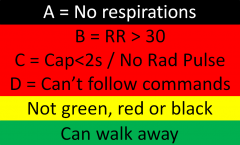
Use the START (Simple Triage And Rapid Treatment) Protocol |
|
|
PERC Rule |
Apply if clinical gestalt = low risk for PE |
|
|
CATCH Rule - High Risk Criteria |
WIGS |
|
|
CATCH Rule - Medium Risk Criteria |
SDH |
|
|
Causes of Seizure |
STATUS EPILEPsy |
|
|
Signs of Delirium (for Confusion Assessment Method, CAM) |
AIDA D - isturbance perceptually Not caused by dementia |
|
|
What is needed for a safe discharge plan? |
No RISKS |
|
|
Diagnostic criteria of major depressive disorder |
MDD classified as 5 or more of these symptoms occurring most days over a 2 week period along with a change in function. MUST have depressed mood or loss of interest/function.
SIGECAPS
Not a mixed episode, due to anxiety, caused by a general medical condition, or consistent with bereavement (<2 months from loss) |
|
|
Approach to the Alarming Ventilator |
D - Disconnect the patient from the ventilator +/- provide gentle pressure to the chest (assess for and treat breath Stacking and Equipment failure) |
|
|
Ottawa SAH Rule |
A - Age > 40y |
|
|
Ring enhancing lesions on Head CT |
MAGICAL DR A - Acute Disseminated Encephalomyelitis L - Lymphoma
D - Demyelinating disease |
|
|
Anterior cord syndrome; causes |
Like a car - if it smashes the front the engine won't work (paralysis and pain) but instruments/GPS will (vibration/proprioception)
Causes - Dissection and aorta surgery (Artery of Adamkiewicz), hypotension, vasospasm, thrombosis |
|
|
What are the NEXUS Criteria? |
2 Exam |
|
|
Unstable C-Spine Fractures |
Jefferson Bit Off A Hangman's Tit And Pinky A ny fracture-dislocation P osterior neural arch of C1 |
|
|
Approach to bradycardia |
DIE! |
|
|
Rashes that start on the palms |
Sifting Rocks Scabbed Emma's Hands |
|
|
Rashes with a + Nikolsky sign |
Stevie got scalded by TEN PV'd nickels |
|
|
Rashes with vesicle / bullae |
Old man with BPPV fell into a pool of necrotizing gonorrhea |
|
|
Rashes with petechiae / purpura |
Henoch the Tick gave Meningitis to DICk the purple drug addict |
|
|
Exam findings of serotonin syndrome |
CHAARM |
|
|
Depression symptoms |
SIGECAPS |
|
|
Suicidal ideation assessment |
SAD PERSONS scale correlates with the decision to admit to psychiatry. Does not predict risk of future suicidality.
S ex (male) - 1
<6 - Outpatient >6 - ED psych evaluation >9 - Psych admission |
|
|
Major and minor criteria for Rheumatic Fever. Treatment. |
Jones criteria (requires evidence of strep infection + 2 major or 1 major/1 minor)
Evidence of strep infection: 1. Elevated ASOT or other streptococcal antibodies 2. Positive throat culture for Group A beta-hemolytic streptococci 3. Positive rapid direct Group A strep carbohydrate antigen test 4. Recent scarlet fever. MAJOR J - oints - polyarthritis of large joints (knees, elbows, wrist, ankles) <3 - carditis (murmurs, effusions, cardiomegaly, CHF) N - Nodules - subcutaneous on extensor surfaces (wrist, elbow, knees, spine) E - Erythema marginatum (painless, non-pruritic) S - Sydenham's Chorea
MINOR F - Fever A - Arthralgias C - CRP E - ESR P - PR interval increased
Treatment -Penicillin 500mg adults / 250mg peds x 10 days or Benzathine penicillin 1.2million U adult or 600thousand U children IM; long-term prophylactic antibiotics -Aspirin |
|
|
Criteria for diagnosing Endocarditis |
Duke's criteria () BE FIVER (+ if 2 major, 1 major 2 minor, 5 minor)
B - Blood cultures (2 positive with typical pathogens) E - Echo lesions (vegetation, perivalve abscess, prosthetic valve dehisence, new regurgitation)
F - Fever (>38) I - Immunologic (Roth, Osler, rheumatoid factor) V - Vascular (Janeway, septic emboli, conjunctival hemorrhage) E - eccentric blood culture (single positive culture unless organism does not cause IE) and echo (consistent with IE but do not meet criteria) R - risk factors (IVDU, prosthetic valve) |
|
|
San Francisco Syncope Rule |
CHESS CHF Hematocrit <30% ECG SOB Systolic BP <90 |
|
|
Dangerous ECG findings on an ECG of a patient with syncope |
Prolonged QT WPW Brugada HOCM Ischemia |
|
|
Causes of hyperacute T waves |
Ischemia Hyperkalemia Pericarditis LVH LBBB Benign early repolarization |
|
|
Causes of tall R wave in v1 |
Posterior MI RBBB WPW type A Children and adolescents Dextrocardia |
|
|
Causes of ST elevation on ECG |
STEMI Printzmetal's LBBB LVH Pericarditis Hyperkalemia Brugada PE Celebral hemorrhage Pacing BER |
|
|
How can VT be distinguished from SVT with aberrancy? |
Brugada criteria (note: not good enough to use in real life): 1. Absence of any RS complexes in the chest leads |
|
|
What are the common pacemaker malfunctions? |
Failure to capture - lead displacement or break, block or battery Oversensing - sensing T waves or extracardiac stimulus Undersensing - poor lead connection or break, small amplitude, poor contact Inappropriate rate - battery, response to atrial dysrhytmias |
|
|
What is the code for pacemaker type? |
Chamber paced - A, V, D Champer sensed - A, V, D Response to sensing - Inhibit pacing (V or A and V) or Trigger pacing (old) Programming - simple, programmable, rate adaptive, communicating, none Antitachy response - pace or shock or dual |
|
|
Complications of ICD and pacemaker placement |
Infection of wound Infection of pouch Thrombophlebitics Chronic thrombosis |
|
|
Indications for a pacemaker |
High level block (2nd or 3rd degree): -And symptomatic brady -And asystole >3s (AFib pauses >5s) -Following AV ablation -With neuromuscular disease -Intermittently block and bi or trifascicular block -With exercise
|
|
|
Indications for an ICD |
1. Cardiac arrest from VF or VT not caused by a reversible event |
|
|
Etiology of Pericarditis |
-Infectious (Viral - Coxsackie, Echovirus, HIV; Bacterial - Staph, Strep, TB; Fungal - Histoplasmosis; Parasite, Rickettsia)
-Postinjury (Trauma, Surgery, Myocardial infarction [Immediate or Dressler's], Radiation)
-Metabolic diseases (Uremia, Myxedema)
-Systemic diseases (Rheumatoid arthritis, Systemic lupus erythematosus, Sarcoidosis, Scleroderma, Dermatomyositis, Amyloidosis)
-Tumors (Leukemia, Lymphoma, Melanoma, Mets)
-Medications (Procainamide, Hydralazine)
-Aortic dissection |
|
|
List the hypertensive emergencies and their ideal treatment (goal) |
-ACS - nitroglycerine, labetolol (Asymptomatic) -Heart failure - nitroglycerine, furosemide (25% reduction) -Dissection - esmolol & nitroprusside OR labetolol (<140/90) -Ischemic stroke - nicardipine, labetolol (<180/110 for lytic) -Intracerebral hemorrhage - nicardipine, labetolol (25% reduction) -Hypertensive encephalopathy - nicardipine, labetolol (25% reduction) -Kidney injury - fenoldopam, nicardipine (25% reduction) -Preeclampsia - magnesium and labetolol (<160/110 and asymptomatic) -Sympathetic crisis - phentolamine (25% reduction) |
|
|
What features distinguish orbital cellulitis from periorbital cellulitis? |
Proptosis, opthalmoplegia, and visual changes (look for afferent pupillary defect secondary to increased IOP). |
|
|
Differential diagnosis for a NAGMA |
HARDUPS Hyperalimentation / TPN Acetazolamide RTA Diarrhea Ureteral diversion Pancreas Spironolactone |
|
|
Niacin deficiency |
aka Vitamin B3 and results in Pellagra
4D’s: Diarrhea Dermatitis Dementia Death |
|
|
Thiamine deficiency |
aka vitamin B1
Wernicke's Encephalopathy- WACO: ataxia, confusion, opthalmoplegia Korsakoff's Psychosis - irreversible short-term memory loss Beri-beri - high output heart failure secondary to vasodilation and fistula formation |
|
|
Cobalamin deficiency |
aka vitamin B12
Megaloblastic anemia Neurologic changes (paresthesias, ataxia, clonus, memory loss) Psychiatric (depression, psychosis)
Folate looks the same except NO neurologic changes and it happens faster. |
|
|
Causes of non-cardiogenic pulmonary edema |
IS NOT THE HEART
I nhaled Toxins (Ammonia, Chlorine, Phosgene, Nitrous oxide)
N eurogenic (seizure, strangulation, trauma, SAH)
T rauma
H igh altitude pulmonary edema |
|
|
Diagnostic criteria of Multiple Myeloma |
-Monoclonal plasma cells or plasmacytoma in the bone marrow -Monoclonal protein in blood or urine -Organ dysfunction (CRAB criteria) C - HyperCalcemia R - Renal failure A - Anemia B - Bone damage (lesions or osteoporosis) |
|
|
What cancers cause bone mets? |
Painful Bones Kill These Suckers Prostate Breast Kidney Thyroid Skin
Also Lungs |
|
|
Hard signs of vascular injury |
HARD Bruit
Hypotension Arterial Bleed Rapidly expanding hematoma Deficit (pulse/neuro) Bruit/thrill |
|
|
Criteria to call a febrile seizure simple |
Fever >38.5 6 months to 6 years 1 episode in 24 hours / per illness Duration <15 minutes Generalized No neurological history |
|
|
HELLP Syndrome |
Severe form of pre-eclampsia
Labs H emolysis E levated L iver enzymes P latelets (<100)
PE -Jaundice, edema, hypertension, tachycardia, dehydration, tachypnea
Treatment -Consider steroids, BP control -> delivery is definitive |
|
|
Kawasaki Disease criteria and treatment |
Warm CREAM
Warm - fever >5 days C - Conjunctival injection (bilateral, non-exudative) R - Rash (primarily on trunk; erythematous, maculopapular, morbilliform - no crusting or vesicles) E - Erythema of palms / soles; Edema of hands / feet; periungal desquamation A - Adenopathy (cervical, >1.5cm, unilateral) M - Mucous membrane changes (lips and oral cavity dry and fissured; erythematous mucousa; strawberry tongue)
Treatment - ASA and IVIg; watch for Coronary Artery Aneurysm |
|
|
Tetrology of Fallot cardiac anomolies |
-Boot shaped heart (Fall over your own Boots) -Pulmonary hypertension, VSD, RVH, Overriding Aorta -Ductal dependent lesion that crash after PDA closes (2-10 days, treat with PGE1 0.1mcg/kg/m) Tet spells (knees to chest to increase SVR and O2 to decrease PVR)
|
|
|
Congenital Adrenal Hyperplasia abnormalities |
21-hydroxylase deficiency Low Na and High K Virulized females, small penis in boys Treat with glucose and hydrocortisone |
|
|
Abuse fracture patterns |
ANY in a child <1yo Bucket Corner** Diaphysis of humerus, radius, femur, tibia (especially <3yo) Rib** Scapular** Spinous process** Sternum** Skull (stellate)* Vertebral* Digits* Multiple* or Bilateral Different stages of healing* |
|
|
Psychological signs of child sexual abuse |
Very broad definition
Regression Acting out Sexualized behavior Disclosure |
|
|
The crashing neonate |
THE MISFITS T rauma / abuse H eart disease / H ypothermia / H ypoxia E ndocrine (CAH, hyperthyroid) M etabolic (hypoglycemia, hyponatremia, hypocalcemia) I nborn errors (ammonia) S epsis (most common!) F ormula mishaps I ntestinal catastrophes (volvulus, NEC, diaphragmatic hernia) T oxins (home remedies) S eizures |
|
|
Cyanotic heart disease |
Increased lung markings 1-Truncus arteriosis 2-Transposition of the great arteries 5-Total anomalous venous return
Decreased lung markings 3-Tricuspid atresia / pulmonary atresia 4-Tetrology of Fallot
|
|
|
Congenital heart disease: Obstructive Lesions |
Also happen with closure of the duct, but NOT cyanotic. Give them 0.1mcg/kg/m of PGE1
Coarctation of the aorta Hypoplastic left heart syndrome Interrupted aortic arch Aortic stenosis (critical) |
|
|
List some symptoms of lead poisoning; Treatment |
LEADeNN L - Lead lines E - Encephalopathy (Neuro! seizures) A - Anemia with basophilic stippling (Heme!) D - Drop (wrist) i N - Nephro tubular fibrosis; proteinurea; Fanconi syndrome G - G I (Nausea / vomiting / abdo pain / liver damage)
Treatment: WBI, Dimercaprol (BAL) and CaNa2-EDTA (with 2nd dose) for acute/symptomatic; PO Succimer (DMSA) and Penicillamine for chronic |
|
|
Outline the phases of iron poisoning, a rare complication, and treatment |
Remember: Gluconate 13%, Sulphate 20%, Fumarate 30% Fe
I GI effects (hemorrhagic GI effects) x 6 hours II Quiscient x 12 hours III Systemic (vasodilatory shock; negative ionotrope, hepatorenal dysfunction with impaired oxidative phosphorylation) IV Liver failure V Resolution (GI scarring, stomach obstruction)
Complication: Weirdly facilitates growth of Yersinia enterocolitica - can cause sepsis
Treatment: WBI, IVF, Deferoxamine (if GIB, AGMA, shock, AMS, >90umol/L, seen on x-ray; watch for hypotension/ARDS/ATN/Yersinia sepsis) |
|
|
Pelvic avulsion fractures (muscle attachments and bony anatomy) |
|
|
|
Anticholinergic Toxidrome |
Blind as a bat (Mydriasis) Mad as a hatter (Altered mental status) Red as a beet (vasodilation, flushed) Hot as a hare (febrile) Dry as a bone (no secretions/diaphoresis) Bowel and bladder lose their tone Heart runs alone (tachycardia)
Atropine, antihistamines, scopalamine, antipsychotics |
|
|
Cholinergic Toxidrome |
SLUDGE and the killer B's Salivation Lacrimation Urination Defication Gastro upset Emesis Bradycardia, Bronchorrhea, Bronchospasm
Also mioisis and lethargy Organophosphates, carbamates, mushrooms |
|
|
Approach to CT Head |
Blood Can Be Very Bad
Blood Cisterns Brain Ventricles Bone
|
|
|
Approach to CXR |
ABCS
Airway Breathing (lungs) Cardiac (heart) Skeleton and Soft tissues |
|
|
Substances that are radioopaque on x-ray |
CHIPES C hloral hydrate / C alcium carbonate H eavy metals (Zn, Li, Barium) I ron / I odide P henothiazines / P lay dough E nteric coated S olvents (halogenated) |
|
|
Associations with Ciguatera toxicity |
Big fish (grouper, barracuda) -Anticholinesterase (cholinergic) effects -Gastroenteritis -Hot/cold reversal of sensation or cold allodynia -Teeth feel loose -Brady / resp arrest Treat with antihistamines (treat the itch), atropine, amitryptaline (allodynia), mannitol (controversial) |
|
|
Associations with Scombroid |
Poorly refrigerated fish (Tuna, mahi mahi) Histidine in decomposing fish gets broken down into histamine; treat with antihistamine -Rapid flushing to head/face/torso -Gastroenteritis -Metallic taste in mouth |
|
|
What is VATER Syndrome? |
AKA VACTERAL Association, these conditions occur together more commonly than would be expected otherwise Vertebral anomolies Anal atresia Cardiac defects Tracheo-esophogeal fistula Esophageal atresia Renal anomolies Limb defects |
|
|
DDx for febrile and altered mental status patients |
SWEAT Sepsis Withdrawal Endocrine (thyroid) & Environment (heat stroke) Agitated delirium Toxidromes (sympathimetic, anticholinergic, amphetamines, salicylates, SS, NMS, MH, strychnine, hallucinogens) |
|
|
General approach to the intoxicated patient |
ABCDDDEF Airway Breathing Circulation Dextrose Decontamination Diagnosis (ECG, VBG, acetaminophen, ASA, osmolality, EtOH) Exposure (features of toxidrome) Elimination (enhance it) Find an antidote |
|
|
TCA mechanisms of action; OD treatment |
TCA Thinker 1 – Indirect GABA antagonism (seizures) Cardiac 4 - Na channel blockade in phase 0 of cardiac depolarization (wide QRS - associated with Sz & arrhythmia, impaired inotropy) Anti 7 – Anticholinergic (delirium, seizures, sedation, coma, prolonged gastric emptying, anhidrosis)
Treatment: HCO3 alkalinization to competitively inhibit Na blockade and decrease TCA affinity for Na channel |
|
|
Sternbach's criteria for serotonin syndrome |
Recent serotonergic med/med increase, no recent neuroleptics, no other cause, and 3 CAN features Cognitive -Agitation, Confusion, Delirium, Hypomania Autonomic instability -Tachy, HTN, shiver, diaphoresis, mydriasis, diarrhea -Neuromuscular activity Fever, ataxia, tremor, hyperreflexia, myoclonus, muscular rigidity |
|
|
Plants and animals containing cardiac glycosides |
FLOWeRY BF Foxglove Lily of the valley white Oleander Weed of milk Red squill Yellow oleander
Bofo toad Firefly
|
|
|
What are the indications for monitoring/ admission after electrical injury? |
Clinical -Cardiac arrest, LOC, hypoxia, chest pain, suspected conductive injury, other injury requiring admission ECG -Abnormal or dysrhythmia has occurred Risk factors -Known CAD -Risk factors for CAD |
|
|
What is the feathering cutaneous burn caused by a lightning strike called? |
Lictenburg figure |
|
|
How do high voltage electrical injuries differ lightning injuries? |
More often, high voltage electrical injury causes -Rhabdomyolysis -Compartment syndrome -Kissing burns -Mouth burns
But does not cause -Lictenburg figures -Karaunoparalysis |
|
|
How do humans transfer heat? |
Conduction - from a warmer to cooler object through direct physical contact Convection - loss to circulating air and water molecules Radiation - transferred by electromagnetic waves Evaporation - conversion of liquid to gas |
|
|
Contrast heat cramps, heat edema, heat syncope, prickly heat |
Cramps - due to fluid replacement with hyptonic fluids Edema - vasodilation causes pooling which leads to swelling Syncope - vasodilation and dehydration lead to decreased CO and fainting Prickly heat - obstruct the sweat pores, staph infection, vesicular rash - treat with chlorhexidine cream |
|
|
What is the difference between classic and exertional heatstroke? |
Classic is in older people with chronic disease in high temperatures, sweating is absent, rhabdo and ARF are rare, lactate is BAD
Exertional is in young people exerting themselves, sweating is common, rhabdo and ARF are common, and lactate is less bad |
|
|
List the ways that a patient can be rewarmed from hypothemic states |
Active external: -Bair hugger, AV anastomosis, hot water immersion, heating pads, hot water bottles, radiant heat lamp, negative pressure rewarming Active internal -Humidified ventilation, warm IVF, thoracic bladder gastric myocardial or colonic lavage, peritoneal dialysis, ECMO +/- diathermy |
|
|
Causes of syncope |
P ressure (hypotensive causes)
A rrhythmias - Bradyarrhythmias, Tachyarrhythmia's (SVT, NSVT, A.F.), pacemaker malfunctions
S eizures
S ugar (hypo / hyperglycemia)
O utput (cardiac) - AS, PS, MS, IHSS, Cardiomyopathies, Atrial Myxoma, Cardiac Tamponade, Aortic Dissection, MI, CHF
O 2 (hypoxia) - PE, Pulm HTN, COPD exacerbation, CO poisoning
U nusual causes - Anxiety, Major depressive disorder, Panic disorder, Hyperventilation syndrome, Somatization disorder
T ransient Ischemic Attacks & Strokes, CNS dz's |
|
|
Describe the Haddon matrix |
Matrix to assess and modify factors related to injury
HAVE Host Agent Vector/Environment
Before, during, and after injury |
|
|
List 3 strategies used to decrease injuries |
The E's
Education - teaching at risk populations how to prevent injury Engineering - design safety into the environment (e.g. highway design) Enforcement - of laws requiring safer behavior (e.g. seatbelts) |
|
|
Hemiparesis ipsilateral to a pupil blown secondary to increased ICP |
Kernohan's notch syndrome secondary to uncal herniation compressing the contralateral cerebral peduncle. It results in 'false localization' |
|
|
Layers of the scalp and associated hemorrhage |
SCALP Skin Connective Tissue (Caput succedaneum) Aponeurosis galea Loose areolar tissue (Subgaleal hematoma) Periosteum (Cephalohematoma limited by sutures) |
|
|
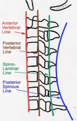
How can we assess for pseudosubluxation on pediatric c-spine x-rays? |
Most commonly C2-C3 Look at spinolaminar (Swischuk's) line drawn from anterior cortex of the C1 to C3 spinous process. If the line is >2mm anterior to the anterior cortex of C2 suspect a posterior element fracture. |
|
|
Differences in the pediatric versus adult airway |
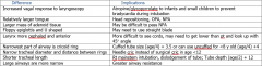
Also note that kids often desat shortly after intubation - be sure to provide PEEP to maintain their smaller than normal FRC |
|
|
Anatomic differences in pediatric patients that change response to trauma |
-Small size = more multitrauma -Less protective fat/muscles = more internal organ injuries (liver, spleen, kidneys) -Elastic chest wall = lung injury without # -Open growth plates = different fracture patterns -Large surface area = quicker hypothermia -Faster metabolic rate = quicker desat, hypoglycemia -Better at maintaining BP = tachy as only sign of shock -Bigger head-to-body, thin skull, less myelin = more head injuries -More elastic vertebral column = more SCIWORA -Bigger head = higher fulcrum = C2-3 versus C6 injuries more common |
|
|
Anatomic difference in pregnant patients that change response in trauma |
Airway - more friable and edematous mucosa, lower esophageal sphincter tone, increased abdominal girth
Respiratory - higher RR = greater minute ventilation and lower CO2; higher diaphragm = lower FRC (quicker desat) and req's higher chest tube
Cardiac - Increased blood volume, tachycardia, decreased PVR, increased venous congestion/pressures, lots of blood to uterus, aortocaval compression when supine
Heme - dilutional and Fe-deficiency anemia; hypercoaguable
Abdomen - displaced contents; decreased sensitivity of exam for peritonitis; ALP doubles; decreased GB contractility (increased gallstones; weight gain
Nephro - bladder is extrapulvic after 12 weeks; decreased GFR; polyuria and hydropnephrosis due to bladder compression
MSK - widened pubic symphesis (4 -> 8mm) |
|
|
How can we assess for atlanto-occipital dislocation on pediatric c-spine x-rays? |
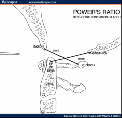
Use power's ratio (should be <1): Basion to anterior cortex of C1 spinous process Opisthion to posterior cortex of dens
Also Basion-Dens (BDI) & Basion to posterior axillary line (BAI should be <12mm
|
|
|
Tube sizes in pediatrics |
Broselow tape ETT = (age/4) + 4 (uncuffed - drop 0.5-1 size for a cuffed tube) Chest tube = ETT size x 4 Foley / NG tube = ETT size x 2 |
|
|
Anatomic difference in elderly patients that change response in trauma |
General - on medications Cardiac - decreased reserve, can't increase HR Pulmonary - decreased compliance and increased chest wall rigidity, brittle bones Neurologic - brain atrophy increases mobility and shearing of bridging veins (SDH); dura is fused so less EDH Derm - skin is thin and brittle, easier to lacerate and tear, forms ulcers quicker MSK - osteopenia so increased fractures, decreased joint mobility, spinal stenosis |
|
|
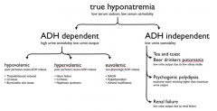
Approach to hyponatremia |
|
|
|
Effects of typical antipsychotics |
HOT DAMN
Fever Dopamine receptor blockade Alpha blockade Muscarinic blockade Na/K channel blockade (wide QRS and long QTc) |
|
|
Addictions that can kill in withdrawl |
ABBA Alpha blockers (clonidine) Benzo's Barbiturates Alcohol |
|
|
Nicotinic stimulation effects |
Monday - mydriasis Tuesday - tachycardia Wednesday - weakness tHursday - hypertension Friday - fasciculations Saturday - seizures
And the 3 C's Confusion Convulsions Coma |
|
|
Oxygen toxicity symptoms |
VENTIAC V ertigo E uphoria N ausea T innitus I mpaired judgement A LOC (Altered LOC) C onvulsions |
|
|
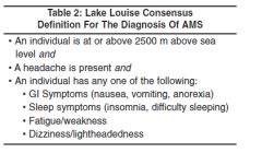
Lake Louise criteria for AMS |
|
|
|
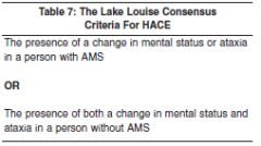
Lake Louise criteria for HACE |
|
|
|
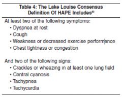
Lake Louise criteria for HAPE |
|
|
|
The TIMI (NSTEMI) Score |
2 or more episodes of angina in past 24h 7 days history of ASA use
C AD (known and >50%) A ge > 65 R isk factors (>3) T roponin S T changes
Gives 14 day risk of death, MI, or need for revascularization (0-1 = 5%; 6-7 = 40%) |
|
|
Predictors of difficult BVM |
B eard O bstructed / O bese / O SA N eck stiffness / N eck mass E xpecting (pregnant) S tridor / S nores |
|
|
Predictors of difficult intubation |
L ook externally E valuate 3-3-2 M allampati O bstruction / O besity N eck mobility (decreased) |
|
|
Predictors of difficult cric |
S urgery H ematoma / H ave infection (abscess) O besity R adiation T rauma / T umor |
|
|
Predictors of difficult LMA |
R estricted mouth opening O bstruction D istored airway anatomy S tiff lungs / Neck |
|
|
What are the lines of the cervical spine? |
|
|
|
How do you assess for pseudosubluxation in the pediatric C-spine? |
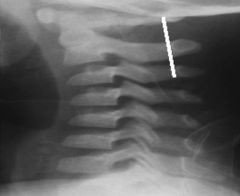
Swischuk's line (anterior arch of C1-C3 is within 2mm of C2)
|
|
|
Cervical spine fracture mechanisms |
All are flexion, except:
Vertical Compression - Burst & Jefferson
Extension - C1 neural arch, Hangman, Extension teardrop
Flexion-rotation - Unilateral facet, Rotary atlantoaxial |
|
|
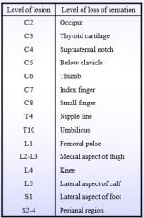
Sensory spinal levels |
|
|
|
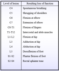
Motor spinal levels |
|
|
|
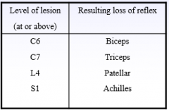
Reflex spinal levels |
|
|
|
Describe the motor deficit in central cord syndrome, causes |
It is MUDdy!
Motor > sensory Upper > lower Distal > proximal
Causes - hyperextension injury of neck, elderly with spinal stenosis |
|
|
Signs of aortic dissection on CXR |
Wide CHAPPLA1N
Wide mediastinum (8cm AP, 6cm PA, >25% chest width at aortic knob) C alcium sign H emothorax A ortic knob obscured P aratracheal stripe widened P leural cap L eft mainstem bronchus depressed A ortic window lost 1 st rib fracture N G deviates to the right along with trachea |
|
|
Occlusive and nonocclusive arterial injuries |
Occlusive -Transection -Thrombosis -Arterial spasm (reversible)
Nonocclusive -Intimal flap -Pseudoaneurysm -AVM -Compartment syndrome |
|
|
Diagnostic aid for a migraine |
Migraine is likely with two or more of the POUND criteria: -P ounding -hO urs lasts (4-72) -U nilateral -N ausea and vomiting -D ebilitating |
|
|
International Headache Society Migraine Definition (without aura) |
1 --> 4 to 72 hours
2 --> At least two of the following: -Aggravation by or causing avoidance of routine physical activity -Moderate or severe pain intensity -Pulsating quality -Unilateral location
3 --> During headache, at least one of the following: -Nausea and/or vomiting -Photophobia and phonophobia
4 --> Not attributed to another disorder
5 --> History of at least five attacks fulfilling above criteria |
|
|
Causes of pancreatitis |
I GET SMASHED I diopathic
G allstones E thanol T umors (pancreas, ampula, choledochal)
S corpion stings M icro - Bacterial (Mycoplasma, Camylobacter, TB), Viral (Mumps, Coxsackie, Rubella, Varicella, CMV, hepatitis, EBV), Parasites (ascaris, echinococcus) A utoimmune (SLE, PAN, Crohn's) S urgery / trauma H yperlipidemia / H ypercalcemia (hyperparathyroid) E mboli / ischemia D rugs / toxins (ethanol, azathioprine, lasix, HCTZ, estrogens, valproic acid, tegetrol, APAP, ASA, sulfonamides) |
|
|
Types/causes of diarrhea |
MMISO
M alabsorption (short gut, CF, IBD, celiac, lactose intolerant) M otility (DM, neuromuscular, scleroderma) I nflammatory - cellular damage causing secretion; can be hemorrhagic (enterohemorrhagic E Coli, Salmonella) or IBD, autoimmune, chemo S ecretory (Toxin-mediated chloride secretion: Enterotoxic E Coli, Shigella, Salmonella, Vibrio, C Diff; does not decrease with fasting) O smotic (altered gut flora from Noro or Rotavirus; ingestion of sorbitol or lactulose; decreases with fasting) |
|
|
Causes of occult irritability in children |
FAT SHIC
F racture A buse T esticular torsion
S urgical abdomen (hernia) H air tourniquet I mproper feeding C orneal abrasion / C olic
Also: diaper rash, anal fissure |
|
|
Dermatologic findings in pediatric seizures due to neurocutaneous disorders |
Cafe au lait - Neurofibromatosis Ash leave - Tuberous sclerosis Port au Wine Staine - Sturge-Weber |
|
|
Appearance assessment of the pediatric assessment triad |
TICLS Tone Interactivity Consolability Look/gaze Speech/cry |
|
|
Lateral soft tissue x-ray findings of epiglottitis |
AAA PBL on TV A ir fluid level A ryepiglottic fold swelling A rytenoid swelling
P revertebral tissue swelling B allooning of the hypopharynx L oss of L ordosis
T humbprint epiglottis V allecula obliteration |
|
|
Approach to the striderous child |
Supraglottic -Congenital (Micrognathia, Macroglossia, Choanal atresia) -Acquired (Retropharyngeal abscess, Epiglottitis) Glottic -Congenital (Laryngeal web, Vocal cord paralysis, Laryngeomalacia) -Acquired (Laryngeal papilloma) Subglottic -Congenital (Subglottic stenosis, Hemangioma) -Acquired (Croup, Subglottic stenosis) Tracheal -Congenital (Tracheomalacia, Tracheal stenosis, vascular ring) -Acquired (Bacterial tracheitis, Foreign body) |
|
|
Signs of retrobulbar hemorrhage and indications for lateral canthotomy |
DIP A CONE (DIP is primary indications; A CONE is secondary)
D ecreased VA I ncreased IOP (>40) P roptosis
A fferent pupillary defect
C herry red macula O pthalmoplegia N erve head pallor E ye pain |
|
|
Medical treatment of increased IOP |
ABCDPS A lpha 2 agonist (Apraclonidine 1% - decrease production and increase outflow) B eta blocker (Timolol 0.5% - decrease humor production) C holinergic (Pilocarpine 1% - constricts pupil and opens trabecular meshwork) D iuretics (acetazolamide decrease production and increase flow / mannitol - increased drainage) P rostaglandins (Latanoprost - increase outflow) S teroids (Prednisone acetate 1% - decrease inflammation) |
|
|
Indications for referral to opthalmology of an eyelid laceration |
The 5 L's
L id margin L acrimal system L evator or canthal tendons L oss of tissue L eaking of fat |
|
|
DDx for sudden visual loss |
Anatomic
Anterior chamber - hyphema, hypopiom, glaucoma Iris/lens - lens dislocation, iritis Posterior chamber - posterior vitreous detachment or hemorrhage Retina - Retinal detachment, central venous occlusion, central arterial occlusion Neuro-opthalmologic - pre-chiasm (optic neuritis due to ischemia/compression/toxin), chiasmal (tumor), post-chiasm (CVA, tumor, AVM, migraine), visual cortex (CVA) |
|
|
Kanavel signs of flexor tenosynovitis |
Fingers held in slight flexion Fusiform (symmetrical) swelling Pain to palpation of flexor tendon Pain on passive extension |
|
|
Hand motor testing |
Radial nerve - wrist extension (get wrist drop)
Posterior interosseous branch of the radial nerve - thumb extension (get Monkey hand)
Median nerve - thumb opposition to fingers
Anterior interosseous branch of the median nerve - OK sign (thumb IP flexion)
Ulnar nerve - Froment's paper sign; finger abduction and adduction (get Claw hand)
Axillary nerve - Shoulder abduction |
|
|
Hand sensation testing |
Radial nerve - dorsal 1st web space Median nerve - volar tip of D2 Ulnar nerve - volar tip of D5 Axillary nerve - deltoid distribution |
|
|
Back pain red flags |
Infectious - fever, IVDU Fracture - history of trauma Cancer - weight loss, history of cancer Cauda equina - urinary retention, fecal incontinence, saddle anesthesia, distal weakness
Nocturnal pain
|
|
|
Indications for lumbar spine x-rays |
M alignancy A ge (<18 or >50)
F ever I mmunocompromised N euro deficits (progressive) D uration (>4-6 weeks)
W eight loss I VDU T rauma |
|
|
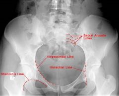
Lines of the pelvis x-ray |
|
|
|
One pill can kill |
Alpha blocker (clonidine) Antihyperglycemic agents (sulfonylureas like gliclazide, glyburide) Beta blockers Barbiturates Calcium channel blockers / Camphor / Chloroquine Digoxin
Hypoglycemic agents (sulfonylureas)
MAO-I Methadone Methyl salicylate
Theophylline TCA Toxic alcohol
Iron Lomotil |
|
|
Drugs that cause seizures |
WITH LA COPS W ithdrawal / Wellbutrin I NH T heophylline / TCA L ithium / L ocal anesthetics / L ead A nticholinergics C holinergics / C amphor O rganophosphates P CP S alicylates / Sympathomimetics |
|
|
Drugs MDAC is appropriate for |
Please Quit Drinking the AC Dummy Phenobarb Quinine Dapsone Theophylline / TCA (maybe) ASA (concretions) Digoxin (maybe) / Dilantin (maybe) / Dabigitran
Consider more in sustained release formulations |
|
|
Dialyzable drugs |
A BIT SLIME
A rsenic / ANTS (BB's Atenolol / Nadolol / Timolol / Sotalol)
B arbiturates I soniazid T heophylline
S alicylates L ithium I ctogenic drugs (Valproate, Phenobarb, Carbamezapine) M ethanol E thylene glycol / E thanol D abigitran |
|
|
Indications for reduction of a distal radius fracture |
Step >1mm Radial inclination <15 degrees (normal 22) Volar tilt less than 0 degrees (normal 10-25) Decreased radial height (normal 11mm, loss of 2mm relative to other side is short) |
|
|
Clavicle fracture's requiring orthopedic consultation |
The rule of 2's >2 cm displacement 2 or more pieces <2cm from either end of the clavicle >2cm of shortening 2 good 2 be true |
|
|
Shoulder dislocation techniques |
Stimson - prone, arm hanging with weight x 20 minutes Traction-countertraction - sheet under arm for countertraction, abducted arm FARES - supine, slow abduction with flexion/extension until 90 degrees then external rotation Milch - supine at 45 degrees, external rotation and abduction to 90/90 then longitudinal traction Scapular manipulation - can be added to traction/countertraction and Stimson, rotate inferior tip medially Cunningham - seated, shoulders adducted, elbow flexed with shoulder on provider shoulder, massage of bicep at mid-humeral level |
|
|
Open fracture classification and management |
Gustillo classification system I - <1cm, clean, tx with 1st gen cephalosporin II - >1cm, minimal soft tissue damage, tx with 1st gen cephalosporin IIIa - significant soft tissue damage with adequate coverage, 1st gen cephalosporin and aminoglycoside (gentamicin) IIIb - significant soft tissue damage with INadequate coverage, same tx as IIIa IIIc - open # with vascular injury , same tx as IIIa
Add Pen G or Clinda if concern for anaerobes (farm injury) and Cipro if concern for salt water (pseudomonas)
Irrigate, cover, splint without reduction UNLESS N-V compromise
|
|
|
Femoral nerve injury |
Motor - weak knee extension, can't climb stairs or get up from sitting Sensory - varies, most reliable superomedial to patella Reflex - decreased patellar |
|
|
Sciatic nerve injury |
Motor - paralysis of hamstring (knee flexion) and all muscles below the knee Sensory - posterior thigh and below the knee Reflex - decreased Achilles tendon |
|
|
Hip reduction techniques |
Allis - patient supine with hip and knee flexed to 90 degrees, get on bed and provide vertical upward traction while someone holds the pelvis to the bed. Works for posterior and anterior-obturator (femoral head seen over obturator foramen).
Stimson - patient prone with one leg hanging off of the bed, flex hip and knee to 90 degrees, vertical downward traction while someone holds the pelvis/pushes down on the femoral head.
Whistler - patient supine, arm under knee of dislocated hip with arm on opposite knee (both legs flexed at the hip/knee) to use opposite leg as a fulcrum. A modification of this is the Captain Morgan with your leg under the patient's knee instead of your hand. |
|
|
Kocher criteria to distinguish septic arthritis from transient synovitis |
With 0-4 criteria the likelihood is: 2%, 9.5%, 35%, 73%, 93% -Non weight bearing -ESR >40 -WBC >12 -Fever > 38.5 CRP >20 is also predictive |
|
|
Ottawa Knee Rule |
Get x-rays if: Age >55 Inability to transfer weight 4 times at time of injury OR in ED Inability to flex to 90 degrees Patellar tenderness Fibular head tenderness |
|
|
Ottawa Ankle Rule |
Applied to acute ankle injuries with malleolar pain (not hindfoot, forefoot, upper fibula) -Pain to posterior edge of the lateral malleolus from its distal part and 6cm proximal -Pain to the posterior edge of the medial malleolus from its distal part and 6cm proximal -Unable to weight bear 4 steps immediately after the injury and in the ED |
|
|
Ottawa Foot Rule |
-Pain over the navicular bone -Pain to the base of the 5th metatarsal -Unable to weight bear 4 steps immediately after the injury and in the ED |
|
|
How do you calculate Boehler's angle? |
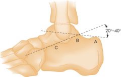
A = Posterior tuberosity B = Apex of posterior facet C = Apex of anterior process |
|
|
Bones at high risk of AVN |
-Head of femur (Legg-Calve-Perthes syndrome in children generally 4-10yo) -Head of humerus -Scaphoid -Capitate -Lunate (Kienbock's disease) -Patella -Talus -Navicular (Kohler's disease) -Second metatarsal |
|
|
DDx for non-accidental trauma in children (fractures and bruising) |
-Osteogenesis Imperfecta -Rickets -Scurvy -Menkes' Kinky Hair Syndrome -Hypervitaminosis A -Hypoparathyroidism -Congenital Syphilis -Pathologic fractures -Birth fractures
-Metaphyseal cupping & spurring (normal variant - bilateral, diaphyseal, smooth) -Periosteal new bone formation (normal variant - especially to the femur)
-Cultural practices (Cupping, Coining, Spooning) -Bleeding disorders (hemophilia, vWD, HSP) -Mongolian spots -Hemangioma -'Tattooing'
-ITP -HSP -Secondary syphilis |
|
|
Diagnostic criteria for staph toxic shock syndrome |
DR FrOH (NO culture needed) D esquamation of the skin (begins during recovery phase after 1-2 weeks; R ash (blanching, macular, erythematous, NOT itchy, fades before desquamation)
F ever (>38.9) r O rgan systems (>3/7 involved: CNS, mucous membranes, GI, renal, hepatic, heme, MSK) H ypotension (sBP < 90 or < 5th percentile in children) |
|
|
Diagnostic criteria for strep toxic shock syndrome |
You going to the strep SHO?
S erology (isolation from a sterile [definite] or nonsterile [non-definite] site) H ypotension (sBP<90 or <5th percentile) O rgan systems (>2/6 involved: Renal, Heme, Liver, Lung, Rash, Soft tissue necrosis) |
|
|
Determining capacity |
C ommunication U nderstanding R easoning V alues E mergency S urrogate |
|
|
When can implied consent be assumed |
-Patient does not have capacity to express their preferences (CURV) -Immediate action is required (E) -No surrogate decision maker (S) |
|
|
Signs of Lithium toxicity |
SNAP MUD S eizures N /V/D A taxia P arkinsonian
M yoclonus U MN D elirium/D ecreased LOC
Chronic: nephrogenic DI and hypothyroidism and SILENT (syndrome of irriversable lithium effectuated neuro toxicity - cerebellar dysfunction, EPS, dementia)
Look for LOW AG |
|
|
Infections requiring airborne precautions |
Respiratory TB Measles SARS +/- Ebola and TB (during aerosolizing procedures) |
|
|
AIDS-defining illnesses |
Heme -CD4<200
Malignancies -Kaposi's Sarcoma -Lymphoma -Cervical cancer (invasive)
Neuro -HIV-associated encephalopathy -Progressive multifocal leukoencephalopathy -Toxoplasmosis of brain
Fungal infection -Candida (esophageal or pulmonary) -Histoplasmosis -Cryptococcus -Coccidiomycosis
Protozoa infection -PJP pneumonia -Isosporiasis -Toxoplasma gondii -Cryptosporidium
Viral -HSV (persistent, pneumonia, esophagitis) -CMV (except spleen/liver/lymphatics)
Bacterial infection -Tuberculosis -Mycobacterium avium complex -Salmonella sepsis -Recurrent bacterial infections |
|
|
SIRS |
HR > 90 RR < 20 OR PaCO2 <32 T < 36 OR > 38 WBC <4 OR >12 OR >10% bands |
|
|
Definition of ARDS |
As per the 2012 Berlin Definition -Respiratory symptoms started or worsened acutely with the last week -PaCO2 / FiO2 ratio 200-300 = mild, 100-200 = moderate, <100 = severe -Bilateral pulmonary infiltrates (CXR or CT) -Not in cardiac failure / no fluid overload
|
|
|
Malaria: Organism, Vector, Incubation, Presentation, Complications, Diagnosis, Treatment |
-Organism: Plasmodium Falciparum is most dangerous (also Ovale, Vivax, Malariae) -Vector: Female anopheles mosquito -Incubation: 8-28 days -Presentation: Fever in the returning traveler, anemia, constitutional (weak, dizzy, N/V/D, lethargy, myalgia, arthralgia, CP, abd pain, SOB) -Complications: cerebral/seizures, encephalopathy, ARDS, ARI, DIC, anemia, acidosis, hypoglycemia -Diagnosis: Thin and thick peripheral blood smears q8-12h x 3d; also PCR/dark field microscopy; U/S shows splenomegaly/papilledema -Treatment: Chloroquine if sensitive; otherwise quinine & doxycycline |
|
|
Lyme disease: Organism, Vector, Incubation, Presentation, Diagnosis, Complications, Treatment |
-Organism: Borrelia Borgdorferi (spirochete) -Vector: Ixodes Tick -Incubation: Tick must attach long enough (>36 hours) or be engorged -Presentation: 1st stage (days-weeks) erythema migrans & flu-like symptoms/HA; 2nd stage (3-5 weeks) with fluctuating meningoencephalitis/bilat Bells palsy, conduction block/pericarditis, arthritis, keratoconjunctivitis; 3rd stage (>1y) with fatigue syndrome, encephalopathy, radiculopathy, acrodermatitis, and arthritis -Diagnosis: Tick bite, IgM+ from 3-6 weeks, IgG+ >1 month (send both) -Complications: Can get Jarisch-Herxheimer reaction when tx started -Treatment: prophylax within 72h in endemic areas (>20% ticks +) if adult tick on for >36h/engorged - use Doxy 200mg x1; treat with Doxy 200mg BID x 28d (Amoxil in <8/pregnant; Ceftriaxone if meningitis) |
|
|
Rocky Mountain Spotted Fever: Organism, Vector, Incubation, Presentation, Diagnosis, Complications, Treatment |
-Organism: Rickettsia Rickettsii -Vector: Rocky Mountain Wood Tick -Presentation: Sudden onset fever followed by N/V/abd pain/HA. Gets into vessels and releases tPA & vWF causing microthrombi and vascular permeability. Petechiae develop on wrists/hands then spread inward. Also cardiac (AVB, myocarditis), pulmonary (ARDS), neurologic (meningismis, transient deficits due to microinfarcts), renal (microinfarcts), heme (DIC). -Diagnosis: Serology not positive for 1/52 but req'd for conclusive Dx. Also PCR+ or skin bx at 4-10 days (immunoflorescence +). Probable if clinical criteria; Confirmed with lab. -Complications: Death due to renal failure then ARDS/myocarditis/DIC in 25% if not treated -Treatment: Doxycycline 100mg po bid until asymptomatic x 3d or 7 days. Steroids if severe. |
|
|
Things that shift the oxygen-hemoglobin dissociation curve |
CADETS turn right and fall down (right shift and decreased oxygen affinity) C - CO2 A - Acid D - 2,3 DPG E - Exercise T - Temperature S - Sickled Hb S
NOTE: CO shifts to the left |
|
|
Diagnostic criteria for delerium |
4 criteria: -Inability to focus/Inattention -Fluctuating course -Cognitive deficit (memory, disorientation, language) or perceptual disturbance not caused by dementia -Evidence that it is caused by a medical condition, ingestion, or withdrawl |
|
|
Diagnostic criteria for dementia |
1 - Memory impairment AND 2 - One of aphasia, apraxia, agnosia, impairment in executive functioning -Causing significant impairment -NOT due to delerium |
|
|
Treatment of active TB |
RIPE (side effects) x 9 months! R ifampin (orange body fluid) I soniazid (INH injures nerves and hepatocytes) P yrazinomide E thambutol (E=eyes - optic neuritis; can't distinguish red/green) |
|
|
Treatment for hyperkalemia |
C BIG K Drop
C alcium (stablize)
B eta agonist / B icarbonate (shift) I nsulin (shift) G lucose
K ayexalate (eliminate)
D iuretics - Furosemide (eliminate) R enal dialysis (eliminate) o p |
|
|
GBS: Cause, Presentation, Diagnosis, Complications, Treatment |
Cause: idiopathic, often secondary to Campylobacter, Mycoplasma, CMV or EBV - results in antibodies to nerves
Presentation: progressive ascending symmetric weakness and areflexia; Miller-Fischer variant starts centrally (areflexia, ataxia, opthalmoplegia with III/IV/VI affected). Also has autonomic dysfunction (tx brady with atropine; use short acting for hypertension, fluids for hypotension)
Diagnosis: CSF elevated protein, normal glucose and WBC
Complications: respiratory compromise req'ing intubation if FVC <20ml/kg or NIF <30mL/kg
Treatment: IVIg or plasmaphoresis |
|
|
Myasthenia Gravis: Cause, Presentation, Diagnosis, Complications, Treatment |
Cause: Antibodies to post-synaptic ACh receptors (take spots & destroy them); set off by BB/ CCB/ Thyroxine/ Steroids/ Surgery/ Trauma/ Infection
Presentation: Ptosis, Diplopia, Dysarthria, Dysphagia, Blurred vision with spared pupils, resp failure. Treated patients can present with cholinergic crisis.
Diagnosis: Tensilon test, ice to eyes, NIF (<15 intubate)/FVC (<15 intubate); check for anti-AChR antibodies
Treatment: Plasma exchange or IVIg (neostigmine and/or thymectomy for chronic); intubate with cisatracurium (Hoffman degradation) |
|
|
DDx of bulbar neuropathy |
-Myasthenia gravis -Lambert-Eaton myasthenic syndrome -ALS -Miller-Fisher variant GBS -Elapidae (coral snake) or Hydraphidae (sea snake) envenomation -Botulism -Lyme disease -Organophosphate poisoning -Congenital syndrome -Penicillamine toxicity |
|
|
Gram stain results of bacteria |
Staph: Gram+ cocci in singles, doubles, tetrads or clusters Strep: Gram+ paired diplococci (other strep in pairs/chains) Listeria: Gram+ rods single or chains Moraxella caterrhalis: Gram- diplococci Neisseria: Gram- paired diplococci H Flu: Gram- coccobacilli E Coli: Gram- rods Pseudomonas: Gram- rods |
|
|
Angina Classification |
Canadian Cardiovascular Society I - No limitation of ordinary activity II - Mild limitation. Symptoms at >1-2 blocks or >1 flight of stairs. III - Moderate limitation. Symptoms at <1-2 blocks or <1 flight of stairs. IV - Severe limitation. Symptoms at rest. |
|
|
Definition of stable angina |
Predictable Transient (<15m) Reproducible with activity Relieved with rest/nitro |
|
|
Definition of acute MI |
-Rise and fall of troponin with: ischemic symptoms, Q waves, ST/T changes, coronary artery intervention -Pathological evidence |
|
|
Types of myocardial infarction |
I - ischemia due to a primary coronary event (plaque rupture or dissection) II - supply-demand ischemia III - sudden cardiac death with symptoms of MI IV - MI with coronary instrumentation V - MI with CABG |
|
|
At risk for an atypical presentation of MI |
Aunt Jemima with dementia -Elderly -Diabetic (from all the syrup) -Non-white -Female -Dementia -Hyperlipidemia (from all the sausages)
Also: No prior history of MI, history of stroke, /CHF, no family history |
|
|
Liver transplant criteria in acetaminophen-induced and non-acetaminophen-induced fulminant hepatic failure (King's College criteria) |
Acetaminophen induced pH <7.3 or lactate >3 after 12h of resuscitation Lactate >3.5 after 4h of resuscitation
OR all 3 of: -Cr >300 -INR >6.5 -Grade 3-4 hepatic encephalopathy
Non-acetaminophen induced INR >6.5
OR 3/5 of: J aundice >1 week prior to encephalopathy A ge <10 or >40 N on-A non-B hepatitis E tiology: indeterminate or drug reaction B ilirubin >300mmol/L I NR >3.5 |
|
|
Sgarbossa Criteria |
In setting of LBBB, the criteria for calling AMI is >3 points: >1mm concordant STE (OR 25, 5 points) >1mm STD in v1, v2, v3 (OR 6, 3 points) >5mm discordant STE (OR 4.3, 2 points)
Also look at ST (baseline to T) / S (top of S to baseline) ratio <-0.25
|
|
|
Classification of AMI severity |
Killip classes 1 no failure 2 crackles, S3, elevated JVP 3 frank pulmonary edema 4 cardiogenic shock, hypotension, vasoconstriction (oligurea & cyanosis) |
|
|
What are the target times for ACS? |
Door Data (10m) Decision Drug (lytic 30m, PCI 90m in center) |
|
|
What is the Ashman phenomenom? |
-Seen in supraventricular tachyarrhythmias (generally AFib) -Long R-R interval (has long refractory period) followed by a short R-R interval results in part of the right bundle being refractory -Get a RBBB waveform that looks like a PVC |
|
|
Mechanisms for arrhythmias |
-Increased automaticity (ischemia, electrolytes, drugs) -Reentry (req's 2 conduction pathways with different responsiveness and conduction speed) -Triggered (early afterpolarizations in brady/long QTc; treat by increasing HR vs late afterpolarizations in tachy/increased Ca; treat by slowing HR and decreasing Ca) |
|
|
Antiarrhythmic types/actions |
Some Buggers Kill Cats
S odium channel blocker (a block fast, b block inactivated phase, c block both) - procainamide/TCA/cocaine, lidocaine/phenytoin, flecainide/dilantin B eta blocker - propranolol/esmolol K potassium channel blocker - amiodarone/sotalol C alcium channel blocker (slow) - verapamil/diltiazam |
|
|
How does Digoxin work? |
1 - Blocks Na/K ATPase leading to increased intracellular Ca++ (increased inotropy, tachyarrhythmias) 2 - Increases vagal tone (anti-arrhythmic, bradyarrhythmias) |
|
|
Non-compensatory pause vs compensatory pause |
Non-compensatory pause: sinus node is reset and beat following the aberrant beat occurs at the same R-R interval as it would have if it came after a regular beat.
Compensatory pause: sinus node is NOT reset. One sinus beat is not conducted (meets refractory AVN) and the next is. The next beat comes after exactly 2x the standard R-R interval. |
|
|
DDx for irregular SVT |
-AFib -MAT -Atrial flutter/tachy with variable conduction -Parasystole -Extrasystoles |
|
|
Contraindications to ED Cardioversion of AFib |
1 - Lasted > 48 hours 2 - Rheumatic heart disease 3 - Mechanical valve 4 - History of stroke/TIA |
|
|
Risk stratification for AFib - who needs anticoagulation? |
CHADS2 C HF H ypertension A ge > 75 (2 points) D iabetes S troke before (2 points) V ascular disease Age 65-74 Sec (female)
0 = nil; 1 = ASA or anticoagulant (1/3%/y); 2 = anticoagulant (2.2%/y) |
|
|
Strong predictors of VT in a rapid wide-complex tachycardia |
-AV Dissociation -Fusion beats -Capture beats -QRS >0.16 -R to nadir of S >0.14 -Extreme left axis -Josephson's sign (notching near the nadir of the S wave - a smaller R prime than R)
Brugada and Griffith criteria are too unreliable for use and likely cause harm |
|
|
Congenital vs Adult Torsades |
Congenital: precipitated by tachycardia, catacholamine excess, and delayed afterpolarization, treat with beta blockers, associated with Romano-Ward syndrome (LQT1 K channel and LQT3 Na channel) and Jervall & Lange Nielson (LQT1 K channel) syndrome
Adult: precipitated by bradycardia, early afterpolarization, treat with beta agonists, associated with drugs |
|
|
Drugs that prolong QT |
Antidysrhythmics Ia, Ic, III: procainamide, propafenone, amiodarone Antibiotics: azithromycin, ciprofloxacin Antipsychotics: haloperidol Antiemetics: ondansetron, metoclopramide Anticonvulsants: Antihistamines: Antifungals: Antimalarials: chloroquine Antidepressants: TCA, citalopram Analgesia: Methadone
Also, hypoCa, hypoMg, hypoK |
|
|
Effect and indications for use of a magnet on a pacemaker |
Changes a standard pacemaker to VOO mode and turns off defibrillation in an ICD/pacemaker
-Atrial tachycardia with rapid ventricular rate -Runaway pacemaker (re-entry tachycardia) -Bradycardia due to oversensing |
|
|
Causes of ICD malfunction |
Frequent shocks -Shocking SVT -Oversensing T waves -Having frequent VF/VT (hypoK, hypoMg, Ischemia, drug-induced)
Inadequate shocks (dizzy/syncope) -Undersensing VT -Shocks not strong enough -Inadequate backup pacing for brady
Cardiac arrest -Likely VF did not respond to defibrillation -May have not detected VF (change parameters) |
|
|
Anemia differential approach |
Decreased production -Lack of stimulation (renal disease, chronic disease) -Unfunctional marrow (infiltrative disease: amyloid, metastasis; marrow disorders: aplastic, myelofibrosis; blood cancers: lymphoma, leukemia; tox: heavy metals, clozapine) -Lack components (B12, Folate, Fe)
Increased destruction -Intravascular (mechanical: prosthetics and microangiopathic DIC/TTP; transfusion reaction: ABO, antibodies; defects: G6PD, sickling) -Extravascular (abnormal RBC: spherocytosis, thalassemia) |
|
|
Causes of sideroblastic anemia |
Impaired production of porphoryn; leads to anemia and excess Fe in RBC's (Fe ring in sideroblasts)
-Toxins: Lead, Alcohol & INH -Premalignant condition in elderly (often get AML) -Malignancy -RA -Pyridoxime deficiency |
|
|
Paroxysmal nocturnal hemglobinuria |
Definition - Stem cell defect with abnormal sensitivity of RBCs, neutrophils and platelets to complement Diagnosis - Get hemosiderinurea, low RBC/Plt/Neutrophils, chronic hemolysis -Luekocyte alkanine phosphatase levels are elevated -Complications: thrombosis of arteries and hepatic vein. Also MUST transfuse with WASHED RBC's or compliment on them will lead to lysis. |
|
|
Encapsulated bacteria |
Even SSome Nasty Killers Have Capsular Protection
E coli (some strains) S trep pneumoniae S almonella typhi N eisseria meningitidis K lebsiella pneumoniae H aemophilus influenzae C ryptococcus neoformans (yeast) P seudomonas aeruginosa
|
|
|
Equipment required for a neonatal resuscitation |
Be prepared for baby WOBLIES
W armer / polyethylene bag - all babies O xygen (blended) - for persistent hypoxia B ag and mask - if HR<100, gasping, apnea give 40-60 bpm with PPV L aryngoscope (0 or 1 McGill or Mac) and ETT (3.5) - for meconium suctioning, ineffective/prolonged BVM, chest compressions I ncubator for transport E pinephrine 0.01-0.03mg/kg / 0.1-0.3mL of 1:10,000 1:1,000 = 1mg/mL 1:10,000 = 0.1mg/mL S uction |
|
|
Does the baby need resuscitation? |
Term? Breathing or crying? Muscle tone?
If yes, no resuscitation needed |
|
|
Neonatal CPR |
CPR is indicated if the infant's HR is <60bpm despite 30s of adequate PPV.
Chest compression rate is 90/minute Breathing rate is 30/minute (q 3 chest compressions) Epinephrine is used if HR <60bpm after 30s of CPR (dose 0.1-0.3mL/kg of 1:10,000 epi IV) |
|
|
When should an infant not be resuscitated? |
-<24 weeks / SFH < umbilicus -<500g birth weight -Anencephaly -Known chromosomal abnormalities incompatible with life (trisomy 13 or 18) -Stop resuscitation at 10m if there has been no HR or respiratory effort |
|
|
Causes of ascending paralysis |
Goes BOTTOM VP
G BS
B uckthorn / B-virus (Herpes Simiae) O rganophosphate (extremity exposure) T ick paralysis T oxic neuropathies (DM, EtOH, B-vitamin deficiencies, Buckthorn) M etabolic (hyperkalemic periodic paralysis)
V iral (Rabies, CNS VZV/CMV, West Nile) P olio |
|
|
Causes of hemolytic anemia (low haptoglobin, high LDH) |
Intrinsic: -Enzymes (Pyruvate Kinase or G6PD) -Membrane (Spherocytosis, Elliptocytosis, PNH) -Heme (Thallasemia, Sickle Cell)
Extrinsic -Mechanical (Microangiopathic - DIC/TTP/HUS/Vasculitis/Preeclampsia) -Other (valves, march) -Immunologic --> Alloimmune (ABO IgM intravasc / Rh IgG extravasc) --> Autoimmune (Reticular neoplasms [CML, CLL, lymphoma, myeloma], Inflammatory (SLE/RA/PAN/UC), Drugs (quinine, quinidine, methyldopa, PCN/cephalosporins, sulfa), Infectious (CMV/EBV/Mycoplasma/Coxsackie/Hepatitis), Thyroid,
Environmental (hyperthermia, brown recluse bites, freshwater drowning, burns, snakes, malaria)
Abnormal sequestration (hypersplenism) |
|
|
Pentad of TTP |
CRAFTY
C NS changes (fluctuating seizures, paresthesias, altered LOC) R enal failure (ARI, hematuria, proteinuria) A nemia (microangiopathic hemolytic with schistocytes) F ever T hrombocytopenia (Plts 10-50) |
|
|
Erythema nodosum causes |
BELTY SLIPS B ehcets E strogen L ofgran's T B Y = V iral (#2)
S trep (#1) L ymphoma (NHL) and Leukemia I BD P CN S ulpha |
|
|
Define and give a DDx for ALTE. How many kids with ALTE have SIDS? |
ALTE is an acute, unexpected change in an infant's breathing (apnea, choking, or gagging), appearance (color change), or behavior (loss of muscle tone) that frightens the observer. Prevalence peaks at 10-12 weeks but can occur in children <1yo.
ALTE's not correlated with SIDS!
-Neuro - Seizures/Hydrocephalus -Cardiac - Arrhythmia, Congenital heart disease -Respiratory tract infection (Pertussis, RSV) -GI - GERD (Sandifer syndrome) -Metabolic - Hypoglycemia, inborn errors of metabolism, hyponatremia -Sepsis - pneumonia, UTI -Heme - anemia -NAT -Factitious illness -Toxins |
|
|
HUS vs TTP vs DIC |
HUS -Caused by Shiga toxin of O157:H7 -Renal symptoms predominate -Consumptive (elevated DDimer decreased haptoglobin but normal LDH) -Children with bloody diarrhea -Plasmapheresis ineffective
TTP -Caused by lack of ADAMTS13 (? autoimmune) not cleaving vWF precursor -Neuro symptoms predominate -Adults -Non-consumptive (normal DDimer/Haptoglobin/fibrinogen but elevated LDH) -Schistocytes -Treat with plasmaphoresis or plasma exchange
DIC -Consumptive: low fibrinogin and fibrin levels; high DDimer -Bleeding and clotting at the same time; ultimately bleed when factors gone -Schistocytes, anemia, thrombocytopenia -Caused by multiple underlying disorders |
|
|
Treatment options in patients with vWD |
1 - Tranexamic acid or Aminocaproic acid (plasmin inhibitors - 5g po/iv) 2 - DDAVP (releases vWF and F8 from endothelium - 0.3mcg/kg SC/IV or 1.5mg nasal spray x 2) 3 - Humate-P F8 concentrate (need to ensure it has enough vWF) 4 - Cryoprecipitate (not recommended due to potential for viral transmission) |
|
|
Describe how factors should be replaced in Hemophilia A and B |
Generally use F8 & F9 concentrate, respectively. Give 0.5IU/kg/% activity needed for F8 repeat q12h prn Give 1IU/kg/% activity needed for F9 repeat q24h prn
Mild: Laceration, epistaxis, early hemarthrosis, hematuria - want 5-10% - empiric 12.5U F8 Moderate: Traumatic epistaxis/MM laceration, soft tissue/muscle hematoma, latehemarthrosis, hematuria - want 20-30% - empiric 25U F8 Severe: GIB, neck/sublingual bleeding, RP or intra-abdominal bleed, HI, majortrauma, CNS bleed, sx procedure - want >50% - empiric 50U F8 |
|
|
Bleeding reversal agents for Aspirin, Clopidogrel, Ticegralor, Warfarin, UFH, LMWH, Dabigatran, Rivaroxaban, Apixaban, t-PA/lytic |
Aspirin: DDAVP for minor, platelets for major
Clopidogrel/Ticegralor: DDAVP for minor, platelets for major
Warfarin: Depends. Hold if not bleeding. Hold + vit K po if have time. Hold + vit K IV + FFP (15mL/kg or 2-4U) OR PCC 50IU/kg
UFH: Protamine sulfate 1mg per 100U
LMWH: Protamine sulfate 1mg per 1mg
Dabigatran: PCC 50IU/kg, try FEIBA, vitamin K, Tranexamic acid (1g IV), dialysis (only 33% protein bound); send TT (thrombin time to confirm cause)
Rivaroxaban/Apixaban: PCC 50IU/kg; try tranexamic acid (1g IV), NO dialysis; send anti-Xa level to confirm cause
t-PA/thrombolytic: FFP 2U q6h x 4; Cryoprecipitate x 10U; Tranexamic Acid 1g; Platelets 1 adult; DDAVP 0.3mcg/kg IV; Protamine to reverse any heparin; treat ICP; be prepared to treat seizures |
|
|
Causes of heart failure |
HEART FAILED H ypertension E ndocarditis / E nvironment (heat wave A nemia R heumatic heart disease T hyrotoxicosis
F ailure to take meds A rrhythmia I nfection / I schemia / I nfarction L ung (COPD, PE, Pneumonia) E ndocrine (Pheochromocytoma / Hyperaldosteronism) D ietary indiscretions (salt / fluid) |
|
|
Heart failure classes |
NYHA Functional classes for CHF I - Asymptomatic with ordinary physical activity II - Symptomatic with ordinary physical activity III - Symptomatic with less than ordinary physical activity IV - Symptomatic at rest |
|
|
Organisms responsible for endocarditis |
Staph aureus (especially in right sided / IVDU) Strep viridans Strep bovis (association with GI malignancies) Enterococcus (add vanco and watch for resistance)
HACEK - haemophilus atrophilus, actinobacilus, cardiobacterium hominus, eikenella corrdons, kingella kingae (often chronic IE, hard to culture)
Immunocompromised fungal - Candida/Aspirgillus |
|
|
ECG changes of pericarditis and how are they different than MI |
1 - PR depression and diffuse STE (hours to days) 2 - Normalization of ST segments and flattening of T waves 3 - Deep, symmetrical T wave inversion 4 - ECG reverts to normal (sometimes T waves remain inverted)
Different than MI: non-anatomic pattern, concave up, no Q waves, no dynamic worsening |
|
|
Distinguishing murmur of AS vs HOCM |
AS is more likely to have insufficiency on top of other findings.
Valsalva (increased intrathoracic pressure decreases pre and afterload) - HCM louder and AS quieter
Squat (increased SVR increases pre and afterload) - HCM quieter and AS louder |
|
|
Prognostic factors for pancreatitis |
Ranson criteria (on admission) - mortality for 1-2 = 1%; 3-4 = 15%; 5 = 50%
Non-gallstone / Gallstone A GALL A ge >55yo / >70yo
G lucose >11 / >12 A ST >250 / >250 L DH >350 / >400 L eukocytes >16 / >18
BISAP score
Urea > 8.92 Impaired mental status >2 SIRS criteria Age >60 Pleural effusion |
|
|
Transfer to a burn center |
ABA -Partial thickness burn 10% BSA (2nd degree) -Any 3rd degree burn -Burns to face, hands, feet, genitalia, perinium, joints -Electrical (including lightning)/ Chemical/ Inhalational burn -Pre-existing medical conditions that complicate management -Children at a location that can not care for children -Cocomitent burn and trauma where the burn is the greatest danger -Burn injury in patients requiring social, emotional, rehabilitative intervention |
|
|
Dive injuries |
On descent -Ear barotrauma (inner, middle, external) -Mask squeeze (facial barotrauma) -Sinus barotrauma
At depth -Oxygen toxicity -Contaminated gases -Hypothermia -Nitrogen narcosis
On ascent -Alternobaric vertigo -AGE -Pneumothorax/ Pneumomediastinum/ Alveolar hemorrhage -GI barotrauma -Barodontalgia |
|
|
Dive injuries requiring a recompression chamber |
-AGE -DCS I and II -Contaminated gases (CO) |
|
|
Arterial embolism vs thrombosis |
Embolism -Source of emboli -Sharp demarcation (no collaterals)
Thrombosis -History of claudication -Contralateral findings of partial occlusion -Diffuse atherosclerosis (lots of collaterals) |
|
|
Causes of CVL obstruction |
Complete -Clots -Precipitant -Mechanical obstruction
Withdrawl -Against vessel wall -Vein thrombosis -Fibrin sheath -Ball-valve thrombus
Intermittent -Pinching between clavicle and 1st rib |
|
|
Well's DVT Criteria |
DImPLES and the 3 C's (-2 points if an alternative diagnosis is as likely) - Likely if 2 or more
D VT previously I mmobilization (paralysis, plaster) P ain (along deep venous system) L eg swelling (entire leg) P itting edema (to only the affected leg) S urgery (last 3m)
C ancer (palliative or treated in past 6m) C alf swelling (>3cm circumference difference C ollateral veins (visible and nonvaricose) |
|
|
Well's PE Criteria |
Likely if 4 or more
D VT signs and symptoms A lternative less likely M alignancy P revious P E/DVT H emoptysis I mmobilization hR > 100 |
|
|
Contraindications for fibrinolytic in STEMI and PE |
1-Dissection?
Stroke 2-Prior ICH? 3-Ischemic stroke in last 3m?
Bleed 4-Known vascular lesion? (AVM) 5-Known intracelebral neoplasm? 6-Significant head/facial trauma in last 3m?
Can't Clot 7-Active bleeding 8-Bleeding diatheses? |
|
|
Gas laws (Pascal, Boyle, Charles, Dalton, Henry) |
Pascal - Pressure on a fluid is transmitted equally throughout Boyle - P1V2 = P2V2 Charles - V1/T1 = V2/T2 Dalton - Pt = P1 + P2 + P3 ... Henry - The amount of gas dissolved in a liquid (solubility) is proportional to the partial pressure of that gas above the liquid |
|
|
Reasons to modify the dose of adenosine |
-Patient weight (obese, pediatrics), need more or less -Heart transplant (don't use it) -Methylxanthines (theophylline) stimulates receptors, need more -Carbamezapine, needs less -Dipyradamole prevents breakdown, needs less -CVL delivery, need less |
|
|
Causes of priapism |
Penile trauma (high flow) Medical conditions -Sickle cell -Leukemia -Spinal cord injury -G6PD deficiency -Thalassemia
Medications -ED - papaverine and PGE-1 -Phosphodiesterase inhibitors - sildenafil -Antipsychotics - chlorpromazine, clozapine -Antidepressants - SSRI's - trazodone -HTN - HCTZ -Mood/convulsant - Valproic acid -Recreational - alcohol, cocaine, amphetamines, heroin |
|
|
Toxic levels: ASA, APAP, Iron, Digoxin, Lithium, Lead, Methanol, Ethylene Glycol, TCA, Theophylline |
ASA Dose: 200mg/kg dose Levels (rule of 7's): Therapeutic 0.7-2.1; signs/symptoms >2.8; bicarb >3.5; dialysis chronic >3.5mmoL/L and Acute >7mmol/L
APAP Dose: 200mg/kg/24h dose; >150mg/kg/d for 48h; >100mg/kg/d for 72h Level: >1000mmol/L
Iron Dose: 20-40mg/kg (mild); 40-60mg/kg (mod); >60mg/kg (severe) Level: >90mmol/L
Digoxin Dose: 0.1mg/kg Level: >19mmol/L acute; >12mmol/L chronic
Lithium Level: >4mmoL/L acute; >2.5mmoL/L chronic
Lead Acute level: >3.4 (IV chelation) Chronic level: >2.2 (PO chelation)
Methanol Dose: 0.15mL/kg of 100% Level: >6mmol/L toxic; >15mmol/L HD
Ethylene Glycol Dose: 0.2mL/kg of 100% Level: >3mmol/L toxic; >8mmol/L HD
TCA Dose: >5mg/kg
Theophylline Level: >100mcg/mL acute; >60mcg/mL chronic HD |
|
|
Contrast Dilated, Hypertrophic, Restrictive, Takotsubo, Peripartum Cardiomyopathies (cause, treatment) |
Dilated: Mostly idiopathic but caused by ethanol, smoking, HTN, pregnancy, infection (myocarditis). Treated with pre and afterload reduction (ACEi, diuretics, PPV)
Hypertrophic: Caused by HOCM, AS, CAD, HTN. Treated with afterload reduction (BB). Must maintain preload!
Restrictive: Caused by amyloidosis, sarcoidosis, hemochromatosis, scleroderma, radiation, glycoven-storage diseases (Fabry/Gaucher). Treat underlying cause. Optimize preload (fluids).
Takotsubo: Caused by ? stress hormones. Treat as MI (indistinguishable from anterior STEMI) then BB and ACEi until recovery.
Peripartum: Caused by pregnancy (3 months before delivery to 6 months after). Treat afterload (hydralazine/labetolol until delivery, ACEi/BB after), preload (nitro), and contractility (digoxin) until recovery. |
|
|
Arteriosclerosis obliterans vs Thromboangiitis obliterans |
Arteriosclerosis: blue toe syndrome, claudication, ischemic rest pain in an elderly (>50) vasculopath (DM, smoker, HTN, cholesterol). Requires intervention if they have pain at rest. Can have distal ulcers.
Thromboangiitis: aka Buerger's disease, get painful erythematous nodules and decreased pulse in peripheral arteries. Only most commonly in male smokers 20-40yo and cure is stopping smoking completely. |
|
|
Distinguish vasogenic skin ulcers |
Arterial - distal to ankle, shiny, hairless, unswollen skin and thick nails. Less painful when dependent.
Venous - proximal to ankle, ++ swelling, weaping. Less painful when elevated.
Neurotrophic - sites of repeated trauma that they don't feel. Heels, toes, plantar surface. Not painful.
Hypertensive - on lateral malleolus, hemorrhagic bleb becomes an ulcer. Very painful. |
|
|
Vascular complications of IV drug use |
AV fistula and pseudoaneurysms (from 'hitting pink')
Unilateral hand edema (obliteration of superficial venous circulation)
Distal ischemia (severe burning pain distal to injections; possibly FB, talc, precipitate - nothing works to fix it; can need amputation)
Infected pseudoaneurysm (infected mass after hitting artery, reason that we assess abcesses for pulsatility) |
|
|
Extensive ileofemoral DVT: Names and diagnosis |
Phlegmasia Cerulia Dolens - swollen, congested, painful, cyanotic leg due to iliofemoral occlusion. Treat with thrombectomy.
Phlegmasia Alba Dolens - painful white leg secondary to arterial spasm that results from iliofemoral occlusion. Looks like arterial occlusion. Worse then cerulia. Treat with thrombectomy. |
|
|
APGAR Score |
A ppearance (pink, acrocyanosis, cyanosis) P ulse (>100, <100, absent) G rimace (sneeze/cough/pull away, grimace, no response) A ctivity (active, arms/legs flexed, limp) R espirations (good crying, weak cry, absent) |
|
|
Diagnosis and management of oncologic emergencies: febrile neutropenia, SVC syndrome, Tumor lysis syndrome, Hyperviscosity syndrome, Hypercalcemia |
Febrile neutropenia: Oral Temp >38.3 (x1) or 38.0 (x1h) with ANC<1 or expected <0.5 (biggest drop 5-10 days post chemo). NO rectal temps. Treat with Tazocin x 14d if stable + vanco/gent if not stable.
SVC syndrome: Present with periorbital edema, plethora, facial swelling, arm swelling, dyspnea. Diagnose with CT. Treat with radiation/chemo or stent (stent best).
Tumor lysis syndrome: See hyperkalemia, hyperphosphatemia, hyperuricemia, hypocalcemia. Treat with IVF +/- urinary alkalinization if acidic +/- dialysis. Can also try rasburicase with consultations. Allopurinol can prevent but not treat.
Hyperviscosity syndrome: Lab can't run tests. Happens with MM, Waldenstrom's Macroglobulinemia, Leukemia. Present with CNS/vision changes. Treat with exchange transfusion, plasma/leukopheresis.
Hypercalcemia: Due to mets or parthyroid-like hormone. Treat with hydration, furosemide, bisphosphonates, calcitonin. |
|
|
Indications for dialysis in tumor lysis syndrome and risk factors |
Indications for dialysis -Phosphate >3.2 -Potassium >6 -Uric acid >590 -Creatinine >880 -Volume overload -Symptomatic hypocalcemia Risk factors -LDH > 1500 -Advanced disease -Preexisting renal dysfunction -Acidic or concentrated urine -Preexisting volume depletion -Youth |
|
|
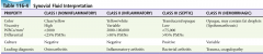
Synovial fluid interpretation (Color, Viscosity, WBC/mm3, Differential, Culture) |
|
|
|
Special tests for the shoulder (Jobe, Drop arm, Neer's, Hawkin's, Painful arc, Lift off, Lift off lag, Yergason's, Speed's) |
Supraspinatus Jobe's: 90 degrees abd, 30 degrees anterior to coronal plane, internally rotated/pronated - weakness or pain = supraspinatus involvement. Drop arm test: passive abduction to 90 degrees. If can't be maintained, possible large supraspinatus tear.
Supraspinatus/Impingement Neer's: Hand stabilizing scapula, passive flexion to 180 degrees. Pain towards 180 degrees indicates impingement. Hawkin's: imagine a hawk being held on an arm (90-90 flexion at shoulder/elbow) then internally rotate and see if there is pain. Indicates impingement.
Subacromial bursitis Painful arc: Abduction with pain from 70-100 degrees indicates subacromial bursitis.
Subscapularis Lift off test: assess for tear by putting internally rotated hand on back, holding elbow, and getting patient to lift off. Lift off lag: assess for rupture by doing same but passively lifting off and seeing if patient can maintain.
Biceps Yergason's sign: Flex elbow to 90 and have patient try to supinate against resistance. Pain is positive. Speed's test: Extend elbow and supinate forearm. Flex shoulder against resistance. Pain is positive.
|
|
|
Types of hypersensitivity reactions |
ACID I A naphylaxis - IgE-mediated degranulation of mast cells and basophils II C ytotoxic - IgG mediated complex fixation III I mmune complex - IgG or IgM antigen-antibody complex deposition IV D elayed - T cell mediated |
|
|
Causes of cavitating lesions |
CAVITY C ancer (metastasis) A utoimmune (Wegener's granulomatosis, Rheumatoid Arthritis, Sarcoidosis) V ascular (PE, septic emboli, infarction) I nfection (TB, MRSA, SA, Klebsiella, Fungal) T rauma (pneumatocele) Y outh (congenital things; bronchogenic cyst)
|
|
|
Treatment of common Tinea (capitis/barbae, kereon, versicolour, unguinum, pedis, other) |
Tinea capitis/barbae: Itraconazole 250mg po od x 4/52; Selenium Sulphide shampoo 2x weekly
Kerion: As per tinea, plus Keflex 500mg po qid (if infected) and Prednisone 1mg/kg/d x 1/52
Tinea versicolour (Malassezia Furfur): Selenium Suphide shampoo (q monthly for prophylaxis) +/- Fluconazole 400mg po x 1
Tinea unguinum: Penlac (antifungal painted on nail) trial; Ketoconazole 200mg po od x 6 months +/- surgical nail removal
Tinea pedis: Clotrimazole 1% bid x 6 weeks
Tinea (other areas): Clotrimazole 1% bid x 3 weeks |
|
|
Treatment of candidiasis (Thrush, Cutaneous, Vulvovaginal) |
Thrush: Adults Nystatin (100,000U/kg) swish and spit 5mL po qid until resolved x 1/52. Infants the same but 'paint the mouth' qid x 7 days. Fluconazole if immunocompromised.
Cutaneous: Dry regularly, zinc oxide prn, 1% hydrocortisone prn, Nystatin (100,000U/kg) cream bid-qid OR Clotrimazole 1% qid x 6/52. Can also use Fluconazole 100mg od x 2/52.
Vulvovaginal: Clotrimazole intravaginal OTC. Can also use Fluconazole 150mg po x 1. |
|
|
Indications for emergent decompression of a subdural hematoma |
-Midline shift >5mm ->1cm thick -GCS decreased by 2 or more since the time of the injury -Fixed dilated pupils -ICP >20mmHg |
|
|
Define SIDS, apnea, pathological apnea, apnea of infancy, apnea of prematurity, periodic breathing |
SIDS: sudden infant death in a child without historical, physical, laboratory, or postmortem findings that explain the death. Peaks at 3-5 months (90% <6 months)
Apnea: cessation of air flow (central, obstructive, mixed)
Pathologic apnea: apnea lasting >20s with bradycardia, cyanosis, hypotonia
Apnea of infancy: pathologic apnea with no identifiable cause
Apnea of prematurity: pathologic apnea associated with pre-term delivery (generally resolves by 37 weeks)
Periodic breathing: breathing pattern with 3 or more pauses each lasting >3s with 20s of normal breathing surrounding them. |
|
|
Gout vs Pseudogout (crystals, risks, treatment) |
Gout -Negatively birefringent needle urate crystals -Risks: obesity, DM, HTN, diuretics, alcohol, meat, seafood, beer, legumes -Treat: allopurinal (production), probenacid (excretion) chronically; colchicine 1.2/0.6/0.6, NSAIDS, steroids acutely
Pseudogout -Positively birefringent rhomboid calcium pyrophosphate crystals -Risks: hyperparathyroid, hypothyroid, hypoMg, hypoPO4, Wilson's, Hemochromatosis -Treat: As for gout except steroids > NSAIDs/Colchicine; also treat underlying cause but does not affect course. |
|
|
What is Still's disease? Treatment? |
Multisystem inflammatory disorder characterized by fever, arthritis, sore throat, myalgias, pericarditis, hepatitis, splenomegaly, and salmon colored rash that occurs ONLY with the fever.
Treat with NSAIDs, Steroids, IVIg |
|
|
What are the seronegative spondyloarthropathies? |
-They are RF NEGATIVE and HLA B27 POSITIVE . Generally involve the axial skeleton (not extremities)
PAIRS - P soriatic arthritis (affects smaller joints, sausage fingers and psoriasis) A nkylising spondylitis (males, back pain, sacroiliitis, bamboo spine) I nflammatory bowel disease R eiters syndrome / reactive arthritis (post GU chlamydia or GI shigella, salmonella, campylobacter, yersinia infection)
|
|
|
Medial and lateral epicondylitis |
Medial - Pitcher's/Golfer's Elbow -Flex wrist then try to pronate against resistance - pain to medial epicondyle
Lateral - Tennis -Extend and supinate wrist then try to flex against resistance - pain to the lateral epicondyle (Cozun test)
Both - treat with rest, RICE, PT |
|
|
Criteria for the diagnosis of lupus |
Require 4/11 ANA is quite sensitive (good rule-out); anti-DS-DNA & anti-Sm are quite specific (good rule-in)
-Malar rash -Discoid rash -Oral ulcers -Photosensitivity
-Nonerosive polyarthritis
-Serositis (pericardial or pleural effusion) -Renal disorder (nephrotic or nephritic) -Neurologic disorder (seizures or psychosis nos) Hematologic disorder (low Hb, WBC, platelets)
-Immunologic disorder (anti-dsdna, anti-sm, LAC, anticardiolpin, false + syphilis serology) -Positive ANA |
|
|
Drugs that cause drug-induced lupus |
Cardiac: procainamide, amiodarone HTN: hydralazine, methyldopa Antimalarial: quinidine Antimicrobial: nitrofurantoin, penicillin, INH, sulfonamides, tetracycline Anticonvulsant: phenytoin Antithyroid: PTU Antipsychotic: lithium, chlorpromazine Gout: allopurinol |
|
|
Diagnostic criteria for giant cell arteritis |
If you have 2 treat and get biopsy, if you have 3 just treat.
1 - >50yo 2 - new onset localized headache 3 - ESR >50 4 - abnormal biopsy with mononuclear infiltration or granulomatous inflammation
Also presents with visual changes, palpable temporal artery, jaw claudication, headache |
|
|
What is serum sickness (pathophys, cause, presentation, treatment)? |
-Type III hypersensitivity response with immune-complex complement fixation in vessel walls. -Associated with penicillin, sulpha, NSAIDs, Dilantin, Procainamide -Get erythema to fingers/toes, then urticaria, lymphadenopathy, arthralgias, constitutional symptoms 7-21 days after exposure. -Give steroids if severe |
|
|
DDx for target lesions |
Pityriasis rosea (herald patch with salmon colored central clearing) Tinea corporis (very well defined) Erythema multiforme (dark center, clearing, dark halo) Urticaria (raised, migratory) Erythema marginatum (dark center, clearing, dark halo similar to multiforme but there is only 1 and it is much bigger) Secondary syphilis
|
|
|
MRSA risk factors |
J ail
S ports H omeless / H ealthcare I VDU R esidence C rowded |
|
|
Antibiotics effective against MRSA |
Clindamycin Septra Doxycycline
Vancomycin Linezolid Cefepime Ceftobiprole
Daptomycin Tigecycline |
|
|
Treatment of pediculitis (lice) and scabies |
Both -Simultaneously treat the patient, sexual partners, family members, clothing, furniture and homes -Clothes should be washed in hot water and dried in a hot dryer. Other things can be frozen for 5 days.
Lice (phthiraptera - pediculosis -Permethrin (Nix) 1% shampoo for 10m on day 1 and 8 while avoiding conditioner for 2 weeks
Scabies (sarcoptes scabiei mite) -Permethrin 5% cream applied for 8-14h on day 1 and 8 |
|
|
Syphilis (Stages, Diagnosis, Treatment) |
Stages -Primary - painless chancre (papule -> 1cm ulcer) with painless lymphadenopathy -Secondary - 6 weeks to 6 months post-exposure, symmetrical non-pruritic macular/papular rash to palms and soles, can have condyloma lata around genitals, fatigue, lymphadenopathy, exanthem, myalgia, pharyngitis -Latent - Nil -Tertiary - gummas, granulomatous ulcerative lesions on skin, liver, bones, brain; Argyll-Robertson pupil; Tabes Dorsalis; Thoracic aortic aneurysm
Treatment -VDRL is positive after primary syphilis. Used for screening (false positives in SLE, thyroiditis, lymphoma, post-vaccine, mycoplasma, mono, hepatitis, measles, malaria, pregnancy) -FTA used for diagnosis (flouresence treponomal antibody test)
Treatment -Primary & secondary: Benzathine penicillin 2.4 million U IM; VDRL goes non-reactive after ~12 months -Latent or tertiary: treat weekly x 3 -Watch for Jerisch Herscheimer reaction |
|
|
Pemphigus vulgaris vs bullous pemphigoid |
PV -Autoimmune reaction affecting patients 50-60yo generally on penicillamine, captopril or rifampim -Present with oral bullae -> painful ulcers then skin bullae -> painful ulcers -Diagnose with history, + Nikolski sign, + Tzank smear -Treat with high dose prednisone (100-300mg/d), immunosuppresants, plasmapheresis
BP -IgG autoimmune reaction of those ~65yo. Less sick than PV. -Present with tense, fluid-filled blisters and a negative Nikolski sign. Mucous membrane involvement is possible but less frequent than PV. -Treat with prednisone and other immunosuppresants for 2-5 years and it generally resolves. |
|
|
Canadian C-Spine Rule |
|
|
|
Indications for dialysis in renal failure |
AEIOU
A cidosis / A lkalosis (note: HCO3 can precipitate tetany/convulsions in the setting of hypoCa) E lectrolyte abnormalities (HyperK, HyperMg, HyperCa - MM) I ngestions that are dialyzable O verload of fluid (CHF, pulmonary edema, severe HTN) U remia (pericarditis, N/V, lethargy) |
|
|
Bacteria causing UTI's |
KEEPPSSS
K lebsiella (institutionalized, newborns) E nterococcus (institutionalized) E coli (>80% of UTI's) P roteus (3-11yo) P seudomonas S taphylococcus saprophyticus (can be normal skin flora in perineum) S erratia S almonella |
|
|
UTI treatment length |
Uncomplicated lower tract - 3 days with nitrofurantoin, cefixime, cipro, septra
Complicated lower tract - 7 days (diabetes, sickle cell, immunocompromised) with cefixime, cipro, septra
Pregnancy lower tract - 10 days with cefixime or nitrofurantoin (avoid near term due to hemolytic anemia) or septra (avoid near term due to hemolytic anemia, jaundice, kernicterus)
Upper tract - 10-14 days with cefixime or ceftriaxone |
|
|
Types of kidney stones and causes |
Calcium oxalate 75% - excess calcium (milk alkali syndrome, high dietary intake, antacids, increased PTH). Oxalate increases in radiation enteritis, IBD, and ethylene glycol ingestion.
Struvite 15% - infection with urea-splitting organisms (pseudomonas, proteus, klebsiella, staph)
Hyperuricemia 10% - gout, tumor lysis syndrome, hematologic malignancies. they are radioluscent.
Cysteine 1% - inborn error of metabolism
Struvite, urate, and cysteine stones can form staghorn calculi. |
|
|
5 locations of urinary obstruction |
Renal calyx UPJ (uretopelvic junction) Pelvic brim UVJ (uretovesicular junction) Vesicular orifice |
|
|
Acute scrotal pain / swelling differential and physical exam features |
Pain -Testicular torsion - negative cremasteric reflex -Torsion of the testicular appendage (appendix testis or appendix epididymis) - blue dot sign -Epididymitis - Prehn's sign -Trauma -Orchitis -Testicular tumor WITH hemorrhage (normally tumor is not painful) -Inguinal hernia (if incarcerated/strangulated)
Swelling -Varicocele (bag of worms) -Ideopathic scrotal edema -Hydrocele
|
|
|
Causes of varicocele |
Venous varicocities of spermatic veins (bag of worms)
-Right spermatic vein -> IVC - generally caused by IVC compression or thrombosis -Left spermatic vein -> left renal vein - generally caused by RCC |
|
|
Approach to priapism |
Determine low (painful) or high (not painful) flow
Treatment of low flow ->4h duration requires treatment -Noninvasive tx - walk up stairs (decrease flow to penis), ice packs, compress -PO treatment - terbutaline 5-10mg PO (beta agonist) -Analgesia with dorsal nerve block -Aspiration of cavernosum -Injection of alpha agonist (phenylephrine) or methylene blue -Sicklers get O2 and hydration as well
Treatment of high flow -Angiography, surgical shunt, if painful can do block |
|
|
Causes of false positive hematuria |
Myoglobin Porphyria Bilirubinuria Munchausen's Menstrual blood
Meds -Nitrofurantoin -Dilantin -Rifampin -Quinine
Foods -Food coloring -Beets -Rhubarb -Berries |
|
|
Nephrotic vs Nephritic syndrome |
Nephrotic - HALEH H ypoalbuminea A lbuminurea (>3.5g/d proteinuria) L ipiduria E dema H yperlipidemia (and clotting - produce increased clotting factors)
Nephritic - PHAROH P roteinuria (<3.5g/d) H ematuria (micro or macroscopic) A zotemia (increased urea/Cr) R BC casts O liguria (<400mL/d) H ypertension |
|
|
Causes of amenorrhea |
-Hypothalamic - exercise, stress, anorexia, hypothalamic tumor, GnRH deficiency
-Pituitary - primary hypopituitarism, Sheehan syndrome, pituitary tumor
-Ovarian dysfunction - PCOS, gonadal dysgenesis (Turner's), menopause, radiation/chemo
-Endocrine - Hyperprolactinemia, hyper/hypothyroidism, Cushing's, hyperandrogenism (PCOS)
-Obstruction - imperforate hymen, cervical stenosis |
|
|
Definition and causes of menorrhagia and metrorrhagia |
Menorrhagia is prolonged (>7d) or heavy (>80cc) bleeding
Metrorrhagia is bleeding at irregular intervals (e.g. between periods
Non-structural (COTIPE) C oagulopathy O vulatory dysfunction (ovulation, anovulation, exogenous steroids) I atrogenic (OCP) / I nfectious (endometritis, cervicitis, vaginitis) P regnancy (implantation, ectopic, abortion, molar) E ndometriosis E ndocrine (Cushing's)
Structural (PLAMT) P olyps L eiomyoma A denomyosis M alignancy (Endometrial / Cervical / Ovarian cancer) T rauma (sexual abuse, foreign body) |
|
|
Treatment of unstable (stable) uterine bleeding |
-Premarin 25mg IV q4-6h until bleeding stops along with an antiemetic (2.5mg PO bid-qid - follow up with progesterone for normal withdrawal bleeding OR 5-4-3-2-1 regular OCP's/day then 1 pill x 7 days, then 4 day period and restart) -Tranexamic acid 1g IV (1g PO tid-qid while menstrating) -Intrauterine foley to tamponade bleeding -OR - D&C / Hysterectomy |
|
|
Causes of false BhCG test |
False positive -Post-menopausal (usually <10) -Abortion (x 60 days) -BhCG secreting tumor (hydratiform mole) -Exogenous source (e.g. to induce ovulation) -Incomplete abortion, abortion with 2nd fetus, abortion with heterotopic ectopic
False negative -Dilute urine early in gestation |
|
|
DDx for hematuria (>5RBC / hpf) |
Hematological/Cardiac -Sickle cell (infarcts) -Coagulopathy -Endocarditis
Renal -Glomerular - primary glomerulonephritis (post-strep) or secondary glomerulonephritis (HUS, TTP, Lupus nephritis, HSP, Beurger's disease, Wegener's, Goodpastures, microscopic polyangitis) -Nonglomerular - trauma, pyelonephritis, AIN, RCC, infarct, AVM, Polycystic Kidneys, Exercise
Postrenal -Ureter - stone, TCC -Bladder - trauma, TCC, cystitis -Prostate - prostatitis, BPH, prostate cancer -Urethra - Foley, urethritis
False -Myoglobin -Menstration -Traumatic cath -Drugs (rifampin, nitrofurantoin, chloroquine/hydroxychloroquine) -Feeds (beets, berries, food coloring) |
|
|
DDx for proteinuria |
Glomerular (can be >10g/d) -Nephrotic syndrome, minimal change disease, membranous GN, focal segmental glomerulosclerosis, Post-strep GN, IgA nephropathy
Tubular (generally <2g/d) -UTI -AIN -Sickle cell
Overflow -Multiple myeloma, Waldenstrom's macroglobulinemia, Amyloidosis
Other -Orthostatic proteinuria -Pregnancy -Exertion, stress
|
|
|
What is ATN and its diagnostic criteria? |
Acute Tubular Necrosis -Death of the tubular epithelium of the kidney -Generally caused by toxins (HHS, rhabdo, hemolysis, aminoglycosides, contrast) and hypoperfusion (shock) -See FENa >1%, Urine Na >40 |
|
|
Requirements for the use of methotrexate in an ectopic pregnancy |
-Patient is hemodynamically stable -Tubal mass is <3.5cm -No FHR -No signs of rupture (FF) -BhCG <1200-5000 |
|
|
Shoulder dystocia: problem, risk factors, diagnosis, treatment |
Problem: vertical (rather than oblique) shoulder orientation of fetus (sacropubic)
Risk factors: maternal obesity, DM; fetal macrosomia; pregnancy post-date, prolonged 2nd stage
Diagnosis: can not deliver either shoulder, turtle sign
Treatment: HELPER H elp (obs, anesthesia, neonatal) E pisiotomy (oblique) / E mpty bladder L egs flexed (McRoberts maneuver) P ressure suprapublically to push the anterior shoulder down and to the side E nter vagina (Rubin - post most accessible shoulder toward fetal chest; Woods - rotate 180 degrees Rubin plus spin the opposite hip the other direction) R emove posterior arm (grab hand and sweep arm across the chest and deliver it with the shoulder; can have humerus and brachial plexus injury)
Other: -Break the babies clavicle -Symphesotomy
|
|
|
BLS Termination of Resuscitation |
No defibrillation by AED No ROSC prehospital Not witnessed by EMS |
|
|
ALS Termination of Resuscitation |
No defibrillation (AED or manual) No ROSC prehospital Not witnessed by EMS or bystanders No bystander CPR |
|
|
Risk factors for death due to asthma |
Asthma history -Intubation/ICU admission for asthma -Hospitalized 2 or more times in past year -To ED 3 or more times in past year -Hospitilization/ED visit in past month ->2 MDI canisters of B-agonist/month -Using or withdrawing from corticosteroids -Difficulty perceiving asthma severity/symptoms
Social history -Low socioeconomic status -Psychosocial problems -Illicit drug use (especially cocaine/heroin)
Comorbidities -Cardiovascular disease -Chronic lung disease -Psychiatric disease |
|
|
Definition and classification of COPD |
Irreversible, progressive airway destruction secondary to an abnormal inflammatory response.
Chronic bronchitis: productive cough for >3 months in the past 2 years; high pCO2 Emphysema: destruction of the lung parenchyma due to imbalance of elastase/antielastase from inflammation; low pCO2 (breath a lot to maintain pO2)
4 classes in the Gold Classification - all have FEV1/FVC < 70% I Mild FEV1>80%; no symptoms II Moderate FEV1<80%; AECOPD and SOBOE III Severe FEV1 <50%; affects QoL IV Very Severe FEV1 <30%; R heart failure 4 |
|
|
Define AECOPD. What decreases mortality in COPD? |
An acute Exacerbation of COPD is characterized by the Antonisen criteria: 1) increased dyspnea, 2) sputum production, or 3) sputum purulence. Generally need 2/3 to treat.
Mortality in COPD is decreased by 1) quitting smoking and 2) chronic oxygen therapy |
|
|
Indications for intubation and mechanical ventilation? |
-Respiratory arrest -Decreased LOC despite maximal therapy -Cardiovascular instability -NIPPV failure or unable to use (exclusion criteria) -Severe dyspnea -Severe tachypnea -Life-threatening hypoxia -Severe acidosis / hypercapnea -Severe illness requiring respiratory support |
|
|
Complications of posterior nasal packing |
BAD NOSE
B radycardia
|
|
|
Distinguish epiglottitis, peritonsillar abscess, retropharyngeal abscess |
Lingual tonsillitis - hot potato voice, pain on tongue depression, scalloped anterior valecula
Epiglottitis - hyoid tenderness and muffled (not hoarse) voice, sniffing position
Peritonsillar abscess - hot potato voice, drooling, hallitosis, trismus, inferomedially displaced tonsil
Parapharyngeal abscess - same as peritonsillar except can have Horner's and oral/nasal/aural bleeding from carotid.
Retropharyngeal abscess - supine with head extended, neck pain, meningismus, cri du canard (duck quack voice) |
|
|
Complications of deep space infections of the posterior pharynx |
Airway compromise Mediastinitis Pericarditis Pneumonia Empyema Lemierre's syndrome (jugular vein thrombophlebitis) Horner's (sympathetic chain) Carotid artery erosion or pseudoaneurysm Cavernous sinus thrombosis Mastoiditis Otitis Meningitis Brain abscess |
|
|
How does O2 increase the speed of PTx resolution? |
Decreases the partial pressure in the blood and as per Henry's gas law this results in the N2 from the PTx being resorbed more quickly into circulation. It increases resorbtion from 1-2%/d to 4-8%/d |
|
|
How do you calculate the size of a PTx in %? |
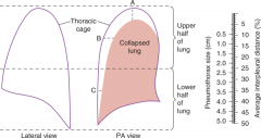
((A + B + C)/3) x 10% |
|
|
What are light's criteria? |
Distinguishes between exudative and transudative effusion
Exudative have at least one of: Pleural fluid >2/3 of upper level of normal serum LDH Pleural LDH/serum LDH >0.6 Pleural protein/serum protein >0.5
Highly sensitive, less specific for exudate |
|
|
Causes of miscarraige |
Two main: -Chromosomal anomolies -Uterine malformations (leiomyoma, bifid uterus, uterine scarring, cervical incompetence)
Other: -Increased maternal/paternal age -Low pre-pregnancy BMI -History of miscarriage -History of vaginal bleeding -Maternal stress -Increased parity -Autoimmune disease -Endocrine disorders (DM) -Maternal infections -Maternal toxin ingestion (cocaine, EtOH) |
|
|
Categories of hypertension in pregnancy |
Hypertension in pregnancy: >140/90 Preeclampsia: A disorder of pregnancy characterized by hypertension and new/worse proteinuria, adverse conditions during pregnancy that is thought to be due to endothelial dysfunction
Types of hypertension in pregnancy Chronic hypertension: dx'd before 20 weeks Gestational hypertension: dx'd after 20 weeks and no proteinuria Pre-eclampsia with chronic hypertension: proteinuria (>300mg/24h) and BP >160/110 in a patient with known hypertension Pre-eclampsia: proteinuria (>300mg/24h) and BP >140/90 |
|
|
Amniotic fluid embolism: pathophysiology, major causes, presentation |
Pathophysiology: release of amniotic fluid into the circulation causing an anaphylactoid reaction
Causes: labor, amniocentesis, uterine manipulation (version), placental separation. Can also occur during miscarriage/abortion and spontaneously.
Presentation: hypoxemia due to plugging of pulmonary vessels, cardiovascular collapse, non-cardiogenic pulmonary edema, DIC
Treatment: aggressive ventilatory and hemodynamic support. Plasma exchange to remove cytokines. Delivery of fetus. |
|
|
Diagnostic algorithm for PE in pregnancy |
If leg symptoms -> compression U/S (treat if pos) If no leg symptoms or U/S neg -> CXR If CXR clear -> V/Q scan If V/Q inconclusive -> CTPE If CXR abnormal -> CTPE If CTPE neg -> stop
|
|
|
Hyperemesis gravidarum: definition, onset, |
Definition: emesis that causes starvation metabolism with weight loss, dehydration, ketonuria, and ketonemia
Onset: 6-20 weeks
Pathyphys: unsure, associated with increased B-hCG, molar pregnancy, and multiple gestation
Management: fluid rehydration, enteral nutrition, diclectin (doxylamine and B6) up to 8 tabs/d then gravol then zofran/maxeran then methylprednisone |
|
|
What is a teratogen? What characteristics of a drug increase its ability to cross the placenta? |
Any chemical, pharmacologic, environmental or mechanical agent that can cause deviant or disruptive development of the conceptus
Characteristics that increase crossing the placenta Size (small), ionization (uncharged), protein binding (free drug), pKa (weak organic acids get caught in fetal base-ness), lipid solubility (more soluble) |
|
|
List 10 teratogens |
Heavy metals/toxins: Lead, CO, Iodine Anticoagulant: Warfarin Antiarrhythmics: Amiodarone, Quinine Anti-inflammatories: NSAIDs, misoprostol Antiepileptics: Phenytoin, VPA, carbamazepine Chemotherapeutics Busulfan, methotrexate, thalidomide Anti-hypertensives: ACEI, ARBs Dermatologic: Retinoic acid derivatives Androgens/Estrogens: OCP, HRT, diethylstilbestrol Antipsychotics: Lithium Drugs of Abuse: EtOH, cocaine Antibiotics: Erythromycin, tetracycline, aminoglycosides |
|
|
Distinguish true labor from false labor |
True labor -cyclic uterine contractions of increasing frequency, duration, and strength -cervical dilation -bloody show
False labor (Braxton-Hicks contractions) -no cervical dilation or effacement -intact membranes -do not escalate in frequency, duration or strength -not sensed by external monitors |
|
|
What is assessed on pelvic exam in true labor? How can fontanelles be distinguished? |
Cervical dilation, cervical effacement, presenting part, station of presenting part, orientation of presenting part
The anterior fontanelle has 4 sutures while the posterior fontanelle has 3. OA is the most common presentation. |
|
|
Steps to breech delivery |
-Get a C-section instead -Call obstetrics -Monitors -Rule-out prolapsed cord -Open pelvis (knees wide) -Episiotomy -When abdomen is through pull out 10-15cm of cord to try to avoid it getting trapped -Grasp neonate by the pelvis and direct face/abdomen away from the symphysis -Keep the next flexed forward (do not let it extend!! Causes spinal cord injury)
|
|
|
Umbilical cord prolapse: diagnosis and treatment |
Diagnosis: see the cord on pelvic, suddenly non-reassuring FHR
Treatment: emergency C-section, mother in knee to chest position with head down, fingers elevate presenting part, Foley to install 500-750 cc of fluid into bladder, replace cord above the presenting part |
|
|
Uterine inversion: risk factors, diagnosis and treatment |
Risk factors: -Primip -Oxytocin use -fundal implantation -forceful traction on umbilical cord -MgSO4 use
Diagnosis -Severe abdominal pain -Visualization of the uterus at the os or in the introitus
Treatment -Do NOT remove the placenta while the uterus is out -Give tocolytics (terbutaline, MgSO4, halogenated anesthetics to relax the uterine ring -Replace uterus -Then start oxytocin |
|
|
DDx for diffuse wheeze |
Pulmonary -Lower airway o Congenital: CF, Bronchopulmonary dysplasia o Trauma: FB, Aspiration o Infectious: Pneumonia, Bronchiolitis, COPD o Inflammatory: Anaphylaxis o Vascular: PE o Degenerative: Sarcoidosis -Upper airway o Congenital: Vascular ring o Trauma: FB, Caustic ingestion o Infectious: Epiglottitis, Croup, Retropharyngeal abscess o Inflammatory: Anaphylaxis, Angioedema o Neoplastic o Vascular ring Extra-pulmonary -CHF -ARDS |
|
|
Asthma severity classifications |
PEFR Mild >70% predicted Moderate 40-70% predicted Severe <40% predicted Life threatening <25% |
|
|
Side effects of steroids |
SHORT TERM o Insomnia o Mood alterations or Psychosis o GI upset o Increased appetite/weight gain o Fluid retention o Hyperglycemia o Hypokalemia
LONG TERM o Hyperglycemia o Osteoporosis o Thin skin, easy bruising, poor wound healing o Rare: HTN, PUD, AVN, Allergic reaction o Adrenal suppression if > 4 courses/year |
|
|
Soft signs of arterial injury in neck trauma |
FOAHHDDS |
|
|
Risk factors for primary and secondary PTx |
Primary: -Tall, skinny, male smokers with Marfan's and Mitral valve prolapse at altitude
Secondary: -Airway: cystic fibrosis, asthma, COPD -Infectious: TB, PJP, lung abscess, necrotizing -Interstitial: sarcoid, fibrosis, pneumoconioses, tuberous sclerosis -Neoplasm: primary or metastatic -Miscellaneous: endometriosis, pulmonary infarction |
|
|
When is a diagnostic thoracentesis indicated? |
Unexplained pleural effusions
Pneumonic and parapneumonic effusions -Pneumonia with a parapneumonic effusion >10mm wide on decubitus films -Loculated pleural effusion -Thickened pleural core
Diagnosis of a possible malignancy |
|
|
Indications for bicarbonate therapy |
-pH <7.1 -HCO3 <12 -Refractory hypotension (to pressors) -TCA toxicity -ASA toxicity -Phenobarb overdose -Ethylene glycol and methanol ingestion
Empiric dose is 1 mEq/kg with 1/2 as a bolus and 1/2 over 4 hours |
|
|
How can bicarbonate cause a paradoxical intracranial acidosis? Other complications? |
Paradoxical CNS acidosis -HCO3 diffuses over the BBB slowly -HCO3 in the plasma is converted by carbonic anhydrase to CO2 which is then blown off to decrease pH -This CO2 can diffuse quickly over the BBB decreasing the CNS pH -With the improved pH the RR is decreased increasing CO2 which again crosses the BBB
Other complications -Hypernatremia -Hyperosmolarity -Hypocalcemia -Hypokalemia |
|
|
Metabolic alkalosis: causes |
Causes: hypovolemia, hypokalemia, hypochloremia
DDx: -Volume contracted (saline responsive, urine Cl<10): Vomiting, diarrhea, NG suction, diuretics -Normal or expanded volume (saline unresponsive, urine Cl>10): primary hyperaldosteronism (Conn's), secondary hyperaldosteronism (CHF, cirrhosis, nephrotic syndrome, Cushing's, Barter's, Licorice, ectopic ACTH) -Other: milk-alkali syndrome, citrate, nonparathyroid hypercalcemia
Treatment -Saline responsive: fluid and acetazolamide -Saline resistant: replace K and spironolactone (aldosterone antagonist) |
|
|
Definition of DKA |
Due to a lack of insulin and increase in glucagon leading to hyperglycemia, osmotic diuresis, and ketoacidosis.
Glucose >13.9mmol/L (peds <11) pH <7.3 HCO3 <18 (peds <15) Serum or urine ketones |
|
|
How does iodide bolus affect thyroid hormone production? |
2 possible effects:
Wolff-Chaikoff effect: excess iodide inhibits ion trapping, thyroglobin iodination, and blocks the release of thyroid hormone
Jod-Basedow effect: in patients with Graves or multinodular goiter who are iodine deficient it can induce hyperthyroidism |
|
|
Thyroid storm: cause, precipitants, and treatment |
Cause: increased T3/T4 over a prolonged period of time increases B receptors and sympathetic surge activates them all at once.
Precipitants: -Trauma (burns, surgery, thyroid trauma) -Vascular (MI, CVA, PE, CHF) -Toxicologic (Iodine, radiocontrast, hormone ingestion, amiodarone, stopping therapy, ASA, chemo, pseudoephedrine, OP's) -Sepsis -Metabolic: hypo or hyperglycemia -Pregnancy -Psych: mania, emotional crisis
Treatment: -Decrease hormone production with PTU 1g po -Decrease release of preformed hormone with Saturated Solution of Potassium Iodide (SSKI) 5 drops 1h after PTU; Li works too -Beta blockade with propranolol 1-2mg IV q15m -Prevent T4->T3 conversion with hydrocortisone 100mg IV -Prevent enterohepatic circulation with cholestyramine -Prevent entry of thyroid hormone into cell with L-Carnitine -Supportive care with cooling, benzos, acetaminophen -Remove thyroid hormones with plasmapheresis, dialysis, plasma exchange -Treat precipitant -Admit to ICU -Thyroid surgery or ablation |
|
|
Myxedema coma: cause, precipitants, and treatment |
Cause: severe longstanding hypothyroidism with a precipitant
Precipitants: -Trauma, burns -Vascular: CVA, GIB, MI -Toxicologic: lithium/iodide (decrease release), narcotics, benzo's, barbiturates -Metabolic: hypoglycemia, hyponatremia, hypoxia, DKA, hypercapnea -Cold exposure
Treatment -ABC's - note macroglossia/mucosal swelling -IVF - watch Na and glucose (often need to be added) -Thyroid hormone - T4 if old/cardiac hx (T4 300-500ug IV bolus); T3 if young (T3 10-20ug IV bolus). Can also give a bit of each. -Hydrocortisone 100mg IV -Rewarming -Treat precipitant |
|
|
Treatment of upper and lower esophageal foreign body |
Upper: -Magill forceps / Glidescope -Foley -Bougienage -Endoscopy
Lower: -Pop -Glucagon -Maxeran -Nifedipine -SL nitro -Midazolam sedation -Endoscopy |
|
|
Indications for immediate endoscopy of an esophageal foreign body |
-Complete obstruction (unable to handle secretions) -Respiratory distress (FB in esophagus can compress trachea) -Sharp objects -Impacted for 24 hours -Coins in the proximal esophagus -Alkaline button batteries -Failure of medical treatment -Coins in children <2yo (relative) |
|
|
Complications of esophageal FB's |
-Abscess -Tracheo-esophageal fistula -Aorto-enteric fistula -Perforation and mediastinitis / pneumothorax / pneumomediastinum |
|
|
Indications for immediate removal of a foreign body in the stomach |
>2.5cm wide >5cm long Sharp Toxic (e.g. lead) >3-4 weeks impaction
90% of objects that make it to the stomach make it all the way through. If past the pylorus things can generally be left alone (then require surgery rather than endoscopy). Remove if hasn't moved in 3-4 weeks. |
|
|
Indications for surgery to remove a foreign body in the small intestines |
>99% of these pass without problem
-Hasn't moved for >7 days -Hasn't passed in >4 weeks ->1 industrial strength magnet (not a fridge magnet) |
|
|
Grades and treatment of hepatic encephalopathy |
Grades I - Depression, irritability, disordered sleep, mild cognitive dysfunction II - Lethargy, disorientation, asterixis III - Somnolence, disorientation, confused speech IV - Coma
Treatment -Stop all sedatives / CNS depressants -Correct hypokalemia (allows ammonia to be excreted renally) -Remove GI protein (treat bleed, decrease protein intake, treat constipation) -Give lactulose 30mL qid (becomes lactic acid and traps NH4+ and decreases transport time) -Flagyl or Clarithromycin to kill NH3 producing gut flora -Acarbose to decrease NH3 production -MARS |
|
|
How does lactulose correct hepatic encephalopathy? What are other treatments? |
Lactulose is converted to lactic acid, acidifying the bowel contents. This converts ammonia (NH3) to ammonium (NH4+) and its positive charge keeps it trapped in the lumen.
Remove other sources of protein (e.g. NG for GIB, protein-restricted diet) Clarithromycin or Flagyl (alter gut flora to decrease ammonia production) Acarbose (changes bacterial activity to decrease ammonia) |
|
|
What is the SAAG? How is it interpreted? |
SAAG = serum-ascites albumin gradient (serum albumin - ascites albumin) This replaces the distinction between transudate/exudate
SAAG<11 = inflammation or decreased oncotic pressure (Carcinomatosis, TB, Pancreatic or biliary ascites, nephrotic syndrome) SAAG>11 = portal hypertension (Cirrhosis, Alcoholic hepatitis, portal-vein thrombosis, Budd Chiari, liver mets) |
|
|
Diagnosis of SBP. Differentiating primary versus secondary bacterial peritonitis |
Diagnosis: -PMN >250 cells/mm3 -Positive culture -Ascites fluid pH <7.34 or a gap between blood pH of >0.10
Primary: -Protein <10 -Prior SBP -Bili >42mmol/L -Platelets <98 -Single bacteria cultured
Secondary: -Protein >10 -Glucose <2.8 -LDH > upper limit of normal serum LDH -Multiple types of bacteria cultured |
|
|
The triad and tetrad of ascending cholangitis |
Charcot's triad: -Fever -Jaundice -RUQ pain
Raynaud's pentad: -Charcot's triad plus altered mental status and shock (hypotension/tachycardia) |
|
|
Sonographic findings of an abnormal TV ultrasound |
-BhCG >3000 and no gestational sac -Gestational sac >13mm and no yolk sac ->5mm crown rump length and no fetal heart tones -No fetal heart tones after 10-12 weeks -Gestational sac >25mm and no fetus |
|
|
Risk factors for ectopic pregnancy |
PMHx - PID, previous ectopic or abortion, tubal surgery, infertility, abnormal anatomy
Patient factors - smoker, age
Pregnancy factors - has IUD, embryo transfer fertility treatments |
|
|
What is a molar pregnancy? What are the types? How does it present |
-Disordered proliferation of chorionic villi
Two types: -Complete hydatidiform mole: absence of fetal tissue -Incomplete hydatidiform mole (much less common): fetal tissue with focal trophoblastic hyperplasia -Can also get choriocarcinoma (responds well to chemo, can metastasize)
Presentation -Hyperemesis gravidarum -Crazy high BhCG -Snowstorm U/S |
|
|
Numbness or pain to the outer side of the thigh associated with pregnancy or obesity |
-Meralgia paresthetica -Due to compression of the lateral femoral cutaneous nerve of the thigh as it passes the inguinal ligament |
|
|
Cause and treatment of postpartum hemorrhage |
Cause: -Tissue - retained products, accreta (placental villi adhere to myometrium) / increta (enter the myometrium) /percreta (through the myometrium) make more likely -Tone - diagnosis of exclusion -Trauma - perineal tears, vulva/vaginal epithelium trauma, uterine inversion, uterine rupture -Thrombin - vWD, coagulopathy, DIC
Treatment Uterine massage Repair lacerations Remove products of conception Oxytocin - run 40U/1L fast; 10U IM Misoprostol (PGE1) - 800-1000mcg PR Hemabate (PGFalpha) - 250mcg IM Pack uterus Foley in uterus Embolize vessels D&C Hysterectomy |
|
|
Types of lactic acidosis |
A - tissue hypoxia B1 - systemic disorders (DM, renal insufficiency, leukemia, sepsis) B2 - substance associated (biguanides, methanol, salicylates, INH) B3 - heritable metabolic disease |
|
|
Risk factors, presentation, and treatment of cerebral edema in DKA |
Risk factors -New onset diabetes -Children <5yo -Extremely ill on presentation -Treated with HCO3 -Excessive fluid replacement -Rapidly dropping serum osmolality
Presentation -HA -Behavioral changes -Incontinence -Seizures -Autonomic (BP and temp) Then coma, respiratory arrest, death
Treatment -Mannitol 1-2g/kg over 15m -Decrease IVF and insulin rate -Intubate, hyperventilate, CT head
NB - cause is unknown - ? idiogenic osmoles |
|
|
4 characteristics that determine the toxicity of a hydrocarbon; Effects of toxicity short term and long term |
VVSS -Viscosity (lower more toxic)
Primary toxicity through aspiration - get bronchospasm, disruption of surfactant, displacement of oxygen, alveolar damage -> V/Q mismatch, hypoxia, alveolar dysfunction, resp failure
Also sensitizes myocardium (arrhythmias & sudden sniffing death syndrome - tx w BB), CNS effects (euphoria acutely, dementia and cerebellar dysfunction chronically), RTA, hepatic necrosis, blood cancers |
|
|
Diagnostic criteria of HHS |
Glucose >33 Sosm >320 pH >7.3 HCO3 >15 |
|
|
Complications of long-term DM |
Infection (immunocompromised secondary to decreased neutrophil and lymphocyte activity) Diabetic foot Insulin allergy (must go desensitization or change type) Cutaneous manifestations (diabetic dermopathy, dermal hypersensitivity at injection sites as well as hypo or hypertrophy, acanthosis nigrans, necrobiosis lipoidica diabeticoricum, xanthoma diabeticorum) Macrovascular complications (CAD, CVD, PVD) Microvascular complications (Nephropathy, Retinopathy, Neuropathy) |
|
|
Causes of rhabdomyolysis |
Traumatic -Crush -Compartment syndrome -Excessive exertion
Non-traumatic (relate to lack of ATP) -Electrolytes (HypoK or HypoP) -Ischemia -Congenital ATP deficiency due to inborn errors of metabolism -Environmental (electrical injury, heat stroke, hypothermia, rattle snake bite) -Endocrine (pheochromocytoma, DKA, HHS, hypo/hyperthermia) -Toxin (SS, NMS, statins, alcohol) -Infections (all types) -Seizures -Rheumatic (polymyositis, dermatomyositis, Sjogren's) |
|
|
Thyroid diseases |
Hyperthyroid -Graves (TSH receptor antibodies) -Toxic multinodular goiter (multiple overactive and big areas, can cause SVC syndrome) -Toxic adenoma -Acute thyroiditis (gland is tender) --> Autoimmune (Hashimoto's antibody to thyroid peroxidase; Postpartum; Sporadic) --> Infectious (De Quervian's viral; suppurative bacterial) --> Drug induced (amiodarone, iodine, interferon, lithium) -Pituitary adenoma -Gestational trophoblastic / germ cell tumors (create TSH-like hormone)
Hypothyroid -Hypothalamic and pituitary underactivity (tumors, Sheehan's, amyloidosis, sarcoidosis, radiation) -Late thyroiditis (as per above) -Iatrogenic (thyroidectomy, ablation, lithium, iodine, amiodarone) -Congenital (causes cretinism)
|
|
|
Primary versus secondary adrenal dysfunciton |
Primary is a disease of the gland itself and affects all 3 functions (glucocorticoids, mineralocorticoids, androgens) -See hyperpigmentation, hyperkalemia, hyponatremia, salt craving, and acidosis from the lack of aldosterone / excess ACTH
Secondary is a disease of the pituitary and does NOT affect mineralocorticoids (regulated by the RAAS) -Still get hyponatremia, but it is due to increased ADH
Both -Hypotension -Depression, delerium, HA, abdominal pain, emesis, hypoglycemia, hyponatremia (differnet reasons), hypercalcemia, fevers, ARF |
|
|
Steroid equivalency (hydrocortisone, prednisone, methylprednisolone, dexamethasone) |
Hydrocortisone = 1 Prednisone/Prednisolone = 4 Methylprednisolone = 5 Dexamethasone = 25 |
|
|
Esophageal narrowings and risk factors for obstruction / dysphagia |
Narrowings -Upper esophageal sphincter (cricopharyngeus) -Aortic arch -Left mainstem bronchus -Lower esophageal sphincter
Causes of obstruction / dysphagia
Poor dentition (they don't chew)
Intrinsic -Esophageal carcinoma -Shatzki's ring -Peptic stricture -Esophageal web
Extrinsic -Cardiomegaly -Aortic aneurysms or anomylous right subclavian -Goiter -Mediastinal tumor -Enlarged lymph nodes -Zenker's diverticulum
Neuromuscular (Neuro - head trauma, brain tumor, CVA, Alzheimer's, Parkinsons, MS, ALS, Myesthenia; Muscular - achalasia, scleroderma)
Toxic (Lead or EtOH)
Infectious (Bacteria - diptheria, botulism, syphilis, tetany OR Viral - rabies, polio) |
|
|
What is Mackler's triad? How can the problem be diagnosed? |
Suggests esophageal rupture
-Subcutaneous emphysema -Chest pain -Vomiting
Diagnose with contrast study. Use gastrograffin if no risk of aspiration (safer but less sensitive test; pneumonitis if aspirated) THEN barium (worse inflammatory response through perforation). Try CT if normal or unsafe to do. |
|
|
Contributors to thte development of GERD and evidence-based ways to get rid of it |
-Decreased sphincter tone (anticholinergics, caffeine, benzo's, nicotine, nitrates, peppermint, chocolate, estrogen, progesterone) -Decreased esophageal motility (DM, achalasia, scleroderma) -Increased intraabdominal pressure (pregnancy, obesity) -Decreased gastric emptying (anticholinergic, diabetic gastroparesis, outlet obstruction)
Evidence-based treatments: weight loss and bed elevation (also try no eating before bed, stop smoking/etoh, change relevant meds, smaller meals) |
|
|
Eradication treatment for H Pylori |
Triple: Clarithromycin 500bid / Amoxicillin 1000bid OR Metronidazole 500bid/ PPI x 10-14d
OR
Quadruple: Bismuth subsalicylate (pepto-bismol) 525qid / metronidazole 250qid / Tetracycline 500qid / PPI x 10-14d |
|
|
Poor predictors of outcome in upper esophageal bleed |
Components of the Rockall score
Age >60 Heart failure Ischemic heart disease Renal failure Liver failure Metastatic cancer Gastric cancer Vigorous bleeding |
|
|
DDx for transaminitis |
-Structural: o Inflammatory/ Autoimmune/ Infiltrative: autoimmune hepatitis (PBS, PSC), NASH (? d/t insulin resistance), amyloid o Vascular: Budd-Chiari (thrombosis of hepatic veins or IVC/SVC), portal vein thrombosis, ischemia, CHF o Congen/Degen: neonatal hepatitis -Toxicology: acetaminophen, EtOH, INH, iron, phenytoin, ecstasy (autoimmune hepatitis) -Infection: o Viral: HAV, HBV, HCV, HDV, HGV, EBV, CMV o Protazoan: amoeba o Toxoplasmosis o Associated with bacterial sepsis -Metabolic: Wilson’s disease (copper overload), Reye’s syndrome, hemochromatosis -Pregnancy: Fatty liver of pregnancy -GI: gallstones, strictures, cholangitis, biliary/pancreatic cancer, annular pancreas (obstructive causes) |
|
|
Hepatitis serology |
Hep A -For acute infection send HAV IgM -For chronic infection send HAV IgG
Hep B -For acute infection send HBV sAg and HBV cAb IgM -For chronic infection send HBV cAb IgG -For vaccine immunity send HBV sAb
Hep C -HCV Ab |
|
|
Hepatitis post-exposure prophylaxis |
A: HAIg to unvaccinated close personal contacts, childcare workers/attendees (people who wipe their bum), similar food-borne source in last 2/52
B: HBIg to: -unvaccinated / low titer recipients exposed to source that is HBV sAg + OR high risk -neonates with HBV sAg + mothers
C: N/A |
|
|
Signs of cholecystitis on ultrasound |
-Stones / biliary sludge -Wall thickening (2-4mm) -Distension of GB (>4cm wide or 10cm long) -Pericholecystic fluid -Air in the GB wall (emphysematous or gangrenous cholecystitis) -Murphy's sign (sonographic) |
|
|
Treatment of hyponatremia with focal neurologic symptoms, seizure, or coma. Complication of rapid correction. |
Hypertonic (3%) saline 100mL over 10m then 100mL over the next hour (approximately 3mL/kg total)
After this aim to correct by 0.5mEq/L/h if chronic, 1mEq/L/h is okay if acute
Complication: central pontine myelinolysis |
|
|
Causes, diagnosis and treatment of SIADH |
Lung masses -Cancer, pneumonia, TB, abscess CNS disorders -Infection (meningitis, abscess), mass (subdural, postop, CVA) Drugs -Thiazides, narcotics, oral hypoglycemic agents, barbiturates, neoplastic agents, vasopressin
Diagnosis - low Sosm (<280), high Uosm (>100) with no other explanation
Treatment - water restriction, treat cause |
|
|
Causes of hypocalcemia |
-Hypoparathyroidism (congenital, maternal, thyroid surgery, radiation) -Tox (chemo, HF, ethylene glycol, furosemide) -Hyperphosphatemia, tumor-lysis syndrome -Malnourished, alcoholism |
|
|
Conditions that can cause a false positive lipase result (not pancreatitis) |
Many false positives at standard cutoff. Quite specific at 5x standard level, but down to 60% sensitivity. 2x cutoff is best for maximal sensitivity/specificity.
-Cholecystitis -Bowel obstruction -Peritonitis -Duodenal ulcer -DKA -Trauma -Post ERCP -Idiopathic |
|
|
x-ray and ultrasound findings of pancreatitis |
x-ray -Pleural effusion -Pancreatic calcification -Free air (? due to perf'd something) -Ileus -ARDS
Ultrasound -Occasionally can see CBD stone and/or enlarged hepatic duct (suggesting distal obstruction) |
|
|
Causes of simple and closed loop SBO |
Top 3: -Adhesions -Hernias -Cancer
Intrinsic - congenital, IBD, radiation enteritis, cancer, intussusception, hematoma
Extrinsic - hernias, adhesions, volvulus, compressing tumors, abscesses, hematomas
Intraluminal - FB, gallstones, bezoar, barium, ascaris |
|
|
X-ray signs of intussusception |
Crescent sign Target sign Abdominal mass (no air in one area - usually RUQ) / No liver edge sign Air fluid levels (SBO) Dilated loops of bowel (SBO)
NOTE: In adults (as opposed to children) you do not want to reduce this with enema as it is often caused by cancer and this can result in seeding |
|
|
X-ray signs of mechanical SBO, closed loop obstruction, ileus. Normal measurements of bowel. |
Mechanical -Dilated proximal loops and flattened distal loops -Sharply angulated or step-ladder loops of small bowel -Multiple air-fluid levels -'String of pearls' (pockets of gas trapped in the plicae semicircularis when the bowel is full of fluid)
Closed-loop -Coffee bean sign (U-shaped bowel loop also seen in sigmoid volvulus) -Pseudotumor sign (fluid filled loop resembling a mass)
Ileus -Dilated loops throughout the entire bowel including the colon -Dilation less prominent -Air fluid levels less prominent
Bowel measurements: -Small 3cm -Large 6cm -Cecum 9cm |
|
|
Causes of mesenteric ischemia and risk factors |
-Arterial embolism ~50% - mostly SMA (CAD, valvular disease, AF, aneurysms, dissections, coronary angiography) - needs embolectomy -Arterial thrombosis >15% - mostly SMA and have h/o abdominal angina (elderly, PVD, hypertension) - needs revascularization, heparin -Venous thrombosis <15% (same risk factors as DVT/hypercoaguability; Factor V Leiden most common) - needs heparain, thromboplasty -Non-occlusive - 20% (all shock states, cocaine, vasopressors; >50yo)
Lactate is highly sensitive CT angiography is most helpful diagnostic test; Angiography is gold standard and early angiography decreases death |
|
|
Modified Alverado score, WBC/CRP, U/S and CT for Appy |
Alverado score History: -Migration of pain to the RLQ -Anorexia -N or V PE: -T>37.3 -RLQ tender (2 points) -Rebound tenderness, Labs -Leukocytosis (2 points) -Left shift Interpretation: -Treat if >7; Image if 4-7; Unlikely if 4 or less
Labs -WBC<10 and CRP<12 have a -LR of 0.09 (very sensitive) but less helpful in peds.
U/S -75-95% sensitive, 85-95% specific - operator dependent but 1st choice for kids/women -See non-compressible, thick-walled (>2mm), dilated (>6mm), thickened mesentary, pain with compression, appendicolith
CT -95% sensitivity and specificity |
|
|
Rome III criteria for IBD |
Recurrent abdominal pain/discomfort for at least 3d in the past 3m associated with 2/3 of: -Improvement with defecation -Onset associated with a change in stool frequency -Onset associated with a change in stool appearance |
|
|
Causes of large bowel obstruction |
1 - Colorectal cancer 2 - Volvulus 3 - Diverticulitis 4 - Extrinsic compression from mets
Also: Abscess, stricture due to chronic ischemia, fecal impaction, IBD, CF, Hirschsprung's, body packers/stuffers, Ogilvie's (pseudo-obstruction) |
|
|
AXR findings of large bowel obstruction |
Distended colon Air-fluid levels Cecal dilation >12cm has increased risk of perforation |
|
|
Volvulus types, risk factors, x-ray findings |
Gastric: hiatal hernia, either between 40 and 50yo or <1yo. Often have diaphragmatic defects, gastric ulcer or cancer, adhesions, paralyzed diaphragm. Can't pass NG tube!!
Cecal: pregnancy, 'coffee bean sign' pointing to LUQ, also can have air-fluid levels in the small bowel, paucity of colonic gas. Treatment surgical.
Sigmoid: elderly, psych/neuro disease, institutionalized, constipation, high fiber diet, 'coffee bean sign' pointing to RUQ, bird's beak contrast. Treatment endoscopic detorsion or surgery. |
|
|
Crohn's versus colitis |
Crohn's affects mouth to anus / Colitis large colon and rectum only
Crohn's commonly found in terminal ileum and colon / Colitis starts at rectum and moves proximally
Crohn's is transmural / Colitis is superficial mucosa
Crohn's has skip lesions / Colitis is continuous
Crohn's gets primary sclerosing cholangitis / Colitis gets colon cancer |
|
|
Extra-intestinal manifestations of IBD |
ULCERATIVE U rinary stones L iver cirrhosis / sclerosing cholangitis C holelithiasis E rythema nodosum / erythema multiforme / pyoderma gangrenosum R etardation of growth A rthralgias / arthritis / ankylosing spondylitis T hrombophlebitis I atrogenic (steroids) V itamin deficiency E yes (uveitis, episcleritis)
Also pulmonary fibrosis |
|
|
Intestinal manifestations of IBD |
COLITIS C ancer O bstruction L eakage / perf I ron deficiency T oxic megacolon I nanition (wasting) S tricture
Also: abscess, fistula |
|
|
What is toxic megacolon? What causes it? What's the treatment? |
Inflammation of the smooth muscles of the colon leads to dilation and perforation if untreated. Patients look toxic and have dilated colon on AXR (>6cm).
Often due to infection (C Diff gets po vanco or po/iv flagyl; other gets ceftriaxone/flagyl), IBD (gets tazo and steroids), antimotility agents (anticholinergic or opioid - stop them). May need OR. |
|
|
What is required to prove negligence in a malpractice suit? |
-Health care provider has a duty of care -That duty of care is breached by breaking the standard of care -The patient is harmed -There is a direct link between the breach and the harm |
|
|
Mimics appendicitis Backpacker's diarrhea Raw/undercooked poultry Associated with GBS Diarrhea and seizures Associated with Reiter's Raw oysters/shellfish Prolonged diarrhea Dysentry without fever Fried rice GI and neuro Cold allodynia / hot/cold reversal Worse after EtOH ingestion Mayo/potato salad Eggs |
Mimics appendicitis: campylobacter and yersinia Backpacker's diarrhea: giardia lamblia, campylobacter Raw/undercooked poultry: campylobacter Associated with GBS: campylobacter Diarrhea and seizures: shigella Associated with Reiter's: salmonella Raw oysters/shellfish: vibrio parahemolyticus, plesiomonas Prolonged diarrhea: yersinia, aeromonas, parasite Dysentry without fever: e coli O157:H7 Fried rice: bacillus cereus (toxin mediated) GI and neuro: ciguatera toxin Cold allodynia / hot/cold reversal: ciguatera toxin Worse after EtOH ingestion: ciguatera toxin Mayo/potato salad: staph aureus Eggs: salmonella |
|
|
Bacterial causes of diarrhea; antibiotic treatment |
CSS Yalk Constantly - So Believe Every Child Vomiting And Pooping
C ampylobacter S higella - treat dysenteriae for public health S almonella - treat typhi and all food handlers for public health Y ersinia C lostridium jejuni S taph aureus (toxins) B acillus cereus (toxins) E coli (toxins) C difficile and perfringins (toxins) V ibrio cholera (toxins) and parahemolyticus A eromonas P lesiomonas
Toxin-producing generally do not respond to antibiotics
Antibiotics for severe infectious diarrhea with no evidence of HUS with cultures pending - children cefixime/azithro x 3-14d; adults cipro x 3-14d |
|
|
Causes of free fluid in the abdomen |
-Blood -Urine -Peritoneal dialysis fluid -Ascites > Liver disease - cirrhosis, alcoholic hepatitis, portal vein thrombosis, Budd-Chiari, liver mets (SAAG>11) > Abdominal or ovarian malignancies / carcinomatosis (SAAG<11) > TB peritonitis (SAAG<11) > Pancreatitis (SAAG<11) > Nephrotic syndrome (SAAG<11) > CHF > Hemodialysis |
|
|
Internal hemorrhoid classification and treatment |
1st degree - sense of fullness, no prolapse, medical management 2nd degree - prolapse during defecation and spontaneously reduce, medical management 3rd degree - prolapse spontaneously and during bowel movement, reduce spontaneously, medical or surgical management 4th degree - permanent prolapse with risk of thrombosis, surgical repair |
|
|
Medical and surgical hemorrhoid treatment |
Medical: WASH W arm water A nalgesia (topical nifedipine, lidocaine for external; internal controversial) S tool softeners H igh fiber diet
Surgical: sclerotherapy, hemorrhoidectomy, banding |
|
|
What is the anal fissure triad? Treatment? |
Deep ulcer Sentinal pile (hypertrophic edematous skin tag) Enlarged anal papilla
Treatment with WASH (warm water, analgesia with nitro/lidocaine/nifedepine, stool softeners, high fiber diet) as per hemorrhoids. |
|
|
Types of rectal abscess |
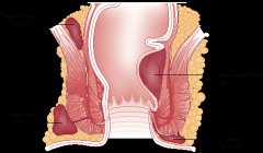
-Supralevator (high and deep) -Intersphincteric (internal, above pectinate line) -Ischiorectal (lateral; may be able to drain in ED - controversial) -Perianal and Perirectal (only ones we'd drain in ED)
Always tx with tetanus; Abx if DM/ immunocompromised/ valvular disease |
|
|
Causes of fecal incontinence |
Pediatric -Congenital (meningocele, myelomeningocele, spina bifida) -Post-op imperforate anus -Sexual abuse
-Neuro (demential, spinal cord injury, autonomic neuropathy from DM, pedental nerve damage from surgery/obstetrics, Hirshsprung's) -Trauma to sphincter -Mass (colorectal cancer, foreign body, hemorrhoids, fecal impaction) -Medical (rectal prolapse, diarrhea, IBD, laxatives) |
|
|
Gastroenteritis bugs that require treatment |
1) Culture positive, 2) immunocompromised, and 3) not improving
Also: -Shigella dysentariae (even if asymptomatic - public health) -Yersinia (even if asymptomatic - public health) -Salmonella typhi in food handlers, healthcare workers, severe colitis, <3m/o, >50yo -Bacillus anthracis -C difficile -Giardia Lambia -Entamoeba histolytica |
|
|
When can anti-diarrheal medications be given? |
AVOID in <2yo and those with fever or dysentery (blood +/- pus or mucous)
Consider in patients with severe symptoms along with antibiotics |
|
|
Diarrhea history - key questions |
Travel - parasites Antibiotics - c diff Ingestions - food poisoning Well-water - parasites Infectious contacts - virulent bacteria Pets at home - salmonella |
|
|
DDx for bilateral CNVII palsy |
Gosh, bilateral CNVII isn't just B2E2LLS3
GBS (Millar-Fischer variant) Ethylene glycol toxicity
|
|
|
Characteristics of self-induced knife wounds |
-Multiple superficial incisions to trunk/arms/face -Multiple superficial stabs to trunk/arms/face -Parallel incisions on the non-dominant side of the body in close proximety to each other -Sparing of sensitive areas -Linear or curved incisions toward the hand inflicting the wound -Intact clothing covering the wound -Evidence of similar prior wounds |
|
|
Types of abuse |
3 categories: -Domestic -Institutional -Self
Multiple types -Physical -Emotional -Sexual -Neglect -Abandonment -Financial -Factitious disease (Munchausen's by proxy) |
|
|
Risk factors for child abuse |
Child -Premature -Difficult temperament -Developmental delay or chronic medical condition -Social isolation
Caregiver -EtOH or substance abuse*** -Abused as a child -Intimate partner violence -Mental illness -Single parent
Demographic -Low SES -Ethnic minority |
|
|
Shaken baby syndrome; imaging studies |
-Generally <1yo; can be <3yo -No evidence of impact -SDH and SAH -On fundoscopy see retinal hemorrhages (>75%), papilledema due to increased ICP
CT is better for SAH, imaging of intracranial injuries, easier to perform. MRI is better if subacute/chronic, deel cerebral injuries, extraaxial fluid, smaller SDH's |
|
|
Historical indicators of child abuse |
-Magical injuries -Inconsistent story -Inconsistent with childhood development (can't bruise if can't cruise; 3 week-old 'rolling' off of a table) -Unexplained delay in seeking care -History does not explain the injury |
|
|
Risk factors for HIV transmission via sexual intercourse |
Victim -Anal > vaginal -Coexisting STD's or genital lesions -Trauma evident -Ejaculate on mucous membranes -Cervical ectopy -Active menstration -Currently pregnant
Assailant -Foreskin -Primary infection -Late stage infection -Viral load in genital tract -STI's or genital lesions -Not on HAART -Multiple offenders -Incarcerated, homosexual, bisexual |
|
|
Risk factors for interpersonal violence |
Victim -Demographics (<35yo, female, immigrant, separated or divorced) -Environment (low SES, homeless, previous exposure to violent caretakers) -History (disabled, previous physical or sexual assault)
Perpetrator -Demographics (young) -Societal (low income, unemployed, low SES, low academic achievement, criminal behavior) -Psych (low self-esteem, personality disorder, emotional dependence, insecure) -Substance abuse -History (abused as a child, violence in family of origin, history of TBI) |
|
|
Medical problems on DDx with interperesonal violence |
-Depression, PTSD, suicidal ideation -Headaches -Stress-related illnesses -EtOH / substance abuse -Trauma in pregnancy -Chronic pain -STI/HIV |
|
|
Historical indicators of elder abuse |
Implausible mechanism of injury Inconsistent history between patient and caregiver Delay to presentation
Unexplained injuries Elder being called 'accident prone' Past history of frequent injuries
Noncompliance with meds, appointments, directions Caregiver does not know patient's history/meds Caregiver answers all questions Caregiver/patient reluctant to give answers
Strained patient/caregiver interactions Poor living situation |
|
|
Diagnostic criteria for a manic episode |
Manic episodes are characterized by a >2 week period with elevated/irritable mood and >3 of the following
GST PAID G randiosity S leep (decreased) T alkative
P leasurable activities / P ainful consequences A ctivity I deas (flight of) D istractable
Is not mixed, causes marked impairment or requires hospitalization, not due to a general medical condition. |
|
|
Anxiety definitions: Anxiety, Panic attack, Agoraphobia, Social phobia, Phobia, OCD, Generalized anxiety, PTSD, acute stress disorder |
-Anxiety: a specific unpleasurable state of tension that forewarns the presence of danger (uneasiness stems from the anticipation of some imminent danger, the source of which is unknown or unrecognized)
-Panic attack: discrete period of sudden onset of intense apprehension, often associated with feelings of impending doom
-Agoraphobia: Anxiety about place or situations from which escape might be difficult (fear of being along in public places).
-Panic disorder with agoraphobia: Pts have recurrent unexpected panic attack and become fearful of situations where they might occur
-Specific phobia: irrational fear of something that is perceived as dangerous (normal in children)
-Social phobia: anxiety d/t social or performance situations
-Obsessive-Compulsive Disorder: Obsessions → stress or anxiety which is relieved by a compulsive behaviour
-Generalized anxiety disorder: persistent, excessive anxiety or worry for > 6 months
-Post-traumatic stress disorder: Heightened arousal and avoidance of stimulus following a significant traumatic exposure
-Acute stress disorder: similar to PTSD occurring immediately in the aftermath of an extremely traumatic event |
|
|
Predictors of organic anxiety disorders |
Predictors -Onset after 35yo -Lack of childhood, personal, or family history of anxiety/phobias -Lack of avoidance behavior -Absence of live events that would exacerbate anxiety -Poor response to anxiolyticsiD |
|
|
Disorders that can manifest as anxiety |
Substance abuse: sympathomimetics (caffeine, amphetamine, cocaine), hallucinogens (LSD, PCP, Ecstasy, marijuana) Withdrawl: depressants (benzos, barbiturates, EtOH) Cardiac: arrhythmias, mitral valve prolapse Endocrine: hypo/hyperthyroid, hypoglycemia, pheochromocytoma, hyperadrenocortism Resp: asthma, PE Medications: alpha agonists, theophylline, corticosteroids, thyroid hormone |
|
|
What is somatization disorder? |
Somatization disorder -Unexplained physical symptoms beginning before 30yo -At least 4 sites of pain, 2 GI symptoms, 1 reproductive/sexual symptom, 1 neurologic symptom -Not explained by another medical condition -Not intentionally feigned/produced
Risk factors -Women, low SES, alcoholism, addictions, poor education, interpersonal problems |
|
|
What is conversion disorder? |
-A somatoform disorder -Sudden onset of a single symptom not under the patient's control and often associated with la belle indifference -Generally neurological (motor: tremors, paralysis, pseudoseizures, aphonia, ataxia; sensory: anesthesia, blindness, tunnel vision) -Often a psychiatric coping mechanism |
|
|
What is somatization? List the somatoform disorders. |
Somatization The unconscious experience and communication of psychological distress through physical symptoms.
-Somatization disorder -Conversion disorder -Pain disorder -Hypochondriasis |
|
|
What is hypochondriasis? Treatment? |
4 key features: -Symptoms are more than the organic disease that is evident -Fear of disease and conviction that one is sick -Preoccupied with their body -Persistent and unsatisfying pursuit of medical care
Treatment -Reassurance, legitimize, share diagnostic uncertianty, assure ongoing care, avoid drugs that cause dependency, come up with realistic treatment goals focused on symptom control, arrange single-physician follow-up |
|
|
Compare factitious disorder, Munchausen's Syndrome, Munchausen's syndrome by proxy, Malingering, |
Factitious disorder: symptoms and signs produced or feigned in the absence of external benefit to take on the sick role, IS a mental disorder, unmarried educated women <40yo with healthcare background.
Munchausen's: a form of factitious disorder, wide variety of illnesses with intent of gaining hospital admission, hospital shoppers, believe they are very important, initially praise care -> become disruptive -> rage and AMA
Munchausen's by proxy: a form of factitious disorder where illness produced/feigned in a child. Persistent presentations with symptoms that stop when perperator is removed. Parents work in healthcare. Notify protective services and consult psych for mother.
Malingering: Malingerers ARE motivated by external incentives! Not a mental disorder. Assume somatization unless otherwise proven. Often medicolegal context, discrepancy between findings and disability, poor cooperation, antisocial behavior. Don't want to get better; gaming the system. |
|
|
Diagnosis of schizophrenia |
>2 of these symptoms for >1 month -Delusions (if delusions bizarre counts as 2) -Hallucinations (if running commentary counts as 2) -Disorganized speech -Disorganized or catatonic behavior -Negative features (avolition, poverty of speech, flat affect)
As well as: -Sharp deterioration -Disturbance for >6 months (with prodrome) -Other causes ruled out |
|
|
Complications of neuroleptic use and treatment |
-Orthostatic hypotension - alpha blockade, give fluids -Acute dystonia - cholinergic, treat with anticholinergic benztropine (cogentin) 1-2mg IV/IM +/- benadryl -Akathisia - motor restlessness, decrease dose or try beta-blocker -Parkinsonism - can be indistinguishable from Parkinson's, tends to resolve over time, decrease dose or start parkinson's meds -Tardive dyskinesia - occurs over years, bad, choreathetoid movements (tongue, grimace, writhing), no known treatments, try switching to atypical or benzo's -NMS |
|
|
Symptoms (in order) of NMS, medications that cause it, treatment |
MR HA altered M ental status (agitated or catatonic) R igidity (lead pipe, tremor)** H yperthermia** A utonomic instability
**rigidity and increased temp are necessary for diagnosis**
Medications: typical and atypical antipsychotics, lithium, withdrawl from Parkinson's medications, maxeran
Treatment: Benzo's, stop neuroleptics, bromocriptine/dantroline/amantidine, cool, ICU, electroconvulsive therapy (Seriously? Seriously??) |
|
|
Simple vs complex skull fracture |
Simple: -Linear not crossing suture lines -<2mm of separation
Complex: -Linear crossing suture lines ->2mm of separation -Stellate -Comminuted -Depressed -Compound -Diastatic |
|
|
Triad of shaken baby syndrome |
-Subdural hematoma -Cerebral edema -Retinal hemorrhages |
|
|
DDx for retinal hemorrhages |
-Vaginal delivery (resolve in 10-14 days) -Bleeding disorders -AV malformations -Meningitis -Severe accidental head injury |
|
|
Physical exam signs of sexual abuse |
-Unexplained vulvar bruising -Hemorrhage -Hymenal or vulvar tears -Loss of hymen out to the margin of the vagina -Signs of STI's (gonorrhea, chlamydia, hsv2, syphilis, trichomonas)
Can get HPV, HSV1, Gardnerella vaginosis, Hepatitis B/D and AIDS without assault. |
|
|
Bronchiolitis treatments |
-Oxygen - yes if hypoxic
-IVF - yes if dehydrated
-Beta agonists - not generally recommended (perhaps 10% responders, atopic people more likely)
-Steroids - no
-Epinephrine - some bad evidence that it can help prevent hospitalization, but not enough evidence to use it
-Epi and steroids together - may be a synergistic response, but more evidence needed. NOT recommended.
-Nebulized hypertonic saline - evidence moving towards its use, but it is still not in the guidelines |
|
|
Inadequate view of prevertebral soft tissue in children |
-View taken on EXpiration -Flexed or neutral (rather than extended) neck |
|
|
Esophageal button battery - mechanisms of injury |
-Current from the battery forming a circuit -Release of hydroxide -Pressure necrosis due to esophageal foreign body |
|
|
What are the goals / indications of PSA? |
Analgesia Anxiolysis Sedation Immobility Amnesia |
|
|
Crisis vs psychiatric emergency |
Crisis: acute emotional upset arising from situational or developmental problems that results in temporary inability to cope
Psychiatric emergency: acute behavioral disturbance related to severe mental or emotional instability or dysfunction requiring medical intervention |
|
|
HEADSS social history |
H ome E ducation A fter school D rugs S exual history S uicidal thoughts/attempts |
|
|
Mental status exam |
Appearance Attitude Behavior Mood Affect (appropriateness, lability, eye contact) Orientation (date/time/place) Speech Thought process (disorganized) Thought content (delusions) Perceptions (hallucinations) Cognition (memory, content of thought, preoccupations, coherent speech, ability to reason, insight, judgement) Insight Judgement Suicidal ideation Homicidal ideation Capacity (CURVES)
|
|
|
Substance dependence |
WITHDraw IT
W ithdrawal I nterest or Important activities neglected T olerance H arm to physical and psychosocial are known but they continue to use D esire to cut down, control it
I ntended time using exceeded T ime spent to acquire it is too much |
|
|
Pediatric vs adult bones |
-Thicker and more stable periosteum -Faster healing with less immobilization -Better remodeling capability and vascularity -Growth plates weaker than ligaments -More porous and pliable |
|
|
Toddler's fracture characteristics |
-Minimal or no history of trauma -Red flags for NAT are: more transverse fracture with an associated fibular injury -Generally 9m to 3y of age -On physical exam spiral oblique axial load provokes pain (put axial load and twist ankle) -Generally treat with an above knee backslab - sometimes don't need anything. |
|
|
Seven pulmonary complications of pneumonia |
-Pleural effusion / empyema -Pneumothorax -Lung abscess -Bronchopleural fistula -Necrotizing pneumonia -Pneumatocele -Acute respiratory failure |
|
|
How long are these pediatric rashes congagious for? (varicella, rubella, measles, parvovirus) |
-Varicella - 2 days before until lesions are all crusted over -Rubella - 1-2 weeks before they present with rash -Measles - 5 days before and 4 days after -Parvovirus - a week before until the rash starts |
|
|
DDx for poor R wave progression on an ECG |
-Old anterior MI -Lead displacement -LVH -LBBB -LAFB -WPW -Dextrocardia -Tension PTx with mediastinal shift -Congenital heart disease |
|
|
Define delusion, hallucination, and disorganized speech |
Delusion: Firm, fixed, false belief not in keeping with a person's cultural upbringing that are often religious, somatic, or persecutory.
Hallucination: Sensory experience that only exists to the person experiencing it.
Disorganized speech: Loosening of associations with shifts between topics. Can be circumstantial, tangential, neologisms, perseveration, or word salad. |
|
|
Schizophrenia negative symptoms |
Alogia Affect (flat) Avolition |
|
|
Types of dystonic reaction and treatment |
-Buccolingual (tongue protrusion) -Torticollus (Head deviation) -Oculogyric (upward eye deviation) -Opisthotonus -Laryngospasm
Treat with benztropine (cogentin) 1-2mg +/- benadryl 50mg both IM/IV - continue for 48h with q6h po doses |
|
|
Indications for psychiatric admission |
Psychiatric condition that makes the patient: -Significant risk of harm to self or others -Lack of capacity to cooperate with outpatient treatment -Inadequate psychosocial support for safe outpatient treatment -Comorbid condition/complication makes outpatient treatment unsafe (withdrawl, acute psychosis, bizarre behavior) |
|
|
Criteria for involuntary admission for mental health reasons |
1 Suffering from a mental disorder 2 Likely to harm themselves or others or substantially deteriorate physically or mentally 3 Unsuitable for admission other than as a formal patient (not willing to come voluntarily) |
|
|
Delirium vs Dementia vs Psychosis (vitals, onset, course, hallucinations, orientation, delusions, speech, movements, psychomotor) |
vitals - delirium ABnormal, dementia normal, psychosis usually normal
onset - delirium acute, dementia slow/insidious, psychosis acute
course - delirium fluctuates, dementia slowly progresses, psychosis stable
hallucinations - delirium visual & auditory, dementia, dementia no, psychosis auditory
orientation - delirium no, dementia maybe, psychosis usually
delusions - delirium transient, dementia yes, psychosis yes
speech - delirium incoherent, dementia normal, psychosis usually coherent
movements - delirium asterixis/tremor, dementia apraxia/intention tremor, psychosis absent
psychomotor - delirium variable, dementia variable / agitated, psychosis variable agitation |
|
|
Physiologic changes in the elderly leading to altered pharmacodynamics |
GI Decreased motility / absorption Decreased hepatic metabolism / albumin for binding Decreased hepatic blood flow / metabolism
MSK Increased adipose and decreased lean body mass / smaller Vd Decreased total water / altered Vd
GU Decreased GFR / decreased elimination
|
|
|
Selected physiologic changes of aging and effects |
-Neuro: Altered autonomic/neurotransmitter function leads to orthostatic hypotension and slowing mental function -Skin atrophy: decreased insulation, increased injury, increased infection -Decreased sweat glands: increased hyperthermia -Bone loss: increased fractures -Decreased antibodies and cell mediated immunity: increased infections -Decreased Tv, compliance, resp drive, diffusion capacity: increased CO2, decreased O2 -Decreased hepatic function, blood flow and enzyme function: altered metabolism of drugs -Decreased renal function, total water, vasopressin response: decreased renal elimination, drug excretion -Decreased GI mucosa and HCO3: increased gastric ulcers and perf
|
|
|
Reasons Cancer patients are at increased risk of infection |
Decreased immune function -Physical barrier breakdown (mucositis, indwelling catheters, cytotoxic effects on GI cells) -Functional asplenia / splenectomy (heme cancers) -Neutropenia (chemo, radiation, bone marrow suppression) -Decreased T and B cell function (disease and chemo)
Increased exposure -Invasive procedures -Prophylactic abx decreases normal flora |
|
|
Neutropenic infections and associated pathogens |
Ulcerative oral lesions: Strep viridans, herpes, candida Necrotizing skin lesions: pseudomonas, aeromonas, aspergillus, mucormycosis Black eschar: mucormycosis, aspergillus Abd pain, distension: typhlitis (neutropenic enterocolitis) due to pseudomonas, e coli, clostridium Perineal pain: Gram - bacilli, anaerobes |
|
|
Infections associated with decreased cell mediated immunity |
Important in transplant and HIV
Bacteria - Listeria, Legionella, Nocardia, TB Viruses - CMV, HSV, VZV, EBV Fungi - Coccidiomycoses, Blastomycoses, Histoplasma, Cryptococcus Parasites - Toxoplasma, Strongyloides |
|
|
Phases of transplant rejection |
Hyperacute - periop or immediate postop, relates to ABO or other antibody mismatch, get organ failure and SIRS Acute - first months after transplant, host vs graft disease, mild systemic symptoms and minimal pain, dysfunction of organ Chronic - gradual deterioration due to inflammation over years |
|
|
How can alcoholism be screened for? |
CAGE questionnaire C ut down? A nnoyed? G uilty? E ye opener needed? 2/4 require further investigation |
|
|
Define alcoholism |
No good precise definition. Multifactorial chronic disease with various presentations affecting health, function, relationships
At least 3/6 of WITHDraw IT (withdrawal / tolerance lumped together to make 6) |
|
|
Zero order vs First order kinetics |
-Zero - constant amount per unit of time (alcohol at low levels, acetaminophen at high levels, salicylates at high levels)
-First - constant proportion per unit of time (CO, alcohol at high levels due to Microsomal Ethanol Oxidizing System, acetaminophen at low levels, salicylates at low levels) |
|
|
Why does EtOH withdrawl occur? |
-Regular use decreases glutamate and increases GABA causing increased glutamate receptors and decreased GABA receptors -CNS is hypersensitive to glutamate when EtOH stops |
|
|
Physiologic effects of long term EtOH abuse |
Heart - HTN, CHF, cardiomyopathy, arrhythmias Lung - pneumonia due to aspiration, decreased airway reflexes GI - esophagitis, mallory-weiss/boorhaave's, fatty liver/hepatitis/cirrhosis, pancreatitis, diarrhea, vitamin deficiency CNS - symmetrical polyneuropathy, Wernicke encephalopathy, Korsakoff dementia, cerebellar degeneration ID - immunosuppression, neutropenia Metabolic - insulin resistance, electrolyte abnormalitiies, AKA Heme - anemia, neutropenia, thrombocytopenia due mostly to malnutrition; decreased clotting factors due to liver failure |
|
|
Drugs that cause disulfiram-like reaction with EtOH |
-Inhibit aldehyde dehydrogenase leading to buildup of acetaldehyde ABx - metronidazole, nitrofurantoin, sulfonamides, some cephalosporins DM - sulfonylureas (gliclazide) |
|
|
Acyanotic heart diseases |
CAP VAP C oarctation A S P S
V SD A SD P DA
|
|
|
Causes of hydrocephalus |
Obstructive - proximal to arachnoid granulations -Masses (abscess, granuloma, tumor) -Aquaduct stenosis -Congenital (Dandy Walker, Chiari malformations)
Non-obstructive - arachnoid granulations -Infection (meningitis, cysticercosis) -Hemorrhage (SAH, IVH, traumatic) -Choroid plexus papilloma
Normal pressure hydrocephalus (idiopathic) |
|
|
Amiodarone side effects |
P eripheral neuropathy P hotosensitivity P ulmonary alveolitis P igmentation of skin P eripheral conversion of T4 to T3 inhibited (hypothyroid) |
|
|
At risk or hazardous drinking (men, women, elderly) |
Men: 14/week or 4/session Women: 7/week or 3/session Elderly: 7/week or 1/session |
|
|
Pathophysiology of AKA |
-Alcoholics don't eat and use all of their NADH metabolizing EtOH so they can't make glucose -With glucose low insulin isn't produced -Ketones are produced but, without NADH, beta hydroxybutyrate can't be converted to acetoacetate/acetone -BHB gets elevated (~3-6:1 ratio) -Treat with fluid, thiamine, glucose, potassium - as corrected serum ketones will increase (acetoacetate is detected) |
|
|
AHA chain of survival |
5 links 1 Early recognition of arrest and activation of EMS 2 Early CPR 3 Rapid defibrillation 4 Effective ALS 5 Integrated post arrest care |
|
|
On-line vs off-line medical control |
Off-line -Protocols (standing orders, practice guidelines, treatment protocols) -Education (initial and ongoing for all provider levels including dispatch; curriculum, evaluation, administration, revision) -Quality improvement (review/observe performance, remediation, develop time standards) -Other (medico-legal, research, debriefing, complaints)
Online -Concurrent direction of field team by protocol or consultation -Has medico-legal responsibility for orders -Can be centralized (one site handles all) or decentralized (hospital receiving patient) |
|
|
When can EMS not transport a patient |
-Patient with capacity that refuses -Obvious signs of death -Danger to crew |
|
|
Treatment of a crush injury in search and rescue |
-Patients tend to decompensate 4-6h after being extricated -Circulation reaches crushed limbs releasing toxins systemically causing hypovolemia, hyperkalemia, dysrhythmia -Require aggressive early fluid resuscitation (prior to extrication) and treatment of hyperK/rhabdo while preventing hypothermia |
|
|
Principles of TCCC?
What are the leading causes of preventable death in tactical trauma?
How are the combat zones classified and how does this affect care? |
TCCC = Tactical Combat Casualty Care 1- prevent additional casualties 2- accomplish mission 3- save maximum lives 4- minimize morbidity of injured
Causes of death -Airway compromise (cric) -Hemorrhage (tourniquets) -Tension PTx (decompress)
Zones -Hot - tourniquet (no airway or C-spine management) -Warm - not under direct fire - cric, decompression, IV and hemorrhage control, CPR, analgesia, abx -Cold - evacuation area - standard ATLS treatment |
|
|
Components of surge capacity and critical substrate for hospital operation |
Surge capacity: S taff S tuff S tructure (physical location and infrastructure)
Hospital operations: 3 S's plus: Communication Transportation Managerial support |
|
|
Describe the PICE nomenclature |
PICE = Potential Injury Creating Event
Assign 3 prefixes: A - static, dynamic (stable vs unknown/escalating number of casualties) B - controlled, disruptive, paralytic (local resources not overwhelmed, overwhelmed requiring augmentation, overwhelmed requiring reconstitution) C - local, regional, national, international
Assign a PICE stage based on the projected need for outside aid: 0 - none I - small (on alert) II - moderate (on standby) III - large (on dispatch)
|
|
|
Components of the incident command structure |
I FLOP
I ncident command (overall management)
F inance (records on personal/resources, payment to vendors, costs alternatives) L ogistics (provision of facilities, services, materials) O perations (tactical law enforcement, fire, EMS, triage; manages staging areas) P lanning (collection, evaluation and dissemination of operation/resource status; coordinates meetings) |
|
|
Phases of disaster plan |
Mitigation - reduce impact of any hazards Preparedness - training exercises, resource catalog Response - assessment of situation and coordination of resources Recovery - debrief and return to normal operations |
|
|
TWhy are children at increased risk from weapons of mass destruction? |
-increased RR = increased susceptibility to airborne agents -short = heavy chemicals travel low to the ground increasing exposure -greater surface area to volume ratio and decreased skin thickness = increased proportional absorption -decreased fluid reserves and increased metabolic rate = increased dehydration from V/D and increased toxicity from some exposures (I131) |
|
|
Types of weapons of mass destruction |
Chemical - nerve agents (sarin/vx), mustard gas Biological - anthrax, plague, botulism, tularemia, smallpox, ricin Radiation - simple or dispersal Nuclear - BOOM Explosive - BOOM |
|
|
Types of radiation exposure |
Irradiation - no need to decontaminate / threat to staff
Internal contamination - isolate patient, secretions, body fluid, staff
External contamination - decontaminate by removing clothes, soap and water, PPE until cleared
Additional management - involve radiation safety officer, appropriate decontamination PREhospital, triage based on condition not exposure, in nuclear fallout give everyone potassium iodide |
|
|
Bacillus anthracis (Anthrax) - transmission, types, treatment |
Gram + spore forming bacilli
Transmission - inhalational (high mortality), cutaneous, oropharyngeal, gastrointestinal
Inhalation - flu like symptoms (fever, cough) over 2-10 days then abrupt deterioration (sepsis, shock, hemorrhagic mediastinitis, dyspnea, stridor); diagnose with CT chest (cultures are late); 50% die with treatment; no human-to-human spread
Cutaneous - spores into open wound; after 1-5 days get a papule, vesicle, black eschar; need to prevent dissemination; can culture or do serology
GI - rare; eating contaminated meat; nausea, vomiting, fever, lymphadenitis, acute abdomen; 50% die
Oropharyngeal - sore throat and neck swelling; dysphagia and respiratory distress
Treatment - cipro OR doxy PO if cutaneous and non-toxic; if toxic cipro/docy + 2 of rifampin/clindamycin/impipenem IV x 60 days along with Vaccine |
|
|
Yersinia pestis (plague) - types. ddx for buboes, treatment, prophylaxis |
Gram - bacilli
Can be pneumonic (infectious! from inhalation), bubonic (buboes!! from flea bite), or septic (release of endotoxin; bubonic can become septic in 50% and septic can become pneumonic)
DDx for buboes - tularemia (francisella), cat scratch disease (bartonella), staph/strep
Treatment - IV or PO cipro and doxy
Prophylaxis - PO cipro or doxy |
|
|
Variola virus (smallpox) - infectious period, quarantine, diagnosis, types, DDx, treatment, prevention |
Aerosolized highly infectious virus
-Infectious from time of rash to when the scabs fall off (1-2 weeks)
-Exposed people must be quarantined x 17 days
-Diagnosis - all lesions are the SAME age! Classic look. Febrile prodrome (major criteria)
-Types - Major (30% mortality; severe rash/toxicity), minor (1%; minor rash/toxicity), hemorrhagic (>90%; petichial), and malignant (>90%; no pustules; no scabs if patient recovers)
-DDx - varicella, monkeypox, HSV
-Treatment - cidofovir
-Prevention - variola vaccine within 3 days in healthy population; VIG IM also given to high risk people |
|
|
Classes of chemical warfare agents |
Nerve gas (sarin - highly volatile and acts within seconds, tabun, VX - liquid only, requires dermal exposure, manifests up to 18h later) - organophosphate so treat with Atropine to dry secretions and pralidoxime to prevent aging
Vesicants (blistering agents - mustard gas gives bullae resembling 2nd degree burns; airway/mucosal injury is dose dependent; decontaminate with 1:10 hypochlorite (bleach); death from secondary infection)
Blood agents (cyanide - treat with hydroxycobalamin)
Pulmonary agents (phosgene and chlorine - cause choking and inflammation, no treatment except supportive) |
|
|
When would it be reasonable to send a physician with an EMS flight crew? |
-Complicated, undifferentiated patient -Challenging airway -Obstetrical case -Procedure (chest tube, CVL) -Mass trauma (for help!) -ICU to ICU transfers |
|
|
START Triage Diagram |
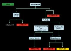
|
|
|
Toxins with characteristic odors: Freshly cut hay - Garlic - Bitter almonds - Rotten eggs - Pears - Glue - Fruity - Wintergreen - |
Freshly cut hay - phosgene Garlic - arsenic, organophosphates Bitter almonds - cyanide Rotten eggs - H2S Pears - chloral hydrate Glue - toluene, solvents Fruity - EtOH, acetone, isopropyl alcohol Wintergreen - methyl salicylate |
|
|
Indications and contraindications for whole bowel irrigation |
Body packers Sustained release Charcoal doesn't work High-risk hydrocarbons (CHAMP) -Camphor
Contraindicated in bowel obstruction, unable to protect airway, hemodynamically unstable |
|
|
Toxic DDx for pinpoint pupils |
Organophosphates Opioids
Clonidine
Phenothiazines PCP
GHB |
|
|
Antidotes
Acetaminophen Anticholinergics Arsenic, Lead, Mercury Benzo Black Widow Spider Bite Beta Blockers Calcium Channel Blockers Cyanide Digoxin Ethylene Glycol Hydrofluoric acid Iron Isoniazid Lead Methanol Methemoglobin forming agents Opioids Organophosphates, Carbamates Rattlesnake bite Serotonin Syndrome Sulfonulureas TCAs Valproic Acid
|
Acetaminophen - N-acetylcysteine Anticholinergics - Physostigmines Arsenic, Lead, Mercury - British anti-Lewisite, D-Penicillamine Benzos - Flumazenil Black Widow Spider Bite - Lactrodectus antivenin Beta Blockers - Glucagon, HIE, Lipids Calcium Channel Blockers - Calcium, Glucagon, HIE, Lipids Cyanide - Hydroxycobalamin, Sodium thiosulfate, Sodium nitrate Digoxin - Digibind Ethylene Glycol - Fomepizole, Pyridoxine, Thiamine Hydrofluoric acid - Calcium gluconate paste (make with lubricant), IM, intra-arterial, with Bier block, IV +/- dialysis Iron - Deferoxamine Isoniazid - Pyridoxine Lead - DMSA (succimer), EDTA Methanol - Fomepizole, Folic Acid Methemoglobin forming agents - Methylene blue Opioids - Naloxone Organophosphates, Carbamates - Atropine 2-4mg q5m, Pralidoxime 1g q1h; decontaminate with 5% hypochlorite Rattlesnake bite - CroFab antivenin Serotonin Syndrome - Cyproheptadine Sulfonulureas - Octreotide (inhibits insulin release), Glucagon, Glucose TCAs - Bicarbonate Valproic Acid - L-Carnitine
|
|
|
Stages of acetaminophen toxicity; how it is metabolized |
Stages 1 0– 24h - NV, anorexia, diaphoresis & malaise; May be completely asymptomatic 2 24-48h - NV, RUQ and epigastric pain, Transaminitis 3 48-96h - Fulminant hepatic failure – encephalopathy, coma, coagulopathy Hypoglycemia, Metabolic acidosis MODS: Sepsis, renal failure (25% of pts with severe hepatotoxicity), cerebral edema 4 - 4-14d - Liver enzymes return to normal; recovery
Mechanisms of metabolism -Glucuronidation 40-60% -Sulfation 20-40% -Direct renal excretion 5% -CYP450 2E1oxidation pathway 5-15% (toxic pathway) |
|
|
When can the Rumack-Mathew nomogram not be used? |
* Time of ingestion cannot be established |
|
|
Acid-base disturbances in salicylate toxicity; treatment |
-Respiratory alkalosis (early) - stimulates respiratory center in medulla
-Respiratory acidosis (late) - prolonged high ASA levels depress drive / LOC (preterminal)
-Metabolic alkalosis - dehydration, emesis, diaphoresis (contraction alkalosis)
-AGMA - early salicylates are a weak acid; late uncoupling of oxidative phosphorylation, increased lactate
Treatment: IVF rehydration, K replacement (needed to alkalinize urine), HCO3 to alkalinize urine to trap salicylates, dialysis for AMS, hepatic/respiratory/renal failure, rising salicylate, failure to respond/unable to use (fluid overload) to conservative tx |
|
|
TCA OD treatment and treatments that are contraindicated |
-AC / MDAC -HCO3 if QRS >100, Terminal RAD >120, Refractory hypotension, Ventricular dysrythmias, Seizures - bolus until improvement or pH >7.5 -Benzo's for seizures -Consider lidocaine for arrhythmias (all others contraindicated) -Cool prn -MgSO4 / overdrive pacing for long QT -Consider lipid emulsion in refractory cases
-Physostygmine, flumazenil and most antiarrhythmics are contraindicated |
|
|
Drugs that can cause serotonin syndrome |
Increased 5HT can occur through a number of different mechanisms: * ↑ 5HT release: ecstasy, mirtazepine
Also lithium |
|
|
Compare NMS, SS and MH problem / treatment |
NMS * Lack of dopamine
SS
MH |
|
|
Mechanism and 3 presentations of MAO-I toxicity; treatment |
Mechanism - prevents breakdown of NE / Epi
Presentations: Overdose - latent for 12-24h, then neuromuscular and cardiovascular excitation, then deplete catecholamines and crash
Food/MAO-i interaction - quick onset sympathomimetic crisis with HTN/HA that is short-lived (cheese, meat, fish, wine, beer, sauerkraut)
Drug/MAO-i interaction - quick onset sympathomimetic/serotonergic crisis that lasts as long as the drug (serotonin and sympathetic drugs)
Treatment - OD (benzo's for agitation, cool, phentolamine for early; IVF and pressors for late), Food (phentolamine), Drug (same as OD with extras if serotonin syndrome) |
|
|
Digoxin: mechanism, ECG findings, toxic presentation, treatment |
Mechanism - 1) increased automaticity due to increased intracellular Ca due to blockage of Na/K ATPase and 2) increased vagal tone causing decreased HR
ECG Findings - see U waves with scooped ST and shortened QTc Common: PVC's, AVB with increased ventricular automaticity, atrial tachy and vent brady due to ABV Rare: AF with slow ventricular rate, bidirectional VT NEVER: AF with rapid ventricular response
Toxic presentation - Metabolic: Hyperkalemia (accumulates due to all of the blocking of Na/K ATPase - predicts outcome!!) GI: Nausea, vomiting, anorexia CNS: lethargy, confusion, weakness (acute) and headache, delirium, yellow-green halos, snowy vision
Treatment - digifab! Lidocaine as antiarrhythmic if necessary (increases AV conduction and decreases automaticity), decrease shock energy and don't TV pace, avoid calcium (stoneheart)
|
|
|
Indications and dosing of DigiFab |
Indications -Levels of Dig (19nmol/L acute, 12nmol/L chronic) or K (>5mmol/L) -Rhythms (ventricular or hemodynamically unstable brady) -Ingestion of 10mg or >0.1mg/kg (child), cardiac glycoside with dysrhythmia, co-ingestion of cardiotoxic drug
Dosing -Empiric - Acute 10 vials; Chronic 5 vials; Arrest 20 vials; Plant 10-20 vials -Amount: Amount ingested (mg) x 0.8 (bioavailability) / 0.5 (vials needed / mg) -Steady state
Note that digifab WRECKS the levels! (Bound is counted the same as unbound) |
|
|
Treatment for BB and CCB OD |
Charcoal Atropine Calcium Glucagon Catecholamines (Isoproterenol, dopamine for BB; norepi or epi for CCB) Vasopression Insulin (1U/kg then 1U/kg/h) / Glucose (D50 amp then 0.5g/kg/h) Lipid emulsion (1.5cc/kg bolus then 0.25cc/kg/h infusion) Balloon pump ECMO |
|
|
How does lipid emluslion work? |
3 theories: -Lipid sink -Lipids as an energy source -Increased intracellular calcium |
|
|
Grades of caustic esophageal injury by endoscopy, contraindications to endoscopy |
Grade 1 (1°): edema and hyperemia
Grade 2 (2°): 2a = noncircumferential 2b = near-circumferential superficical ulcers, friability, white membrane, hemorrhage
Grade 3 (3°): transmural involvement with deep injury, necrotic mucosa, perforation
Increased grade = increased stricture
Contraindications - perforation! Do scope at 12-24h to optimize avoidance of this in significant ingestions. Can do earlier in ? ingestions. Surgery for perf! |
|
|
Complications of caustic ingestion |
Acute Airway burn -> Laryngeal edema Perf'd esophagus -> Mediastinitis Perf'd stomach -> Peritonitis GI bleed
Delayed -Esophageal stricture -Pyloric obstruction -Esophageal cancer (1000x) |
|
|
Effects and treatment of cocaine. Who needs admission? Why does it cause MI's? |
Releases DA, NE, Epi and 5HT and decreased pre-synaptic reuptake of NE, DA, 5HT -NE: vasoconstriction by stimulating alpha receptors (alpha agonist) -Epi: increases myocardial contractility and heart rate by stimulating B1 receptors (B agonist)
Fast Na channel blockade → local anesthesia + ↑ cardiac depolarization (↑ QRS)
Similar to TCAs; acts as a class Ia antidysrhtymic (blocks the fast Na channels; binds quickly during phase 0)
Vasoconstriction and platelet aggregation d/t ↑ endothelin production and ↓ nitric oxide production
Treatment: Benzo's (no haldol due to anticholinergic), Cooling, Phentolamine (1mg prn), IVF, intubate/paralysis prn, ASA/heparin/nitro if ACS
Admission: CAD or CAD risk factors, chest pain, cardiogenic shock, elevated cardiac markers, ECG changes, arrhythmias
Why does it cause MI's? Vasoconstriction, increased cardiac demand, decreased filling time, increased platelet aggregation |
|
|
Stimulant induced chest pain |
Cardiac -Endocarditis -Pericarditis -Ischemia/infarction -Stent thrombosis
Non cardiac -Pneumothorax -Pneumomediastinum -Pneumopericardium -Aortic dissection -Pulmonary infarction -Infection
Foreign body |
|
|
HEART score |
History - slight 0/ moderate 1 / high 2 ECG - normal 0 / nonspecific 1 / ST depression 2 Age - <45 0 / 45-65 1 / >65 Risk factors - none 0 / 1-2 1 / >2 2 Troponin - < limit 0 / 1-3x limit 1 / >3x limit 2 |
|
|
Methanol ingestion: metabolism, complications, treatment |
Metabolism: to formaldehyde then formic acid (detoxified with folate)
Complications: putaminal necrosis (in basal ganglia -> parkinsonism), optic neuropathy (blindness), increased free radicals, AGMA (formic acid and lactate due to cytochrome oxidase dysfunction)
Treatment: block conversion with fomepizole, HCO3 to keep formic acid trapped in serum; Folate to aid in decontamination, HD (if suspect ingestion, ph<7.3, HCO3<20, OG>10, >15mmol) |
|
|
DDx of osmolar and anion gap |
Methanol Ethylene glycol DKA SKA AKA Lactic acidosis Uremia |
|
|
Ethylene Glycol ingestion: metabolism, complications, treatment, stages of toxicity |
Metabolism: to Glycoaldehyde -> Glycolate -> Glyoxylate -> Oxalate -> Calcium Oxalate
Complications: AGMA (due to glycolic acid and lactate due to cytochrome oxidase dysfunction), hypocalcemia (chelation), crystal nephropathy, CNS punctate hemorrhages and aseptic meningoencephalitis, myonecrosis
Treatment: Block with fomepizole, HCO3 to keep ethylene glycol trapped, give Pyrodoxine, Thiamine, Magnesium to aid detoxification; correct hypocalcemia, HD > 8mmol
Stages of toxicity: I acute neurologic, II cardiopulmonary, III renal, IV delayed neurological |
|
|
Ethylene glycol vs methanol |
Methanol gives visual symptoms more often Methanol targets basal ganglia/putamen (EG more diffuse) Ethylene glycol causes hypocalcemia Ethylene glycol causes calcium oxylate crystals |
|
|
Serotonergic agents Entactogens Dissociative agents
|
Serotonergic agents: LSD, psilocybin (mushroom); panic attacks, psychosis, flashbacks
Entactogens: -Amphetamines, MDMA; hyperthermia, hyponatremia, SIADH -Bath salts; halucinogen and sympathomimetic -Mescaline; like LSD but with N/V
Dissociative agents: -PCP, ketamine; seizures, hyperthermia -Dextrometorphan; dissociative, opioid, and serotonergic (SS) |
|
|
Arsenic (gas, acute, chronic) Mercury (elemental, inorganic, organic) |
Arsenic -Acute salts give encephalopathy, ARDS, dysrhythmias, N/V/D -chronic salts give sensory neuropathy, Mee's lines, sideroblastic anemia, cancer. >Tx with WBI, BAL (IM), DMSA (PO)
Mercury -Elemental vapor (ARDS, pneumonitis) or injection (CNS/renal toxicity) or ingestion (nil) -Inorganic ingestion (ATN, gastroenteritis) or chronic (neurasthesia, nephritic syndrome, gingivostomatitis) > BAL (IM) / DMSA (PO) -Organic ingestion/dermal (neurotoxicity, ataxia, tremor, dysarthria) > DMSA (PO) but NOT BAL |
|
|
Sudden sniffing death syndrome |
-Happens in HC use. Blocks the delayed rectifier-K channel resulting in long QTc and ventricular arrhythmias. -Upregulation of catecholamine receptors on myocardium and sensitization; sudden surges of catecholamines from adrenaline surge. -Consider avoiding epi in this type of a code. -After resuscitated give benzo's, Mg, beta blockers, overdrive pacing |
|
|
List simple asphyxiants, pulmonary irritants, cellular toxins |
Simple asphyxiants: NO, CO2, N2, methane
Pulmonary irritants: Low solubility (Phosgene, NO2 - mucous membranes - upper airway) Medium solubility (Chlorine) High solubility (Ammonia, HF, HCl, H2S, SO2 - lower airway); considered nebulized HCO3
Cellular toxins: CN, H2S, CO
Thermal injury: smoke inhalation |
|
|
Cyanide toxicity and treatment |
Get AGMA with a crazy high lactate; MSOF
Treatment: hydroxycobalamin to form cyanocobalamin (B12) 5g empirically. Historically used amyl nitrite (if no IV) or sodium nitrite (if IV) to make methemoglobin and then Na thiosulfate to make excretable thiocyanate. ONLY thiosulfate or hydroxycobalamin in fires |
|
|
CO treatment; hyperbaric indications |
t1/2 on room air 4.5h; 100% 1.5h; hyperbaric 30m
Indications for hyperbaric oxygen: COHb >25% (anyone), >15% pregnant/child; symptoms (syncope/seizure/AMS/AMI/focal neuro/dysrhythmia); consider in other cases -> Does NOT decrease mortality; may decrease neuro sequelae |
|
|
Drugs that naloxone can reverse |
Opioids (all) Clonidine Tramadol EtOH Valproic acid |
|
|
Cholinergic drugs |
Organophosphates Carbamates Edrophonium/Tensilon for Myesthenia Gravis Donepizil/Aricept for Parkinsons Urocholine for bladder spasms Physostigmine |
|
|
Toxic seizures: effect of glutamate, GABA, benzo's, barb's, EtOH, pyridoxime
Treatments for benzo, barb OD |
-Glutamate excites; GABA inhibits w Cl channel, Benzo's potentiate GABA (need it!), Barb's and EtOH keep Cl channel open longer (no GABA needed!). GABA is made by Glutamine using Pyridoxime
Barb OD: respiratory depression! Intubate and treat with MDAC
Benzo OD: flumazenil generally not recommended especially if ictogenic coingestants, seizure disorder, withdrawl, paralyzed |
|
|
Gamma hydroxybutyrate uses, effects, presentation |
-Used recreationally, for date rape, and for bodybuilding -Affects dopamine release (inhibits and releases) and binds GABA -Present with decreased LOC, respiratory effort, emesis and mioisis. Rapidly fluctuating LOC and agitation. |
|
|
What is DRESS? Treatment? |
Drug Rash with Eosinophilia and Systemic Symptoms
Found in anticonvulsant hypersensitivity (carbamezapine, phenytoin, lamotragine, phenobarb). Usually in first 2months of therapy. Can cause MOSF (nephritis, carditis, pneumonitis) as well as tight blisters, morbilliform rash, facial edema.
Treatment Withdrawl of the causative drug. Steroids are controversial. Consider NAC. |
|
|
Phases of cold injury / frostbite and treatment |
Prefreeze: <10 degrees, loss of sensation, endothelial leak
Freeze-thaw: extra-cellular ice crystal formation, extracellular shift of fluid, cells can begin to die
Microvascular collapse: intravascular sludging, hyperviscosity, thrombosis, ischemia, necrosis
Treatment: rapid warming in 37-39 degree water, do NOT let refreeze or partial thaw; pain control; watch for afterdrop; tetanus; consider abx and splinting; stop smoking; consider thrombolytic or sympathectomy |
|
|
Non freezing injuries |
Frostnip - superficial injury, no tissue death, resolves with warming
Chilblains - repetitive dry cold, cold 'sores' to face and hands (erythema, pruritis, edema), can ulcerate, more likely in Raynaud's & APLS, can tx with nifedipine
Trench foot - prolonged wet cold >0 degrees. Wet socks. Rubor when dependent, pallor when elevated (vasomotor paralysis). Painful. Can get bullae. |
|
|
Sequelae of frostbite |
Neuropathic -CRPS -Dysesthesia /paresthesia /anesthesia /hypesthesia -Heat/cold sensitivity -Hyperhidrosis -Raynaud's
MSK -Atrophy -Compartment syndrome -Rhabdomyolysis -Stricture -Necrosis -Amputation
Derm -Edema -Lymphedema -Ulcers
Miscellaneous -Afterdrop -Electrolyte abnormalities -ATN -Sepsis |
|
|
Dive injuries on descent, at depth, on ascent, and require decompression therapy |
Descent: IEBT, MEBT, EEBT, Facial barotrauma, barosinusitis
Depth: Contaminated gases, Nitrogen narcosis, Oxygen toxicity
Ascent: Arterial gas embolism, Pulmonary edema, Barotrauma (alternobaric vertigo, pneumothorax, pneumomediastinum, barodontalgia, GI barotrauma)
After surfacing: DSC
Decompression therapy: DCS I, DCS II, Arterial gas embolism, contaminated gas (CO) |
|
|
Acute mountain sickness / high altitude cerebral edema treatment |
-Descent or discontinue ascent -Hydration -No smoking -Tylenol / Advil -Oxygen -Prochlorperazine - stimulates hypoxic ventilatory response -Acetazolamide - prevents periodic breathing at night; helps adapt by excreting HCO3 -Dexamethasone - decreases swelling
|
|
|
High altitude pulmonary edema treatment |
-Descent -Salbutamol -Oxygen -Nifedepine - decreases pulmonary vasoconstriction -Sildenafil - decreases pulmonary vasoconstruction -Hyperbaric oxygen or CPAP |
|
|
Physiological changes associated with high altitude acclimitization |
-Increased HR, BP, and venous tone due to catecholamine release -Increased hemoglobin (due to increased Epo and fluid shift) -Increased 2,3 DPG and right shift of O2 dissociation curve (more O2 for tissues) -Increased minute ventilation (decrease PaCO2 and increased PaO2) -Increased renal excretion of HCO3
Combined these last two are: -Hypoxic ventilatory response (hypoxia induces hyperventilation which blows down PaCO2 causing kidneys to compensate by excreting HCO3) |
|
|
Clinical features of heat stroke |
-Temperature >40.5 -CNS dysfunction (delirium / coma) -High output CHF -Centrilobular necrosis in liver (AST / ALT >10,000) -Elevated lactate -Rhabdomyolysis -ATN -Coagulopathy / DIC -Hypocalcemia and Hyponatremia |
|
|
Differentiating features between heat stroke and heat exhaustion |
-Heat stroke has neurological symptoms -Heat stroke has a temperature >40.5 -Heat stroke AST and ALT are much more elevated (in the 10000's) - elevated LFT's are a common lab abnormality in both |
|
|
Temperature of hypothermic physiological changes |
28 - VF 30 - ACLS meds effective 32 - shivering loss 34 - ataxia and apathy |
|
|
Compare DCS I vs II |
DCS I (the bends) affects MSK, skin, lymphatics. Get cutis marmorata, periarticular pain, peau d'orange,
DCS II (any other organ system: CNS (spinal or cerebral) Inner ear (staggers - similar to IEBT) Pulmonary (chokes) Fetal |
|
|
Signs of hypothermia on ECG |
-Osborne waves -Shivering artifact -Bradycardia -Prolonged PR, QRS, QTc -Ectopic ventricular beats -Atrial fibrillation -AVB's -VT/VF/Asystole |
|
|
What effect does warming the blood of a hypothermic patient have on the patient's ABG results? (pH, pO2, PCO2) |
pH - lower pO2 - higher pCO2 - higher |
|
|
Contraindications to high altitude travel |
4 -COPD -Pulmonary hypertension -CHF -Sickle cell
Relative contraindications -Pregnancy -Radial keratotomy -Seizure disorder -Previous altitude sickness |
|
|
Treatment of frostbite |
-Pain control -Warm the limb in 37-39 degree water (do not allow partial rewarming or refreezing) -Warm the patient with monitors
Consider thrombolytics, heparin, tetanus, antibiotics, smoking cessation, debridement |
|
|
Hypothermia stages |
Mild 34-36 Moderate 30-34 Severe <30 |
|
|
Indications for active rewarming in hypothermia |
Moderate or severe hypothermia No shivering Cardiovascular instability Co-morbidities (DM, trauma, endocrine) Failure to rewarm Toxicologic hypothermia Septic hypothermia Infants and elderly |
|
|
Risk factors for decompression sickness |
-dehydration -prolonged dive -inexperienced diver -not using dive tables -multiple dives in a short period -depth of dive -flying after diving -exceeding no-decompression limits -elderly -obesity -cold after diving |
|
|
Define: Drowning, Immersion syndrome, Diving reflex |
Drowning Respiratory impairment from submersion/ immersion in liquid that can cause morbidity and death
Immersion syndrome Syncope or cardiac arrest following sudden immersion in water with a more than 5 degree change from core temperature (results in a vagal response +/- vasoconstriction)
Diving reflex The immersion of the face in cold water shunts blood to the heart and brain producing apnea and bradycardia and prolonging the duration of submersion tolerated |
|
|
What factors indicate a poor prognosis following drowning / near-drowning? |
-Temperature of patient -Age (young) <3 -Duration of submersion > 5 min -Pulse (presence of) -Neurologic status at time of arrival to ED -Bystander CPR delayed > 10 min -Acidosis -Fixed, unreactive pupils -GCS 3 |
|
|
Pathophysiology of arterial gas embolism following scuba diving |
At depth there is an increased solubility of nitrogen which accumulates in the body while the diver is at depth (Henry's law). Upon rapid ascent, the nitrogen comes out of the solution resulting in bubbles which collectively form a gas embolism (Boyle's law) |
|
|
Stages of wound healing |
Coagulation - hours - 2 days Collagen - days - peaks at 7 days; 60% strength at 4 weeks Contraction - begins at 4 days Epithelialization - new skin; can complete over sutured lacerations within 48 hours |
|
|
Indications for admission of a fight bite |
Patient -Immunocompromised -DM -Unreliable patient
Wound -Signs of infection -Open >24 hours -Penetration of joint/tendon sheath -Bone involvement -FB
Treatment -Clavulin as an outpatient; Tazocin as an inpatient |
|
|
Drugs that can cause methemoglobinemia and its treatment |
Nitrates (NTG, nitroprusside) Local anesthetics (eg benzocaine, lidocaine, prilocaine) Dapsone Primaquine Antimalarials (quinine, chloroquine) Paraquat Naphthalene Methylene blue
Treatment: Methylene Blue - reduces Fe3+ back to Fe2+ |
|
|
Drugs that require quantitative measurement for treatment in overdose |
Acetaminophen Ethylene glycol Methanol Digoxin Carbon monoxide Lithium Aspirin Theophylline Valproic acid
|
|
|
Contraindications to succynilcholine |
PMHx -MH (any time) -Pseudocholinesterase deficiency (anytime)
Recent PMHx -Burns (>10%, >5 days until healed) -Crush injury (>24h) -Stroke/denervation with paralysis (>5 days to 6 months) -NM disease (>6 months)
Current presentation -HyperK (any time) -Cholinergic toxicity |
|
|
Consideration for deciding to give AC |
(1) Toxin can be bound by AC (2) Ingestion <1h generally, sometime <2h or longer (especially if ingestion slows gastric motility) (3) Protecting airway (4) No known antidote (5) No bowel obstruction / ileus (6) Ingested substance / amount likely to be toxic |
|
|
Pathophysiology of carbon monoxide toxicity
Indications for hyperbaric oxygen in carbon monoxide toxicity |
Pathophysiology -CO binds to cytochrome 4 of ETC causing cellular asphyxiation -displaces O2 and shifts oxyhemoglobin curve to left -binds to myoglobin causing rhabdomyolysis
HBO >25% carboxyHb level >15% carboxyHb level in pregnant or children CVS instability Neurological symptoms |
|
|
Mechanisms of NAC in acetaminophen toxicity |
(1) Glutathione precursor (2) Glutathione substitute (3) Enhances sulfation pathway leading to non-toxic metabolites (4) Antioxidant effects and free radical scavenger (5) Enhances hepatic microcirculation |
|
|
Treatment of box jellyfish sting |
-Box jellyfish antivenom -Verapamil IV -Remove nematocysts with razor / credit card after deactivating with vinegar -Analgesia |
|
|
How is volume of distribution calculated? |
Ingestion (mg) / Concentration (mg/L) = Distribution (L)
Distribution (L) / Patient weight (kg) = Vd (L/kg)
Very low (<1) = dialyzable High = not dialyzable |
|
|
Drugs that most commonly cause adverse reactions in elderly patients |
-Cardiovascular -Diuretics -Non-opioid analgesics -Hypoglycemics -Anticoagulants -Sedatives |
|
|
Drug side effects: Delirium, Syncope, GIB, Tinnitus |
Delirium -Benzodiazepines (lorazepam, diazepam) -Opioids (morphine, fentanyl) -Hypoglycemics (glyburide, insulin) -CVS (BB, CCB) -Anticholinergics (benadryl)
Syncope -Antiarrythmics (IA, IC, II, III, IV) -Antibiotics (quinolones, macrolides) -Antipsychotics (chlorpromazine, haloperidol), --Diuretics via electrolyte disturbance -Antihypertensives (Clonidine, nitrates) -Hypoglycemics (glyburide, insulin)
GI bleeding -Anticoagulants (coumadin) -Antidepressants (SSRI) -NSAIDs (toradol) -Steroids
Tinnitus -Salicylates -Lithium -Diuretics (Furosemide, Acetazolamide) -Aminoglycosides -Antimalarials (chloroquine, quinine) |
|
|
Theophylline (mechanism of action, presentation in toxicity, cause of death, treatment) |
Mechanism: 1) PDE inhibitor - raises cAMP 2) Adenosine antagonism → cardiac effects 3) direct beta- and alpha-adrenergic agonism
Presentation: -GI - Nausea/Vomiting -CVS - Tachydysrhythmias -Resp - Tachypnea & resp alkalosis, ALI -CNS - Seizure, anxiety / Agitation -MSK - Rhabdomyolysis -Metabolic - AGMA (lactate), hypoK, hypoMg, hyperglycemia
Cause of death: -Arrhythmia in chronic -Status epilepticus in acute
Treatment: -MDAC if early -Dialysis or charcoal hemoperfusion |
|
|
Heroin: drug complications, IVDU complications, adulterant complications |
Drug - Seizure, Coma, Death - Apnea/hypoxia, Non-cardiogenic pulmonary edema - Bradycardia, Hypotension - Hypothermia
IVDU - HIV, Hep B, Hep C - Sepsis, Endocarditis - Cellulitis, Abscess - DVT, Thrombophlebitis
Adulterant - Botulism - Enterobacter agglomerans (cotton fever) - Agranulocytosis (levamisole) - Sepsis/infection |
|
|
Name 3 amide and ester local anesthetics |
Amides Bupivicaine, lidocaine, prilocaine
Esters Tetracaine, Benzocaine, Cocaine |
|
|
ECG findings of lithium toxicity; drugs that cause lithium toxicity |
ECG findings -QT prolongation -Sinus Bradycardia -T-wave inversion or flattening -ST depression -U wave -SA block -First degree AV block
Drugs that cause lithium toxicity -ACE inhibitor - Ramipril -ARB - Valsartan -NSAID - Naproxen -Diuretics - Lasix, thiazides |
|
|
Indications for laparotomy in penetrating abdominal trauma |
DIE Unstable PIG!!
D iaphragmatic injury I mplement in situ
P eritoneal signs G I hemorrhage / G SW to abdomen |
|
|
Most common abdo injury in stab wound
Most common abdo injury shooting
Most common abdo injury blunt trauma |
Most common abdo injury in stab wound Liver
Most common abdo injury shooting Small bowel
Most common abdo injury blunt trauma Spleen |
|
|
Indications for reimplantation of amputation; contraindications to reimplantation; relative contraindications to reimplantation |
Indications of replant -Multiple Digits -Single digit between PIP & DIP (insertion of tendons between there) -Thumb -Wrist/forearm -Sharp amputation proximal to elbow -All pediatric amputations
Absolute contraindications to reimplantation -Crush injury -Unstable patient
Relative contraindications -Severe multilevel -Self inflicted -Extremes of age -Serious comorbidity -Proximal to PIP |
|
|
Signs of urethral injury. Most reliable in female? |
-high riding prostate -blood at the meatus -open pelvic fracture -Scrotal hematoma -Penile Hematoma -perineal hematoma -failure to pass foley x 1 -Fractured penis
In female the most reliable sign is inability to pass a Foley catheter |
|
|
False positive free fluid on FAST |
-peritoneal dialysis -ascites -physiologic free fluid in female -urine from bladder rupture -Ruptured Ectopic Pregnancy -PID -fluid in bowel or stomach -mesenteric fat -Operator inexperience |
|
|
False negative free fluid on FAST |
250-500mL of fluid must be present to be visible on FAST (Sn 60-99%; Sp 80-99%)
-small volume -Early in trauma -Adhesions -Timing (not enough preceding time supine) -Operator inexperience |
|
|
Most common fractures in the elderly (UE and LE) |
UE -Distal radius (50%) -Proximal humerus (30%)
LE -Hip |
|
|
Clinical and radiographic findings of an orbital fracture |
Clinical findings -Enophthalmos -Diplopia -Inability to gaze upward; Pain or Restrictions of EOM -Step deformity palapable -Subcutaneous emphysema -Infraorbital nerve anesthesia
Radiographic -Tear drop sign (opacification in the shape of a tear at the bottom of the orbit) -AFL in maxillary sinus -eyebrow sign (lucency in the shape of an eybrow at the top of the orbit) |
|
|
Clinical findings of anterior chamber trauma |
-Hyphema -Flare -Iridodialysis -Ciliary flush -Sluggish Pupil (cyclitis) -Deep anterior chamber from posterior displacement of iris -Seidel's sign |
|
|
Slit lamp findings of blunt trauma to the sclera or lens |
-Abrasion - Positive Fluroscein uptake -Scleral laceration -Positive Siedel’s -Subconjunctival hematoma -Iridodonesis (iris movement with eye movement - due to lens dislodgement) |
|
|
Slit lamp findings of penetrating globe injury |
-Decreased Anterior Chamber depth -Positive Siedel’s -Hyphema -Scleral laceration -Bloody chemosis -Retained implement (FB) -Corneal Laceration -Teardrop pupil -Iris prolapse -iris transillumination defect due to vitreous hemorrhage |
|
|
ECG changes seen in massive head injury |
-Bradycardia -Deep T-wave inversion -ST seg Elevation -ST seg depression -QT Prolongation -PR Prolongation -RBBB / incomplete RBBB -Junctional Rhythm, Sinus Dysrhythmias, Sinus Tachy -U wave > 1mm |
|
|
Physical exam findings of basilar skull fracture |
-Racoon Eyes -CSF Otorrhea -CSF Rhinorhea -Hemotympanum -Battle Sign -Acute hearing loss (CN8 injury) -Bilateral CN 7 palsy (looks like ‘bilateral bells’) -blood in the external auditory canal |
|
|
Systemic manifestations of hydrofloric acid exposure |
-Metabolic Acidosis -Hypocalcemia -Dysrhythmias -Seizures -Tetany |
|
|
Clinical criteria for massive hemothorax |
->1500 mL immediate output blood from chest tube ->200mL/h drainage x 2-4h -Absent breath sounds, dullness to percussion, and shock with or without tracheal deviation |
|
|
Causes of persistent air leak on a chest tube |
-Tracheobronchial transection -Bronchopulmonary fistula -**Incomplete insertion of chest tube** -Leak in the chest tube system |
|
|
Canadian CT Head Rule (high risk, low risk, inclusion criteria, exclusion criteria) |
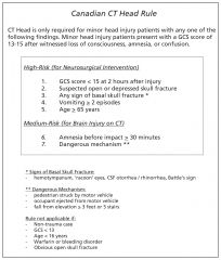
|
|
|
Contraindications to cricothyrotomy |
-Laryngeal # -< 8 yoa -anterior neck hematoma |
|
|
Physiologic changes that affect the pregnant trauma patient |
-Gravid uterus compresses IVC (supine hypotension) -Baseline diastasis of pubic symphysis -laxity of pelvic ligaments -Physiologic anemia -Increased blood volume allowing for greater compensation for blood loss but rapid deterioration when reach maximal compensation -increased minute ventilation -Relative hypocarbia -Higher resting HR -Higher resting RR, blood gas with PCO2 30, HCO3 21 -Lower FRC -Abdominal viscera protected by uterus and difficult to examine (abdo exam unreliable) |
|
|
2 additional management considerations in the initial resuscitation of the pregnant trauma patient |
-Supine hypotension due to compression of IVC by uterus (lean to the left 30 degrees) -Feto-maternal hemorrhage - do type and Kleihauer-Betke test and treat with WinRho prn |
|
|
Complications and non-pharma treatments of rib fractures |
Complications: -Pneumothorax -Hemothorax -Pain -Pneumonia
Non-pharma treatments: -Incentive spirometry -Coughing / Deep breathing exercises -IPPV -Internal Fixation
|
|
|
Factors affecting the rate of heat transfer between objects |
-Duration -Transfer Coefficient of objects -Temperature Differential -Heat Capacity -Conductivity of tissues |
|
|
Risk factors for falls in the elderly |
-Poor vision -Poor coordination -Position hypotension -Poor hearing -Cachexia |
|
|
Reasons for non-compliance with medications in the elderly |
-Dementia -Polypharmacy -Increased side effects -Financial constraints |
|
|
NEXUS Rule |
NSAID
-No N euro deficits -No S pinal tenderness -No A ltered LOC -No I ntoxication -No D istracting injury |
|
|
Low to high dose trauma imaging |
Least
CXR Abdo Flat Plate KUB RUG CT Abdo
Most |
|
|
How to perform a retrograde urethrogram |
-Flex hip/knee -Displace penis from midline -Shoot KUB -Toomey syringe with water-soluble contrast (60ml) -Infuse over 1 min, KUB during last 10ml |
|
|
Treatment of arterial air embolism |
-Trendelenburg or LLD position -Needle aspiration -Thoracotomy -Aspiration via central line -Compression of source vessel -IVF |
|
|
What is the appropriate imaging study for penetrating flank trauma? |
CT-Chest/Abdo/Pelvis with Contrast (triple phase - oral / IV / rectal) |
|
|
Hyphema grading system |
Micro - just anterior chamber RBC's I - <1/3 II - 1/3 - 1/2 III - >1/2 IV - full (eight ball hyphema) |
|
|
Complications and management of hyphemas |
Complications -Traumatic Glaucoma -Synchiae -Staining of cornea -Rebleed -Blindness
Management 1) Conservative Mgt - HOB up, metal shield 2) Long acting cycloplegic (homatropine, Atropine) 3) Stop anticoagulants, ASA; avoid NSAIDs 4) Manage IOP (traumatic glaucoma) 5) R/O globe rupture |
|
|
Components of informed consent |
CAPACITY -Capacity assessment (CURVES)
DISCLOSURE -Nature of the treatment -Benefits and risks of the treatment -Common/dangerous side effects of the treatment -Alternatives to the treatment (including not having anything done) and their likely outcome
VOLUNTARY -Consent given voluntarily and without coercion |
|
|
Zone 1 of the neck and associated injuries |
Angle of mandible to occiput (upper neck)
jugular Lung Trachea superior mediastinal vessels Spinal cord Thoracic duct Thyroid Subclavian artery Carotid artery Vertebral artery Esophagus |
|
|
Zone 2 of the neck and associated injuries |
Zone II - Angle of jaw to cricoid cartilage
Recurrent Laryngeal N. Larynx Pharynx Thyroid cartilage Esophagus Spinal Cord Carotid Jugular Vertebral Vagus nerve Recurrent laryngeal nerve |
|
|
Zone 3 of the neck and associated injuries |
Zone I - cricoid to clavicles and suprasternal notch
Brain Stem (IX-XII) Carotid Jugular Parotid gland Vertebral Artery Spinal Cord Salivary glands |
|
|
Indications for operative management of penetrating neck trauma |
-Hard signs in zone 2 -Unstable -Transcervical gunshot wound -Platysma violation in zone 2 (relative) |
|
|
Anatomic locations commonly involved in traumatic aortic injury |
-Isthmus (Ligamentum Arteriosis) -Root (Ascending distal to the Aorta) -Distal descending at the diaphragm |
|
|
Immediate management of a pressure gun injury |
Cleanse Splint Td Analgesia Antibiotics Examine for compartment syndrome Plastics for admission |
|
|
What key feature distinguishes spinal and neurogenic shock? |
-SPINAL SHOCK – bulbocavernosus reflex lost; refers to neuro symptoms -NEUROGENIC SHOCK - shock that occurs due to disruption of sympathetic outflow; bradycardic |
|
|
Intra vs Extraperitoneal bladder injury (mechanism, location, treatment, clinical, cystogram) |
Mechanism Intra: Blunt Extra: Penetrating
Location Intra: Dome Extra: Anterior
Treatment Intra: OR Extra: Foley unless bladder neck, rectal, vaginal injury (then OR)
Clinical Intra: hematuria, anuria, pain, meatal blood, peritonitis and abdominal distension Extra: hematuria, anuria, pain, meatal blood, pelvic fracture
Cystogram Intra: enhances colon and other intraperitoneal structures Extra: remains in pelvis |
|
|
Uncal herniation (anatomy, pathophysiology, physical exam) |
Anatomy - Ipsilateral uncus of the temporal lobe herniates down through the tentorium
Pathophysiology - Compresses ipsilateral CN III and cerebral peduncle of midbrain
PE - decreased LOC, ipsilateral blown pupil, ipsilateral 'down and out' gaze, contralateral paralysis (opposite in Kernohan's notch syndrome) |
|
|
Descending transtentorial herniation (anatomy, pathophysiology, physical exam) |
Anatomy - expanding lesion in the vertex / frontal / occipital poles or diffuse swelling
Pathophysiology - Cerebrum and midbrain pushed down through the tentorium
PE - decreased LOC, small but reactive pupils, posturing, sunset eyes (can't move up) |
|
|
Cerebotonsillar herniation (anatomy, pathophysiology, physical exam) |
Anatomy - tonsils herniate through the foramen magnum
Pathophysiology - cerebellar or large cerebral mass push brain down compressing the brainstem
PE - decreased LOC, flaccid quadriplegia, posturing, respirations cease |
|
|
Define flail chest and how it causes respiratory compromise |
A flail chest has at least 3 continuous ribs, 2 breaks each. This results in paradoxical breathing and inadequate inspiration / expiration. It is also very painful |
|
|
PE findings of tension pneumothorax |
Mediastinal shift Decreased AE to affected side Tracheal Deviation Subcutaneous emphysema Hypotension JVD |
|
|
Benefits and drawbacks of ultrasound in trauma |
Benefits -No transfer required -Less resource intensive (Less expsenive) -Available in trauma suite (Bedside) -Easily repeated -No contrast -No radiation -No need for IV Access -Quick -Non-invasive -Sensitive to ~250mL (variable)
Drawbacks -Does not visualize parenchyma, retroperitoneum -Poor at identifying hollow viscous -Compromised by subcut air -False negatives - ascites -User-dependent |
|
|
Injuries associated with lap belts |
-Chance # -Pancreatic Injury -Duodenal Injury -Mesenteric Injury -Bladder injury |
|
|
Common blast injuries |
-Hollow viscous -Dismemberment -TM rupture -Air embolism -Pneumothorax -Blast lung |
|
|
Lab changes in pregnancy |
Leukocytosis Relative anemia Low Hematocrit PCO2 down Elevated D-Dimer Increased fibrinogen |
|
|
PDA - when does it close? PGE1 - indications and complications |
Closure -Physiologic at 10-15 hours -Anatomic at 30 days
PGE1 indications -Cyanosis refractory to oxygen -Refractory shock -Refractory cyanosis AND shock
Complications -Apnea -Fever -Brady -Seizures -Flushing -Hypotension -Decreased platelet aggregation |
|
|
NRP algorithm - indications for entering and 6 steps |
Enter algorithm if problem with Term / Tone / Breathing 1 - Warm, position airway, dry, stimulate x 30s 2 - If SpO2 not at target - supplemental O2 prn 3 - If HR<100, gasping, apnea, O2 not rising - Assist breathing with PPV x 30s 4 - If HR<60 - ETI and Chest compressions (rate of 90; 3:1 compression:breath ratio) 6 - If HR still <60 - Epinephrine, consider hypovolemia or PTx
|
|
|
Targeted preductal SpO2 after birth |
1m - 60-65% 2m - 65-70% 3m - 70-75% 4m - 75-80% 5m - 80-85% 10n - 85-95% |
|
|
Indications for neonatal intubation, tube size |
6 reasons
HR< 60 Meconium aspiration in a flat baby Ineffective or prolonged PPV Congenital diaphragmatic hernia <1000g Delivery of meds
Tube size 3.5 if TERM |
|
|
The H's and T's |
5 H's H ypoxia H ypovolemia H ydrogen ion (acidosis) H ypo / H yper kalemia H ypothermia
5 T's T ension pneumothorax T amponade T oxins T hrombosis (pulmonary) T hrombosis (coronary) |
|
|
Doses of inotropes / vasopressors (epi, norepi, dopamine, dobutamine, phenylephrine) |
Epinephrine - 0.05-0.5mcg/kg/m Norepinephrine - 0.05-0.5mcg/kg/m (5-20mcg/m push) Dopamine - 5-20mcg/kg/m Dobutamine - 5-20mcg/kg/m
Phenylephrine - 5-20mcg/m push |
|
|
Mixing push-dose epinephrine and phenylephrine |
Epinephrine 1mg / vial 1mcg/mL in 1L 10mcg/mL in 100mL 100mcg/mL in 10mL
Phenylephrine 10mg / mL 100mcg/mL in 100mL 40mcg/mL in 250mL |
|
|
BLS: trained provider vs untrained provider |
Both -AED -911 -Trade
Untrained provider -Hands only CPR (2 inches or 1/3 of chest) at 100/m
Trained provider -30:2 compressions:breaths if 1 OR 2 providers at 100/m in adults -15:2 compressions:breaths if 2 providers at 100/m in children |
|
|
CPR with advanced airway |
Both adults and children -8-10 breaths/m (q 6-8s) with each breath lasting 1s and not synchronized with compressions (don't stop!) |
|
|
BLS Choking |
<1y -Alternate 5 back slaps and 5 chest thrusts until unresponsive
1 to puberty -Abdominal thrusts until unresponsive
Puberty to adult -Abdominal thrusts UNLESS pregnant or obese (then chest thrusts) until unresponsive
All -If unresponsive with no or agonal breathing begin CPR -Check mouth for FB q breath stop |
|
|
Key etCO2 readings during CPR
Uses in CPR |
Key readings: If <10 improve CPR or patient dead If >40 expect ROSC
Key uses: -Monitor quality of CPR -Monitor tube placement -Monitor for ROSC during CPR (will see increase >= 40mmHg) -Determine if ROSC unlikely (persistently <10mmHg) |
|
|
Characteristics of benign early repolarization |
Diffuse Temporally stable Highest in V2-5 Concave up QRS notched at J point Concordant T's |
|
|
DDx of inverted T's |
Normal in children / persistent juvenile pattern Intracranial hemorrhage Myocardial infarction BBB Strain in ventricular hypertrophy Hypertrophic cardiomyopathy PE |
|
|
Indications for rescue PCI |
4 indications CP not resolved Persistent ST elevation (especially of >50%) Hemodynamically unstable |
|
|
Indications to transfer a patient for primary PCI |
-First medical contact to needle time can be <120m -Contraindications to lytic -Presented >12h post onset of CP -Ventricular dysrhythmias -Requires rescue PCI (CP, hemodynamically unstable, persistent STE >50%) |
|
|
Risk of bleeding on an anticoagulant |
HAS BLED
H ypertension A bnormal liver/renal function S troke history
B leeding predisposition L abile INR E lderly (>65) D rugs / EtOH usage |
|
|
Causes of high output heart failure |
AV fistula Pregnancy Cirrhosis Anemia Beriberi Thyrotoxicosis Paget's disease |
|
|
Echo findings of tamponade |
-RA compression -RV collapse -Hepatic vein dilation -Dilated IVC (no collapse with respiration) -Abnormal TV and MV flow velocities |
|
|
Causes of culture negative endocarditis |
HACEK
H aemophilus aphrophilus A ctinobacillus C ardiobacterium hominus E ikenella corrodens K ingella Kingae |
|
|
IJ central line complications and treatment |
1) Air embolism - supportive care, fluids, aspirate air, thoracotomy 2) Dysrhythmia - pull back insulting device (wire, line) 3) Pneumothorax - chest tube, O2, aspiration, Hiemlich valve 4) Arterial puncture with finder needle - with draw 5) Hematoma - pressure 6) Bleeding - pressure 7) Right atrial or ventricular perforation - Cardiac surgery consult |
|
|
3 causes of no detectable etCO2 in cardiac arrest |
-Dislodged/misplaced tube -Obstruction of tube (massive PE) or pulmonary edema -Prolonged arrest or downtime -Severe asthma -Large glottic air leak -Equipment failure -Transient decrease expected after administration of epinephrine (vasoconstricts pulmonary blood flow) |
|
|
Indicators of inadequate blood flow during CPR |
-Carotid or femoral pulse not palpable -CPP <15mmHg -EtCO2 <10mmHg -ScvO2 <30% -Art line DBP <20mmHg |
|
|
Contraindications to CPR |
-DNR -Danger to bystander -Irreversible death (Rigor mortis, decomposition, decapitation, lividity)
In neonates (anencephaly, trisomy 13/18, <400g, <23 weeks, SFH < umbilicus) |
|
|
Contraindications to IO line at a specific site |
-Osteoporosis -Osteogenesis imperfecta -Fractured bone -Recent IO site -Needle insertion through areas of cellulitis, infection, burns |
|
|
Complications of an IO line |
-Fracture -Fat embolism -Pain with infusion -Compartment syndrome -Skin sloughing -Osteomyelitis -Epiphyseal damage |
|
|
Physical exam findings of aortic dissection |
-Hypertension -Pulse Deficit -BP differential between two limbs -Signs of cardiac Tamponade -Stroke like syndrome |
|
|
Complications of aortic dissection |
-Pericardial Tamponade -Inferior MI -Aortic valve insufficiency -Ischemia -> spinal (Art of Adamkiewcz) -> ischemic stroke -> mesenteric ischemia -> renal ischemia |
|
|
Signs of SAH on CT scan |
-Blood in the basilar cisterns >> Interpeduncular [anterior to pons just superoposterior of the suprasellar] >> Suprasellar cistern [star sign] >> Prepontine cistern [just inferoposterior suprasellar anterior to the pons] >> Ambient cistern [bottom star legs around the midbrain] >> Sylvian fissue >> Anterior interhemispheric fissure
-Isodense basilar cisterns
-Hydrocephalus (temporal horns and infundibulum of 3rd ventricle)
-Reflux of blood into the 4th ventricle |
|
|
Hunt & Hess grading system for SAH |
0-5 (prognosis good for 0-1; intermediate 3; poor 4-5)
0 - Unruptured 1 - Asymptomatic / headache 2 - mod-severe headache, nuchal rigidity, nothing focal 3 - decreased LOC, mind focal deficit 4 - stupor, hemiparesis 5 - coma, decerberate |
|
|
Etiology of nontraumatic SAH |
B CARMEN
B lood dyscrasias
C avernous angioma A VM R uptured saccular aneurysm M ycotic aneurysm E xtension from intraparenchamyl N eoplasm |
|
|
Noncontrast CT findings of stroke |
Acute (cytotoxic edema) -visualization of the clot/embolism (e.g. hyperdense MCA sign) - immediate -loss of grey-white distinction (e.g. lentiform nucleus and insular ribbon) - 1-3h -Low attenuation of grey matter -Effacement of the sulci
Subacute (vasogenic edema) -Wedge shaped area of low attenuation of both gray and white -Mass effect (3-5 days)
Chronic (3 weeks - 1 year) -Resorption of infarcted area (looks like CSF) -Volume loss (negative mass effect dilating the ventricles) |
|
|
Classic stroke neurological findings (AMA, MCA, PCA, Lacunar, Verterbrobasilar, Basilar, Cerebellar) |
AMA - contralateral weak/numb legs > hand/face MCA - contralateral weak/numb hands/face > legs PCA - contralateral visual field and light touch Lacunar - pure motor OR sensory Vertebrobasilar - ipsilateral cranial nerve and contralateral weakness Basilar - 'Locked in' syndrome (normal mental status and blinking, can't move voluntary muscles) Cerbellar - dysmetria, dysdiadokinesia, can't walk, N/V |
|
|
Brown-Sequard Syndrome deficits, causes |
Deficits: ipsilateral proprioception / vibration / motor; contralateral pain and temperature
Causes: penetrating injury, cord compression |
|
|
West Nile Virus: type of virus, vector, diagnosis, presentation |
Type: Flavivirus
Vector: Birds (crows/ravens/jays) and Culex mosquitos
Diagnosis: WNV IgM in serum or CFS (crossreacts with infection / immunization for Japanese encephalitis virus serocomplex - St. Louis encephalitis; dengue)
Presentation: Most often fever, malaise, myalgias, headache, nausea, emesis, rash, lymphadenopathy. Occasionally in the elderly (>70) get weakness -> paralysis; meningitis -> encephalitis; morbidity/mortality.
Treatment: supportive.
Prevention: mosquito repellents, reducing mosquito numbers |
|
|
4 tributaries to Kiesselbach's plexus, causes of anterior epistaxis |
-Anterior ethmoidal artery -Septal branch of superior labial artery -Nasopalatine branch of sphenopalatine artery -Greater palatine artery
Causes -Traumatic -Cocaine -Vasculitis -Platelet -FB -Polyp |
|
|
When do the facial sinuses become aerated? |
Birth - ethmoid and mastoid antrum 3y - sphenoid and mastoid air cells 6y - frontal 10y - maxillary
|
|
|
Distinguish Le Fort I, II, III |
I - through maxilla; maxilla moves forward when pulled II - through nasal bridge, orbits, lacrimal bones, maxilla; nose and maxilla move forward when pulled III - nasal bridge through orbits (ethmoid, maxilla, orbital walls) and zygomatic arches; face moves forward when pulled |
|
|
Zones of the hand |
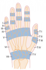
Key zones: I - Mallet finger III - Boutonniere deformity V - Fight bite |
|
|
Acceptable angulation for Boxer's fracture and metacarpal shaft |
Boxer's: 10-20-30-40 rule (D2-5) Shaft: 10 degree in 2-3 and 20 degrees in 4-5 |
|
|
x-ray signs of posterior shoulder dislocation |
Internal rotation Lightbulb / Drumstick sign
Loss of overlap of humeral head on glenoid Increased distance between glenoid and head of humerus
Trough sign (reverse Hill-Sachs lesion) |
|
|
Injury and associated nerve damage: -Elbow fracture -Shoulder dislocation -Sacral fracture -Acetabular fracture -Hip dislocation -Knee dislocation -Lat tib plateau fracture |
Elbow fracture - Median or ulnar Shoulder dislocation - Axillary Sacral fracture - Cauda equina Acetabular fracture - Sciatic Hip dislocation - Femoral Knee dislocation - Popliteal Lat tib plateau fracture - Peroneal |
|
|
LE motor nerve testing
Femoral Saphenous Sciatic Tibial Common peroneal Superficial peroneal Deep peroneal Sural |
Femoral - Knee extension
Saphenous - N/A
Sciatic - Knee flexion
Tibial - Foot plantar flexion
Common peroneal - N/A Superficial peroneal - Foot eversion Deep peroneal - Dorsiflexion of foot / toe extension
Sural - N/A |
|
|
LE sensory nerve testing
Femoral Saphenous Sciatic Tibial Common peroneal Superficial peroneal Deep peroneal Sural |
LE sensory nerve testing
Femoral - anterior thigh / knee Saphenous - medial foot Sciatic - N/A Tibial - sole of foot Common peroneal - N/A Superficial peroneal - dorsum of foot Deep peroneal - first web space Sural - lateral foot |
|
|
Signs of pulmonary edema on CXR |
10 classical findings: 1. Cephalization of Vascular markings (marked pulmonary redistribution) 2. Enlarged Heart Shadow - Cardiomegaly 3. Prominent Azygous Vein 4. Bat wing hilum 5. Kerley A lines 6. Kerley B lines 7. Kerley C lines 8. Peribronchial Cuffing 9. Pleural Effusion 10. Interlobar Fissure lines |
|
|
Differences between the adult and pediatric C-spine |
1. a proportionally heavier head 3. Less neck muscle mass 4. lax ligaments, allowing for more mobility at C1-C2; 5. incomplete ossification / un-fused physes; 6. horizontally inclined articular facets that facilitate sliding. 7. Anterior wedging of vertebral bodies 8. Pseudosubluxation of C2 on C3 9. Secondary ossificaiton centres may mimic avulsions 10. Variable interspinous distances 11. Widening of the pre-dental space (up to 5mm) 12. Lateral displacement of the masses C1 on C2 13. instability of atlanto-axial joint 14. SCIWORA - due to flexibility of bone |
|
|
DDx for elevated troponin |
1) Myocardial Infarction 2) Myocardial contusion 3) Chronic renal insufficiency for Troponin T 4) PE 5) Myocarditis 6) Acute heart failure 7) Perimyocarditis 8) Sepsis, ARDS, end organ strain 9) post-Cardiac procedure (cath, Surg, ablation) 10) Cardiomyopathy (Tako-tsubo) 11) Sympathomimetic drugs (cocaine!) 12) Aortic Dissection 13) Radiation therapy |
|
|
Eponym Aviator’s Barton’s Bennett’s Boxer’s Chance’s Chauffeur’s Clay shoveler’s Colles’ Cotton’s Pseudo-Jones Dashboard Dupuytren’s Essex-Lopresti Galeazzi’s Hangman’s Jefferson Jones’ Le Fort Lisfranc’s Maisonneuve March Monteggia’s Nightstick Pilon Rolando’s Salter-Harris Segond Stener Smith’s Teardrop Tillaux |
Aviator’s - Vertical # neck of talus with subtalar dislocation Barton’s - Intra-articular wrist #-dislocation; dorsal or volar Bennett’s - Oblique intra-articular # through base 1st MC Boxer’s - # neck of 4th or 5th MC Chance’s - Lumbar vertebral # through spinous process, pedicles, and vertebral body Chauffeur’s - # radial styloid Clay shoveler’s - # tip spinous process C6 or C7 Colles’ - # distal radius with dorsal displacement and apex volar angulation Cotton’s - Trimalleolar # Pseudo-Jones - # base 5th MT, < 15 mm from proximal end (insertion peroneus brevis) Dashboard - # posterior rim of the acetabulum Dupuytren’s - #-dislocation of the ankle Essex-Lopresti - Radial head # with dislocation of DRUJ Galeazzi’s - # radial shaft with DRUJ dislocation Hangman’s - # pars interarticularis of C2 Jefferson - Burst # C1 Jones’ - Transverse # 5th MT base, > 15 mm from proximal end Le Fort - Maxillary # Lisfranc’s - #-dislocation of tarsometatarsal joint Maisonneuve - # proximal fibula, disrupted syndesmosis, # medial malleolus March - Stress # of metatarsal Monteggia’s - # proximal ulna, dislocation radial head Nightstick - # of ulna, radius or both Pilon - ankle # & distal tibial metaphyseal #, usually w/ intraarticular comminution Rolando’s - Comminuted # base of 1st MC Salter-Harris -Epiphyseal # in children Segond - Avulsion of lateral tibia associated with ACL tear Stener - Avulsion ulnar corner base proximal phalange in thumb Smith’s - # distal radius with volar displacement (reverse Colles’) Teardrop - Wedge-shaped # of anteroinferior portion of vertebral body Tillaux - Avulsion # anterolateral portion of distal tibial epiphysis in adolescents |
|
|
Complications of fractures |
Hemorrhage Vascular injury Nerve injury Compartment syndrome Volkmann’s ischemic contracture Avascular necrosis RSD Fat embolism syndrome Fracture blisters |
|
|
Complications of immobilization |
Pneumonia DVT/PE UTI Wound infection Decubitus ulcers Muscle atrophy Stress ulcers |
|
|
Life or limb threatening ortho emergencies |
Open fracture Fracture/dislocation with major vascular disruption Major pelvic fracture Hip or knee dislocation Compartment syndrome |
|
|
Grades of acromioclavicular joint separation |
I - AC ligament sprain II - AC ligament rupture; CC ligaments sprained but intact. Joint space widened and slight ↑ displacement of clavicle III - Complete rupture of AC and CC ligaments, muscle attachments. Joint space widened and CC distance ↑ IV - Similar to III, but clavicle → posterior into trapezius V - Similar to III, but clavicle → upwards even more VI - Similar to III, but clavicle → inferiorly |
|
|
Indications for angiography with a pelvic fracture |
-Hemodynamic instability with –ive DPL or FAST -Large pelvic hematoma or active contrast extravasation on CT -Large/expanding RP hematoma seen at laparotomy -≥ 4 units blood transfused for pelvic bleeding in 24 h, or ≥ 6 units in 48 h |
|
|
DDx of the painful hip |
-SCFE -Perthes’ disease
-Bursitis -Tendonitis -Toxic synovitis -Septic arthritis -OA
-Arterial insufficiency -AVN femoral head -DVT
-Ligament injury (hip, knee) -Occult fracture
-Tumour (lymphoma)
-Referred pain (spine, knee) |
|
|
Classification of tibial spine fractures |
I Incomplete avulsion without displacement II Incomplete avulsion with minimal displacement IIIA Complete avulsion with displacement IIIB Complete avulsion with displacement and rotation |
|
|
Causes of traumatic hemarthrosis of the knee |
ACL injury PCL injury Patellar subluxation/dislocation Peripheral meniscal tear Osteochondral fracture Capsular tear |
|
|
DDx of presumed ankle sprain |
-LCL sprain -Peroneal tendon dislocation -Osteochondral talar dome # -Talar post process # -Talar ant process # -Talar lateral process # -Calcaneus ant process # -Midtarsal joint injury -Base 5th MT # |
|
|
Foot injuries requiring ortho consult |
-Open # -Fracture-dislocation -Major talar head/neck/body #s -Lisfranc injuries -Most GSWs -Compartment Syndrome |
|
|
Ankle fractures that require ortho consult |
-Displaced medial, lateral, or posterior malleolus -Bimaleolar fractures and equivalent (Deltoid ligament, syndesmosis rupture) -Trimalleolar -Intraarticular with step deformity -Open # -Pilon # -Fracture dislocation -Open fracture -SH III, IV, V |
|
|
Causes of compartment syndrome |
Increased content: -Bleeding (Vascular injury, Coagulation disorder, Anticoagulant use) -↑ Capillary filtration (Reperfusion post-ischemia, Trauma, Intensive muscle use, Burns, Intra-arterial injection, Orthopedic surgery, Snakebite) -↑ Capillary pressure (Intensive muscle use, Venous obstruction)
↓ Compartment Volume - Closure of fascial defect - Excessive limb traction - Tight cast, dressing, splint - Lying on limb
Miscellaneous -Infiltrated infusion -Pressure transfusion -Leaky dialysis cannula -Muscle hypertrophy -Popliteal cyst |
|
|
DDx solid, lucent bone lesions |
-Metastasis/Myeloma -Aneurysmal bone cyst - Solitary bone cyst -Fibrous dysplasia -Osteoblastoma -Giant cell tumour (EPIPHYSIS adults) -Chondroblastoma (EPIPHYSIS children) -Hyperparathyroidism (brown tumour) -Hemangioma -Infection -Non-ossifying fibroma -Eosinophilic granuloma/Enchondroma |
|
|
Age and associated bony malignancies |
Age (years) Tumour < 1 - Neuroblastoma 1-10 - Ewing’s (tubular bones) 10-30 - Osteosarcoma, Ewing’s (flat bones) 30-40 - Primary histiocytic lymphoma, fibrosarcoma, parosteal osteosarcoma, malignant giant cell tumor, lymphoma > 40 - Metastatic carcinoma, multiple myeloma, chondrosarcoma |
|
|
Splint complications |
-Pressure necrosis – may occur as early as 2 hours -Tight cast => compartment syndrome >> Univalving = 30% pressure drop >> Bivalving = 60% pressure drop >>Also need to cut cast padding -Abrasions / Cellulitis -Loss of reduction -Thermal Burns → avoid plaster > 10 ply and water > 24°C -DVT/PE: ↑ in lower extremity # -Joint stiffness → Leave joints free when possible and splint in in position of function |
|
|
Presentation of fat embolism |
1.Respiratory distress (earliest and most common manifestation) 2.Neurologic manifestation (confusion, ↓ LOC) 3.Thrombocytopenia 4.Petechial rash 5.Fever, tachycardia, jaundice, retinal changes and renal involvement may occur
Fat in urine in 50% within 3 days |
|
|
List of ECG changes to look for in the syncope patient |
QT BRIDE is Hot She makes your heart race and you feel like passing out - arrhythmias (brady, tachy, AFib, AFlutter, blocks, bifasicular/trifasicular blocks VT...etc) any irregular rate or rhythm QT - long/short QT B - Brugada R - Right sided strain (RBBB, s1q3t3, inverted T-waves V1-V4, tachy, Right axis deviation) I - Ischemia D - Delta wave (WPW) E - Epislon wave (ARVD) H - Hypertrophy - HOCM, LVH (AS, DCM) |
|
|
Pertussis incubation, phases, prevention, treatment, complications |
Incubation: 1-3 weeks
Phases: -Catarrhal (1-2 weeks) - rhinorrhea, fever, malaise. Infective. -Paroxysmal (2-4 weeks) - staccato cough (40-50x/d) worse at night/cold. Whoop on inspiration between coughs, Emesis after cough. Infective. -Convalescent (months) - residual coughing
Prevention - immunization and boosters
Treatment - Azithromycin x 3-5days to prevent spread within 3 weeks of onset (6 weeks for pregnant, <1y). Not infective after 5 days.
Complications - pneumonia, encephalopathy, earache, seizures. 1.6% fatality rate in infants <1y. |
|
|
Complications of coughing paroxysms in pertussis |
Periorbital edema Subconjunctival hemorrhage Petechiae Epistaxis Hemoptyis Subcutaneous emphysema Pneumothorax Pneumomediastinum Diaphragm rupture Umbilical or Inguinal hernia Rectal prolapse |
|
|
Tetanus definition, complications, management |
Definition: Acute onset of hypertonia or painful muscular contractions (usually of the muscles of the jaw and neck) and generalized muscle spasms without other apparent medical causes,
Complications: Respiratory failure, autonomic dysfunction, MSK spasms (Long bone fractures; tendon rupture; Subluxations, Dislocations (esp TMJ)), rhabdomyolysis
Management: -Supportive treatment with intubation, GABA blockade (Benzos, Midaz, Propofol +/- Dantrolene, Magnesium), alpha blockade (phentolamine) -Toxin treatment with Human Tetanus IG 500U (administer AWAY from Td shot) -Infection treatment with debridement, Flagyl 500 mg IV q6h (7.5mg/kg for children) |
|
|
Tetanus prophylaxis |
-Clean or dirty wound and immunized (x3 and up to date) - nothing -Clean wound and unimmunized - Td -Dirty wound and unimmunized - Td and TIG |
|
|
Botulism cause, types, diagnosis, treatment |
Cause: toxin produced by clostridium botulinum; inhibits acetylcholine release
Types: infant, food, wound, inhalation
Diagnosis: toxin in blood, toxin or bacteria in stool / wound culture, EMG (supportive)
Treatment: Source control (if infection), Intubate if FVC<15mL/kg; Trivalent equine antitoxin (adults) or Baby BIG (infants), NG tube (for ileus), treat autonomic dysfunction prn |
|
|
Contraindications to LP |
-skin or soft tissue infection at puncture site. -Likelihood of brain herniation (mass lesion, papilledema) -INR>1.5 -Plt < 50 |
|
|
Indications for CT pre-LP |
-Immunocompromised -ALOC (GCS <14) -Seizure in past 7 days -Focal neurologic signs -Papilledema -PMHx of CVA, mass lesion, focal infection, head trauma, CNS Sx
Also consider age >60, severe HA/N/V, suspected brain bleed or lesion. |
|
|
Empiric treatment for meningitis |
<1m: Ampicillin (Listeria) & Cefotaxime (Kernicterus) 1-50y or basal skull: Ceftriaxone & Vancomycin >50y: Ampicillin (Listeria), Ceftriaxone, Vancomycin Instrumentation or penetrating trauma: Vancomycin & Ceftazidime |
|
|
Treatment of toxic shock |
As for sepsis: fluids, vasopressors, O2, ventilation, steroids, source control
Penicillin and clindamycin (stops toxin synthesis) and IVIg (especially for staph TSS) |
|
|
Rabies mechanism, stages and management |
Mechanism: -Travels up nerves at a rate of 8-20mm/d to the brain (Negri bodies) and salivary glands (infect others), causes cytokine storm and serotonin hyperactivity.
Stages: I Incubation II Prodrome (1d-1w) III Acute neurological illness - can be furious (80% - hyperactive then lethargic, hydrophobia, salivation, spasms), dumb (20% - limb weakness, fever, looks like GBS), or non-classic (Thailand; brainstem/ motor/ sensory deficits pronounced; seizures) IV Coma (7-10d following acute neuro) V Death (2-3d following coma)
Prophylaxis -Scrub and clean wound then rinse with povidone-iodine -If vaccinated give additional doses at 0 and 3 days-If unvaccinated give 20U human RIG near bite (and far from vaccine) then vaccine at 0,3,7,14,28 days
Management -Ketamine (inhibits viral replication?) |
|
|
Rabies carriers and PEP |
Most to least common: -Bats, Raccoons, Skunks, Foxes (assume rabid unless none in area or lab neg - give PEP) -Dogs/cats/ferrets (observe x10d if caught; PEP if caught and suspected rabid; consult public health if escaped and not suspected) -Rodents/lagomorphs (no proven transmission - no PEP) |
|
|
Measles incubation, symptoms, complications |
-Exposure -10 days later get cough, coryza, conjunctivitis, koplik spots and fever -4 days later get rash -Contagious 5 days before the rash and 4 days after -Complications include otitis media, encephalitis, pneumonitis and 20 years later Subacute Sclerosing Panencephalitis |
|
|
Mumps incubation, presentation, complications, treatment |
Incubation - 18 days (and lasts 7-10)
Presentation - fever, myalgias, malaise, parotid swelling, orchitis, meningitis
Complications -Orchitis, Meningitis, GBS, Transverse myelitis, Deafness, Pancreatitis, Mastitis, Oophoritis, Myocarditis, Arthritis
Treatment -IVIg if meningitis/pancreatitis |
|
|
Workup for Parvovirus B19 exposure in pregnancy |
-Get Parvovirus IgG and IgM levels -If only IgG+ patient is immune; no acute infection -If only IgM+ patient has an acute infection - refer to OBGYN and U/S for ? hydrops -If neither IgG or IgM no acute infection |
|
|
HIV prophylaxis - bugs and drugs |
1. PCP (Septra) 2. Toxo (Septra) 3. TB (INH) 4. MAC (Azithro) 5. CMV (Gancyclovir) |
|
|
High risk HIV exposures |
Exposures 1. Percutaneous needles (deep injuries, blood on device, venipuncture) 2. Mucous membrane 3. Sexual contact
Contact (Triple therapy PEP) 1. Patient has symptomatic HIV 2. AIDS 3. Acute seroconversion 4. High viral load
|
|
|
How to remove ticks |
1 - “Tweezer” method: Grasp w/ Tweezers close to skin, slow and steady, ensure removal of head; keep for inspection
2 -“Straw Knot” Subtle but constant upward pressure on the string once the tick is "caught" with the knot.
3 - Excisional technique – excise tick with the tissue around it – cut elpitically, close with simple interrupted
4 - Kill with viscus lidocaine a. Grasp with blunt forceps as near to attachment as possible b. Gentle upward traction to remove (no squeezing or jerking) |
|
|
Unilateral CNVII palsy |
Infectious -Lyme -Bell’s Palsy -Ramsay-Hunt -Viral (VZV, HIV) -Otitis Media / Externa / Mastoiditis
Trauma -Middle Ear Barotrauma -Facial laceration
Brain -Schwannoma -MS -Brainstem lesion or mass (aneurysm)
Weird -Diabetic Neuropathy -Sarcoidosis |
|
|
Limping child |
Rheumatic / Inflammatory -JRA – usually polyarticular arthritis -Rheumatic fever – usually polyarticular arthritis -Ankylizing spondylitis -HSP -Gout or pseudogout
Congenital / Mechanical -Sickle cell -Limb length discrepancy -Developmental dysplasia -Legg-Calve-Perthes -SCFE -Osgood Schlatter -Femoro-patellar syndrome Trauma -Toddler's fracture -NAT -Hemarthrosis – traumatic or spontaneous Cancer -Osteochondroma -Ewing's sarcoma
Infection -Osteomyelitis -Transient (toxic) synovitis – 3 mo-6yrs, usually hip -Reactive arthritis – Strep, Chlamydia, Salmonella, Shigella, Lyme, Yersinia, viruses
Referred pain -Appendicitis -Testicular torsion |
|
|
Gas in tissues (infectious) |
-Clostridia (perfringens, septicum) -Gram negatives (E. coli, Klebsiella, Enterobacter) -Anaerobes (Peptostreptococcus, B. fragilis) -Peptococcus -Group A Streptococcus |
|
|
Aquatic skin infections: fresh, salt, tropical |
Fresh - aeromonas hydrophilia (cipro) Salt - vibrio velnificans (cipro) Aquarium - mycobacterium marinum (TB meds - RIPE) |
|
|
Cavernous venous thrombosis: cause, presentation, treatment |
Cause: preceded by trauma, bacteremia, or local (facial, dental, sinus, ear) infection
Presentation: headache, CN III V IV VI VIII findings (deaf, dizzy, EOM, diplopia, facial movements), periorbital edema, exopthalmos, decreased LOC, death
Treatment: antibiotics and anticoagulation |
|
|
Pneumonia definitions: CAP, HAP, VAP |
CAP: no hospital or LTC stays for 14 days before presentation
HAP: new infection >48h after arrival in care facility
VAP: new infection >48h after intubation
Healthcare associated: home IV antibiotics, dialysis, wound care, immunocompromised, chemotherapy, nursing/LTC, hospital for 2 days in past 90 days |
|
|
Pneumonia admission decision score |
CURB 65
C onfusion U remia (BUN > 7mmol/L) R espiratory rate greater than 30 B lood Pressure < 90/60 65 years of age or more
If 2 consider admit; 3 consider ICU |
|
|
High risk for endocarditis |
-Prosthetic heart valve -Hx of endocarditis -Unrepaired cyanotic CHD (including palliative shunts, conduits) -Completely repaired CHD with prosthesis during first 6 months post op -Repaired CHD with residual defect (at or near site) of prosthetic valve -Cardiac valvulopathy in a transplanted heart -HCM -MVP with regurgitation -IVDU |
|
|
Reportable STI's |
CCHAGS
-C hancroid -C hlamydia -A IDS/HIV -G onorrhea -S yphilis |
|
|
Painful vs painless genital lesions |
BlouCH (painful) Behcet's C hancroid H SV
Some Lesions On my Dong S yphilis L GV O ncologic (SCC) D onovanosis |
|
|
Diagnostic criteria for PID |
Major -CMT -Adnexal Tenderness -Uterine Tenderness
Minor -Oral temp > 38.3 -AbN cervical or vag discharge -WBC on wet mount of prep -Elevated ESR -Elevated CRP -Lab documentation of G+C |
|
|
PID requiring admission |
-Cannot exclude surgical emergency (e.g. appendicitis) -Pregnancy -Tubo-ovarian abscess -Severe illness with vomiting or high fever -No clinical response to PO meds in 24-48 h -Unable to FU or tolerate outpatient regimen |
|
|
DDx for dysuria in peds |
Infection: UTI, pinworm, vaginitis (gardneralla, trichomonas, candida, STI), balanitis, pinworm
Trauma: self-induced, sexual abuse, straddle injury
Irritation: bubble bath, soaps, douches, foreign body
Other: labial adhesions, renal stones |
|
|
Peripartum infections that cause maternal or fetal morbidity |
Toxoplasmosis Others (syphilis, Hep B, Coxsackievirus, EBV, VZV, HPV) Rubella virus CMV HSV |
|
|
DDx neonatal hyperbilirubinemia |
Unconjugated -Benign: Physiologic (1st), Breast Feeding (2nd due to dehydration with late milk in 1st week), Breast Milk (due to inhibition of hepatic enzymes in 2-3 weeks) -Hemolysis: ABO, hematoma breakdown, spherocyte, elliptocyte, sickle cell, G6PD, PK -Sepsis: TORCH, UTI -Obstructive: Meconium ileus, Hirschsprung's, Pyloric stenosis, Duodenal atresia -Metabolic/genetic: Hypothyroid, Gilbert, Crigler-Najjer
Conjugated -Sepsis: TORCH, UTI, Listeria, TB, Hep B, VZV, HIV, Coxsackie -Obstructive: Biliary atresia, choledochal cyst, bile duct stricture, primary biliary cirrhosis -Metabolic/genetic: Gaucher's, Galactosemia, Nieman-Pick, alpha-1 antitripsin Miscellaneous: drugs, toxins, TPN |
|
|
Practical approach to the crashing neonate |
SCRAMS
S epsis C ardiac A buse M etabolic S urgical emergencies |
|
|
List of conditions causing immunocompromise |
HIVED ARMS ITCH
H IV I VDU V ascular insufficiency E thanol use D M
A splenia R F M alnutrition S teroid use
I mmunizations lacking T ransplant C ancer H epatic Failure |
|
|
Antibiotics for bites |
Human - Eikenella Corridens - give Clavulin (Clindamycin + [Septra OR Levo] if allergic); resistant to 1st gen cephalosporins
Cat - Pasturella multicoda - give Clavulin (Septra or Levofloxacin if allergic); resistant to Clindamycin/1st gen cephalosporins
Dog - Polymicrobial (staph, strep, anaerobe) and Capnocytophagia Canimorsus - give Clavulin (admit with 3rd gen cephalosporin if elderly, asplenic, immunocompromised) |
|
|
Assessment of infectious risk of a dog bite |
Dog’s Characteristics -immunization status of dog -behavior of dog -endemic area -provoked attack
Patient Characteristics -immunization status of patient (incl. tetanus) -medical comorbidities in patient -immunocompromsie in patient -irrigation of wound |
|
|
Bacterial sinusitis: presentation, causes, complications |
Presentation of sinusitis: -Purulent nasal secretions -Purulent posterior pharyngeal secretions -Mucosal erythema -Periorbital edema -Tenderness overlying sinuses -Air-fluid levels on transillumination of the sinuses (60% reproducibility rate for assessing maxillary sinus disease) -Facial erythema
Bacterial more likely to have: -Severe presentation -Double sickening -Extra-sinus manifestations -Persisting/worsening after 10 days
Causes: -S. pneumoniae -H. influenzae -M. catarhallis
Complications: -Venous sinus thrombosis -Lemierre’s -Meninigitis/abscess -Orbital cellulitis -Facial cellulitis -Optic Neuritis -Periorbital abscess -Blindness -Proptosis |
|
|
Ludwig's angina: presentation, bacteria, treatment |
Presentation -SIRS -Elevation of the floor of the mouth / tongue -Pain to submental, sublingual, submandibular compartments -Poor / infected dentition
Bacteria -Multibacterial: staph, strep, anaerobes, bacteroides
Treatment -High dose penicillin + metronidazole -Ceftriaxone + clindamycin |
|
|
Sinus venous thrombosis: signs and organisms |
Signs: -Presents with fever and headache -Can have proptosis (opthalmic vein), mydriasis, CN 3,4,6 palsy, Periorbital edema and eye pain -Progress to altered mental status, meningeal signs, seizure, coma
Organisms: -Strep, Staph, Moraxella, Hemophlius, Bacteroides, Fungi |
|
|
Risk factors for TB |
1) HIV 2) Close quarters (military, native american, LTC) 3) travel to endemic area 4) homeless 5) IVDU 6) Close contact with patient with TB 7) Occupational exposure 8) Foreign born |
|
|
Needlestick injury: worrysome pathogens, prevalence of transmission |
Worrysome pathogens 1) Hep B up to 30% (if HBs & HBe +ve; only 2-5% if only HBs +ve) 2) Hep C up to 3% (1-2%) 3) HIV up to 0.3% percutaneous / 0.09% mucocutaneous
Prophylaxis: injection of blood, needlestick from known + patient, sexual assault No prophylaxis: random needlestick or spitting |
|
|
Physical exam findings of central retinal artery occlusion; treatment |
PE -Cherry red spot -Pale retina -RAPD -Decreased visual acuity
Treatment 1. Globe Massage 2. IOP Mgt (Acetozolamide, Mannitol) 3. Anterior Chamber Paracentesis 4. Thrombolytics (tPA) 5. Bag hyperventilation or carbogen (increase PC02) |
|
|
Physical exam findings of central retinal venous occlusion |
-Blood and thunder -Disk edema -Dilated tortuous veins -Decreased visual acuity; -RAPD (if ischemic) |
|
|
Physical exam findings of iritis |
- red eye - ciliary flush - flare - consensual photophobia - acutely - decreased IOP |
|
|
Causes of sudden hearing loss |
Infectious: -Mumps -Measles -Influenza -HSV -Herpes zoster -CMV -Mono -Syphilis
Drugs (ototoxic) - Aminoglycosides – Gent, Amikacin - Loop diuretics – Lasix - ASA - Indomethacin - Chemotherapeutic agents (Cisplatin, Methotrexate) - Quinine
Vascular (sludging) -Macroglobulinemia -Sickle cell disease -Leukemia -Polycythemia -Fat emboli -Hypercoagulable
Conductive: - Cerumen - Foreign Body - OM - OE - Barotrauma - Trauma - Neoplasm |
|
|
Optic neuritis: physical exam, conclusive diagnosis, associated condition, Treatment |
PE - see RAPD and red desaturation
Conclusive diagnosis - MRI
Associated condition - MS
Treatment - Solumedrol IV |
|
|
Plexus associated with posterior epistaxis and its tributatries; cause of posterior bleed |
Woodruff's plexus, tributaries: -Sphenopalatine artery -Posterior ethmoidal artery
Causes: -Anticoagulation -Blood dyscrasia -Rupture of carotid aneurysm -HTN -Ca -Trauma |
|
|
Subluxed tooth: management, complication |
Management: -Pain control -Soft diet -Setting and stabilization of the tooth -Dental referral
Complication -Osteomyelitis -Loss of tooth -Alveolar fracture |
|
|
Organic versus functional blindness |
Functional -Can't write own name -Difficulty with finger apposition -Optokinetic reflex intact |
|
|
Causes of RAPD |
-Ischemic optic neuropathy (CRAO or CRVO) -Optic neuritis -Optic nerve compression (orbital tumours or dysthyroid eye disease) -Retrobulbar hematoma -Trauma -Asymmetric glaucoma |
|
|
Conductive vs sensorineural hearing loss |
Conductive -Weber: Lateralizes toward affected side (conduction louder to affected side) -Rinne: BC is > AC -Caused by cereneum impaction, perf'd TM, cholesteatoma
Sensorineural -Weber: Lateralizes away from affected side (conduction louder to normal side) -Rinne: AC is > BC (or normal) -Caused by acoustic neuroma, meniere's disease |
|
|
Admission criteria for epistaxis |
-Posterior bleed/balloon -Bilateral posterior pack -Hemodynamic instability -Associated facial trauma/ polytrauma -Severe coagulopathy -Significant comorbidity -No follow up/psychosocial concern |
|
|
Otitis externa: bacterial cause, treatment, risk factors |
Causes: pseudomonas (!!), staph, gram negatives, fungal
Treatment: Ciprofloxacin / ciprodex + clavulin
Risk factors: immunocompromise, DM, trauma, AIDS, elderly |
|
|
Acute necrotizing ulcerative gingivitis |
Risk factors: immunocompromise, smoking, local trauma, stress/fatigue
Presentation: trench mouth, fever, malaise, LA, ulcerating of papillae, gray pseudomembrane, pain, hallitosis
Cause: anaerobes (fusobacterium, spirochetes)
Management: saline rinses, analgesia, abx (penicillin or erythromycin), dilute hydrogen peroxide rinses, dentistry f/u |
|
|
Indications for hyphema admission |
Rebleed Elevated IOP >50% (grade III) Decreased VA Child abuse Noncompliant |
|
|
Management of otitis media |
<6 months - antibiotics 6 months to 2y - antibiotics if certain; observe if uncertain 2y - antibiotics if severe; observe if uncertain
Observe = reassess in 48-72h |
|
|
DDx of neck masses |
Inflammatory -Adenitis (Bacterial, Viral, Fungal) -Cat-scratch disease -Tularemia -Local skin infection -Sialoadenitis -Thyroiditis -TB
Congenital -Branchial cleft cyst -Thyroglossal duct cyst -Dermoid cyst
Neoplastic -Benign -Malignant (Sarcoma, Salivary gland, Thyroid, Lymphoma) Metastasis (1° ENT cancer, Lung, Esophageal) |
|
|
What is the NRP pyramid? |
-Assess if resuscitation needed, warm, position and clear airway, dry and stimulate -O2 -PPV -Intubation -Chest compressions -Drugs |
|
|
Atropine in pediatrics: indications, mechanism and complications |
Indications: 1.Post-intubation bradycardia 2. Symptomatic Bradycardia secondary to vagal tone 3. Neonatal intubation – as prophylaxis 4. Cholinergic toxidrome
Mechanism - decreases vagal tone speeding sinus/atrial and AV conduction. Onset is 1-2m and it lasts 2-4m.
Complications - can get a paradoxical bradycardia in low (<0.1mg) doses |
|
|
Pediatric vs adult CPR: 1 rescuer ratio, 2 rescuers ratio, rate, depth, monophasic J, biphasic J, Epi dose, Amiodarone dose |
1 rescuer ratio - 30:2 both 2 rescuers ratio - 30:2 adult, 15:2 peds rate - 100 both depth - 2 inches adult, 1/3 of chest AP diameter peds monophasic J - 360J adult, 2 then 4J/kg peds Epi dose - 1mg adult, 0.01mg/kg peds Amiodarone dose - 300mg adults, 5mg/kg peds |
|
|
Treatment of Tetrology of Fallot |
Knee to chest / squat (increased SVR) IVF (increase preload) Anxiolytic (benzo/fentanyl) (decrease RR and PVR) NaHCO3 (correct acidosis - maybe) Phenylephrine (increased SVR) Propranolol (increased preload) 100% O2 (decreased PVR) |
|
|
Abnormal bloodwork in Kawasaki's disease |
-Elevated ESR / CRP -Sterile pyuria -Decreased Hb -Elevated WBC -Plt>450 -Decreased albumin (<30) -Elevated ALT |
|
|
Common causes of renal failure in children |
-Post-strep GN -HSP -Pyelonephritis -Obstructive Nephropathy (VUR) -Lupus Nephritis -Minimal change disease |
|
|
Explain the hyperoxia test |
If a hypoxic child presents and is put on 100% O2 and the sats don't improve, it is likely a cardiac / shunting problem. If it improves to PaO2 >150 or by >10% SpO2 it is likely a pulmonary problem. |
|
|
Exposure prophylaxis (Pertussis, Varicella, Yersinia Pestis, Measles, Hepatitis A, Meningococcus) |
Pertussis - macrolide for close contacts Varicella - Varicella Ig if unimmunized and immunocompromised OR pregnant within 72h of exposure; vaccine if unimmunized Yersinia pestis (plague) - ciprofloxacin or doxycycline Measles - Measles Ig within 6 days if not immunized (and pregnant, immunocompromised, <12months) OR MMR within 3 days Hepatitis A - HAIg to unvaccinated close personal contacts, childcare workers/attendees (people who wipe their bum), similar food-borne source in last 2/52 Meningococcus - Ciprofloxacin for adult close contacts, Rifampin for pediatric close contacts
|
|
|
Causes of pediatric GI bleed |
-NEC -Ingested Maternal Blood (Cracked Nipple) -Milk Protein Allergy -Reflux esophagitis -Meckel’s -Intussuception -Infectious Gastroenteritis -Anal Fissure -IBD -Blood dyscrasia -Child Maltreatment -Polyps -Toxic ingestions |
|
|
Treatment of neonatal unconjugated and conjugated bilirubinemia? |
Unconjugated -IVF rehydration -IV antibiotics -Exchange transfusion / plasmapheresis -Phototherapy
Conjugated -IVF rehydration -IV antibiotics -Exchange transfusion / plasmapheresis -Abdominal ultrasound -Surgical consult -IVIg (if ABO incompatibility) |
|
|
Croup vs bacterial tracheitis |
Bacterial tracheitis is: -Inspiratory AND expiratory stridor -Does not respond to treatment -Toxic appearance -Hypoxia/Cyanosis -Shaggy trachea on x-ray -Copious secretions following intubation |
|
|
Causes of congenital stridor |
- treacher-collins - pierre-robin - laryngomalacia - tracheomalacia - vascular ring - unilateral or bilateral vocal cord palsy - laryngeal web - down syndrome - hypothyroidism - glycogen storage disease - lingual thyroid - choanal atresia - mediastinal mass - tracheal stenosis |
|
|
Causes of HSP |
-Viruses (EBV, Measles, Mumps, Rubella, Chickenpox, Parvovirus B19) -Bacteria (Shig, Salmonella, Campylobacter, Mycoplasma, GAS) -Drugs (ampicillin, erythromycin, penicillin, quinidine, quinine) -Insect stings |
|
|
Post-strep glomerulonephritis: presentation, diagnsosis, treatment |
Presentation: hypertension, hematuria, edema, AKI
Diagnosis: confirm strep with serology, ASOT, Creatinine, U/A
Treatment: treat volume overload (Lasix, Dialysis); hypertension (ACEi, Dialysis), Strep (Penicillin) |
|
|
Causes of descending paralysis |
“Got BOMBED”
G BS (MF Variant)
B otulism O rganophosphate (HEENT exposure) M yasthenia Gravis B rainstem (Pontine infract) E aton Lambert D iphtheria |
|
|
Stroke mimics |
-Todd’s Paralysis (postictal) -Hemiplegic migraine -Bell’s Palsy -Hypoglycemia/DKA/HONK -MS -Hypertensive encephalopathy -Wernicke's -Central venous sinus thrombosus -ICH - SAH/SDH/EDH -Brain tumor -Conversion disorder -Meningitis/Encephalitis/Abscess |
|
|
Define TIA; Risk for stroke following TIA |
Transient neurological symptoms due to ischemic etiology that ‘typically’ resolve within 60 minutes of onset. Must result in complete recovery to qualify as TIA.
ABCD2 score
A ge > 60 B P>140 and/or DBP>90 C linical - Speech (1 point) Unilateral Weakness (2 points) D M D uration 10-60m (1 point) >60m (2 points)
7 day risk score: 0-3 = 1% 4-5 = 6% 6-7 = 12% |
|
|
Central vertigo vs peripheral vertigo |
Central: -Positive HINTS -Insidious onset -No N/V, mild severity -Associated with other neurological symptoms -No auditory symptoms -Nystagmus not fatiguable, vertical or rotatory -No change with head position Peripheral: -Severe -Sudden onset -Associated with Nausea / Vomiting -Normal other neurologic symptoms -Fatiguable Horizontal Nystagmus -Positive Dix-Hallpike |
|
|
Causes of hypoMg |
Redistribution - TPN, refeeding, pancreatitis, DKA correction Extrarenal losses - Vomiting, Hyperemesis Gravidarum, Anorexia, Bulimia, Diarrhea, sweating, burns, fistula, hyperaldosteronism
Renal losses - Diuretics (Loop Diuretics, amphoteracin, EtOH), Bartter’s, Osmotic Diuresis, DKA Inadequate intake - Malnutrition, Malabsorption, Alcoholism, Critically unwell Toxins - Cisplatin, Hydrofluoric acid |
|
|
Post-LP headache |
Needle used -Whitacre -Quincke
Cause -Big needle -Too much fluid drawn -Traumatic needle -Stylet not replaced -Bevel not longitudinal
Treatments -Analgesia -Caffeine -IVF -Blood patch |
|
|
Drugs that cause thrombocytopenia |
1) Septra 2) Quinine 3) NSAIDS 4) Heparin (unfractionated, LMWH) 5) HCTZ 6) Amiodarone 7) Abciximab (GpII b IIIa inhibitor) 8) Rifampin 9) Ethambutol 10) Dilantin 11) Valproic acid 12) Ethanol |
|
|
Transfusion reactions (immune, non-immune) |
Immune -Febrile non-hemolytic 1:300 -Hemolytic (ABO incompatability) 1:40,000 -Delayed hemolytic -Anaphylactic 1:40,000 -Urticarial 1:100 -TRALI 1:40,000 -Transfusion-related immunomodulation (TRIM) - immunocompromised following -Post-transfusion purpura
Non-immune -Citrate toxicity (hypoCa) -HyperK -Hyperthermia -Dilutional coagulopathy -TACO 1:700 -Transfusion of pathogens (Hep B 1/50,000; Hep C 1/2,000,000; HIV 1/2,000,000; bacterial 1/20,000) |
|
|
Transfusion complications (immediate, delayed, massive) |
Immediate -Acute hemolytic reaction -Febrile non-hemolytic reaction -Anaphylactic reaction -Urticarial -TACO -TRALI
Delayed -Delayed hemolytic -GVHD -Infectious transmission (bacterial, Hep B/C, HIV, Syphilis, Malaria)
Massive -Citrate toxicity (hypoCa) -HyperK -Hypothermia -Dilutional coagulopathy |
|
|
Factors contained in FFP |
Factors 2, 5, 7, 8, 9, 10, 11, 12, 13 |
|
|
Infectious agents transmitted by blood products and their risks |
Bacterial - 1:20,000-50,000 Hep B - 1:153,000 Hep C - 1:2.3 million HIV - 1:7.8 million HTLV CMV WNV Rare Prion dz Rare |
|
|
Effect of diseases on clotting: HUS, TTP, DIC, Hemophilia, vWD |
HUS - decreased Plt; increased BT TTP - decreased Plt; increased BT DIC - increased INR/PTT/BT; decreased Plt Hemophilia - increased PTT vWD - increased BT |
|
|
Emergent presentations of sickle cell disease |
-Acute chest crisis -Priapism -Stroke -MI -Splenic sequestration -Infection/sepsis -Aplastic anemia -Pain crisis |
|
|
Hemophilia % classifications |
Mild 6-49% activity Moderate 1-5% activity Severe <1% |
|
|
Mucosal edema: DDx and treatment |
DDx -Anaphylaxis -Hereditary angioedema -ACE-i induced angioedema
Tx -Epi -Benadryl -Steroids -C-1 esterase inhibitor -FFP |
|
|
Options for LA in lidocaine allergy |
-Use code lidocaine (preservative-free) -Use ester local anesthetic (benzocaine, tetracaine) -Use Benadryl -Test dose pre-use -Use skin glue and/or steri-strips to close wound |
|
|
Side effects of sulpha drugs |
-Erythema Nodosum -Erythema Multiforme -SJS -TEN -Hypersensitivity vasculitis -DRESS -Anaphylaxis -Allergic carditis |
|
|
Diabetic drugs: name, mechanism |
Insulin - opens GLUT channels so cells can use glucose Glyburide - stimulates insulin release Metformin - sensitizes cells to insulin +/- decreases intestinal absorption Acarbose - prevents glucose absorption from intestines Rosiglitazone - sensitizes cells to insulin |
|
|
Drugs that cause hypoglycemia |
Diabetic drugs -Glyburide -Insulin -Metformin -Acarbose -Rosiglitazone
HTN / Heart drugs -Beta-blocker overdose (Propranolol) -MAOi -ASA
Abuse -Methanol -EtOH |
|
|
Conn's syndrome |
Primary hyperaldosteronism from an adrenal adenoma or adrenal hyperplasia. Can also get from an aldosterone producing tumor. Secondary hyperaldosteronism is from a renin-producing tumor or renal artery stenosis.
Presentation: hypertension, hypokalemia, and alkalosis.
Diagnosis: CT abdomen, serum aldosterone, serum renin. Can also do urine lytes, TTKG
Treatment: Spinrolactone (aldosterone antagonist) and ACEi |
|
|
Causes of urinary retention |
Obstructive 1. BPH 2. Stone 3. tumour 4. gross hematuria 5. FB 6. Phimosis 7. Paraphimosis 8. Meatal stenosis
CNS 1. Spinal Cord Injury 2. Neurogenic shock 3. Spinal Epidural Hematoma 4. Syringomyelia 5. DM 6. MS
Infectious 1. Prostatitis 2. UTI 3. Tabes Dorsalis (Syphillis) 4.Urethritis 5. Balanophthis
Medications 1.anticholinergic 2.anti histamines 3.Narcotics 4.TCA (anticholinergic + antihistamine) 5.alpha agonists 6. antipsychotics (Haldol) 7. NSAIDs
Other 1. Priapism 2. Penile Fracture 3. Ureteric laceration 4. Lazy bladder syndrome |
|
|
Indications for admission of renal colic |
1. infected stone 2. pregnant 3. septic 4. intractable vomiting 5. severe pain 6. urinary extravasation 7. Hypercalcemic crisis |
|
|
Causes of hypercalcemia |
CHIMPANZEES
C a supplementation H yperparathyroidism I atrogenic (Li, thiazines) M ilk-alkali syndrome P aget’s A cromegaly, A ddison’s N eoplasm Z ollinger-Ellison E xcess Vit A E xcess Vit D S arcoidosis |
|
|
Steps for accessing a fistula if necessary |
1) No tourniquet! 2) Sterile/Clean Prep 3) Firm steady pressure for 10 min after 4) Document thrill before/after 5) continuous infusion to maintain laminar flow and prevent stasis 6) avoid puncturing posterior wall of vessel |
|
|
Post trauma care in the pregnant patient -Observation -Describe test -How much Rhogam |
-Continuous fetal monitoring x 4 hours -Kleihaur Betke test for feto-maternal hemorrhage (if Rh- 50mcg if <12 weeks, 300mcg if <16 weeks; then calculate) -U/S for hemorrhage -NST (best test for abruption - see fetal distress); if >3 contractions in a 1h period observe for 24h; if >12 cxns/h high risk for abruption |
|
|
Risk factors for preeclampsia; Predictors of eclampsia |
Person -Adv. Mat. Age -Younger than 20 -New Partner -Low SES -Obesity -Cocaine
Pregnancy -Primiparous -Molar preg or mult gestation -IVF
PMHx -Hypertension -Hypercholesterol -Previous PIH -Previous GDM -Connective Tissue Disease
FHx -Family hx of PIH -Inherited Thrombocytopenia
Predictors of eclampsia -elevated WBC, ALT, Creatinine |
|
|
Adverse conditions that qualify hypertension in pregnancy as preeclampsia |
Maternal symptoms -persistent or new/unusual headache -visual disturbances -persistent abdominal or right upper quadrant pain -severe nausea or vomiting -chest pain or dyspnea) -maternal signs of end-organ dysfunction (eclampsia, severe hypertension, pulmonary edema, or suspected placental abruption)
Abnormal maternal laboratory testing -elevated creatinine -elevated AST, ALT or LDH with symptoms -platelet count <100x109/L -serum albumin < 20 g/L
Fetal morbidity -oligohydramnios -intrauterine growth restriction -absent or reversed end-diastolic flow in the umbilical artery by Doppler velocimetry -intrauterine fetal death). |
|
|
Treatment of PID |
Oral: Ceftriaxone 250mg IM then Doxy 100mg / Flagyl 500mg po bid x 14d
IV: Cefoxitin 2 q6h IV + Doxycycline 100mg IV q12h |
|
|
Treatment of PE, cholecystitis, appendicitis, pyelonephritis in pregnancy |
PE -LMWH (Enoxaparin) and admission
Chole -Ceftriaxone / Flagyl IV -Gravol / Maxeran / Ondansetron -IVF
Appy -Ceftriaxone / Flagyl IV -Gravol / Maxeran / Ondansetron -IVF
Pyelo -Ceftriaxone / Ampicillin IV -Gravol / Maxeran / Ondansetron -IVF |
|
|
Ultrasound findings in ectopic pregnancy |
-Ectopic fetal heart activity -Ectopic fetal pole -Moderate or large cul-de-sac fluid without IUP -Adnexal mass* without IUP -Indeterminate -Empty uterus -Nonspecific fluid collections -Echogenic material -Abnormal sac |
|
|
Diagnosis and causes of SROM |
Causes: -UTI -Infection (choriaminoitis, Bacterial vaginosis, GBS) -Trauma -Incompetent Cervix -Cigarette Smoking
Diagnosis -Perform sterile speculum exam vaginal exam, pooling in posterior fornix -Nitrazine test (Blue = +) – false pos with semen and urine in vagina -Ferning – false positives with blood (if >10% blood), semen, fingerprints or cervical mucus |
|
|
RUQ pain in pregnancy / RUQ pain and jaundice in pregnancy |
RUQ pain in pregnancy -Hepatitis -Cholestasis of pregnancy -Choledocholithiasis -Cholecystits -HELLP -Acute Fatty Liver of Pregnancy -Capsular hematoma -Appendicitis
RUQ pain and jaundice Cholestasis of Preg Acute Fatty Liver of Pregnancy |
|
|
Indications and Contraindications to tocolysis |
Indications -Preterm premature labour with -High risk for safe transport
Contraindications -Chorioamino -Acute vaginal bleed -Fetal demise -Eclampsia -Sepsis -DIC |
|
|
Reasons for treatment failure of an appropriate antibiotic |
-Wrong dosage -Wrong duration -Noncompliance -Immunocompromise -Local resistance patterns |
|
|
PE Xray, ECG, echo findings PE predictors of mortality |
XRay -Normal -Hampton's hump (wedge shaped opacity) -Westermark's sign (oligemia of distal vasculature) -Pleural effusion
ECG -Normal -S1Q3T3 -RBBB -RAD -P-pulmonale -Anterior ST depression / T wave inversion
Echo -Normal -Dilated RV -Bowing of septum into LV -McConnell's sign -Dilated IVC
Increased mortality -Troponin -BNP -SpO2 <95% on RA -Echo with RV strain / dilation -Shock / hypotension |
|
|
etCO2 for tube placement -False positives -False negatives |
False positives -Carbonated beverage in stomach (should wash out after 6 BVMs) -Recent BVM ventilation -Administration of bicarb (first 5-10 min after administration)
Possible False Negatives - Prolonged arrest - Equipment failure (cuff leak, expired) - Complete obstruction at level trachea or both bronchi - Severe asthma - Massive PE |
|
|
Vent settings for an asthmatic |
RR - 6-10 Vt - 5-8mL/kg Minute ventilation - low PEEP - 2.5-10 (match iPEEP) Flow - 100L/m (high) I:E ratio - 1:4-6 |
|
|
Causes of pleural effusion |
Transudative -CHF -Nephrotic syndrome -Liver cirrhosis -Myxedema -Malnutrition / hypoalbuminemia -Peritoneal dialysis-SVC obstruction -PE Exudative -Malignancy -Pneumonia -ARDS -Pancreatitis -Rheumatic (RA / SLE) -Esophageal rupture -Uremia -PE |
|
|
Mechanisms for hypoxic respiratory failure (examples) |
-Decreased pO2 (climber at altitude) -Decreased minute ventilation (opioid OD, obesity, GBS, MG, hypoMg/PO4) -Decreased diffusion capacity (COPD) -Shunting (congenital heart disease) -V/Q mismatch (PE, pneumonia, pulm edema) |
|
|
Management options for a pneumothorax
Indications for a chest tube
Indicators of treatment failure |
Options -100% Oxygen -Aspirate and re-xray -Heimlich valve -Chest tube
Indications - Bilateral PTX - Traumatic - Hemopneumothorax - PPV - Tension - Large - Transport/HBO - Respiratory symptoms - Failure of conservative treatment
Failure - Persistent or expanding PTX - Persistent airleak - Clinical deterioration (worsening SOB) - Recurrence when CT removed |
|
|
Approach to the alarming ventilator |
DOTTS D – Disconnect the patient from the ventilator +/- provide gentle pressure to the chest (assess for and treat breath Stacking and Equipment failure) |
|
|
Mallampati classification |
1 -> visible tonsillar pillars, fauces, uvula, soft and hard palate 2 -> visible uvula (except tip), fauces, soft and hard palate 3 -> visible soft and hard palate and base of uvula 4 -> visible hard palate only |
|
|
Techniques for airway management after a failed intubation |
- LMA/supraglottic airway - OPA/NPA + BVM - intubating LMA - Cricothyrotomy - Fiberoptic - glidescope, bronch, light wand |
|
|
Pros / Cons of BiPAP in respiratory failure |
Pros - No sedation required - Less risk of VAP/decreased risk nosocomial infection - Preserves airway reflexes - No blunt airway trauma - In CHF, decreases WOB, ETT, LOS, ICU admission, increases CO - In COPD, decreases ETT, LOS, ICU admission, mortality
Cons: - No airway protection - Pressure necrosis - Aerophagia - Claustrophobia - Trauma to eyes (corneal abrasions) - Aspiration |
|
|
General criteria for low-risk outpatient care |
VC SERF
Vitals normal Comorbidities (none)
Support system in place Emergency care accessible Reliable patient Follow-up arranged |
|
|
Reasons abdominal pain is difficult to diagnose in the elderly |
-Abdominal musculature decreases - less likely to show rebound or guarding -Omentum is thinner and less likely to contain intra-abdominal process -Increased rate of atherosclerotic disease - decrease in blood flow leading to increased perforation -Dementia - unable to localize pain and difficult historian -May not present with fever or a WBC - immunosenescence -General physiological changes |
|
|
For pancreatitis: -Management principles -Local complications -Systemic complications |
Management principles 1. Aggressive IV resuscitation, analgesia and antiemetics 2. Initial bowel rest and then early enteral feeding 3. Monitor electrolytes and replace as needed 4. Early Imaging (U/S) to rule out biliary tract source 5. Consider antibiotics 6. Manage complications (local and systemic) 7. Stress ulcer prophylaxis
Local complications 1. Pseudocyst 2. Pancreatic necrosis 3. Pancreatic abscess
Systemic complications 1. DIC 2. ARDS 3. ARF 4. Hypocalcemia 5. Shock 6. Hyperglycemic |
|
|
Causes of non-anion gap metabolic acidosis |
H yperalimentation A cetazolamide R TA D iarrhea
U reto-enteric fistula P ancreatico-duodenal fistula
S aline E ndocrine (hyperparathyroid) A rginine (excess TPN) S pironolactone |
|
|
Potential treatments of upper GIB |
1) Pantoloc 2) Octreotide 3) Ceftriaxone 4) Erythromycin 5) Vitamin K 6) Vasopressin 7) EGD (EsophagoGastroDuodenoscopy) 8) Blakemore 9) Interventional radiology 10) Surgery |
|
|
Acute angle closure glaucoma - definition |
NOBy MICCS
Symptoms (2 of) -N /V -O cular pain -B lurring vision with halos
Signs (3 of) -M id-dilated nonreactive pupil -I OP greater than 21 mm Hg -C onjunctival injection -C orneal epithelial edema -S hallower anterior chamber |
|
|
Complications of physical restraints |
1) Asphyxia 2) Abrasions 3) Compartment Syndrome 4) Aspiration 5) Death 6) Fracture 7) Skin breakdown |
|
|
Anorexia behavioural characteristics |
1. Excessive exercise 2. ‘Type A’ perfectionist oriented personality 3. Restricting 4. Purging 5. Body dysmophia, fear of gaining weight, lying re: intake 6. Laxative abuse 7. Food- refusal, preoccupation, lying re:intake, denial of hunger 8. Depression 9. Social isolation |
|
|
Characteristics that predispose to violent behavior |
-male -hx violence -substance abuse -Poor impulse control or anger control -acute psychosis -Mania -head injury -Dementia -hypoxia -Metabolic disorder |
|
|
Features of drug seeking behavior |
Frequent -Multiple visits for the same complaint -Changes appearance or alias between visits
Focus -Unbearable pain -Focused on getting pain medicine not determining the underlying problem -Allergic to narcotic alternatives -Requesting specific medication
Stories -GP unavailable -Lost prescription -Presents with common unverifiable conditions (toothache, renal colic, abdominal pain) -Creative complaints / explanations |
|
|
Addressing the patient leaving AMA |
-Determine that they have capacity -Get 'consent' for them to leave AMA - explain risks/benefits/alternatives and ensure that they are not leaving under duress -Ensure they know that they will be welcomed back if they choose to return -Document the encounter and discussion -Have others (nurses) corroborate the information in their notes |
|
|
Intoxicated colleague -immediate responsibilities -behavioral characteristics suggesting substance abuse |
Immediate responsibilities -Report to CPS -Relieve them from their duties
Behavioral characteristics 13. Heavy `wastage`of drugs 14. Inappropriate prescription of large narcotic doses 15. Insistence on personal administration of parenteral narcotics to patients 11. Uncharacteristic deterioration of handwriting and charting 18. Alcohol on breath 1. Personality changes 2. Loss of efficiency and reliability 3. Increased sick time and other time away from work 4. Patient and staff complaints about physician`s changing attitude or behaviour 5. Indecision 6. Increasing personal and professional isolation 7. Physical changes 8. Unpredictable work habits and patterns 9. Moodiness, anxiety, depression, suicidal thoughts or gestures 10. Memory loss 12. Unexpected presence in hospital when off duty 16. Long sleeves when inappropriate 17. Frequent bathroom use 19. Wide mood swings |
|
|
Goals of ED triage |
Top priority -To quickly assess (2-3m) all patients as they arrive in the ED -To rapidly identify patients with urgent, life threatening conditions. -To insure the right care at the right time from the right provider -To determine the most appropriate treatment area for patients presenting to the ED. -To reduce morbidity associated with medical conditions through early interventions -To initiate infection control procedures (TB, infectious childhood diseases) Other-To decrease congestion in emergency treatment areas. -To provide ongoing assessment of patients. -To provide information to patients and families about expected care and waiting times -To contribute information that helps to define departmental acuity. -To make a rapid initial assessment of the patients’ needs -To identify patient with subtle presentation with potential for serious outcome -To prioritize treatment in accordance with the severity of their medical condition -To reduce delay in treatment and reduce risk of further injury or deterioration |
|
|
CTAS system -What does CTAS stand for? -How was it derived? -What are the levels and time they need to be seen in? -What is it predictive for? |
Canadian Triage and Acuity Scale - derived from ICD diagnoses with modifiers
1 - Resuscitation - immediate assessment 2 - Emergent - 15m 3 - Urgent - 30m 4 - Less urgent - 60m 5 - Non-urgent - 120m
CTAS predicts: -Need for consultation -Need for CT -Need for admission -LOS |
|
|
Conditions that require mandatory reporting |
Level 1 (immediate call) ABCDeFGHS -Anthrax, Botulism / Bites from suspec animals, Cholera, Diptheria, e, Food poisoning (Shigella, Typhoid fever, E coli with verotoxin), Febrile with travel, Gastroenteritis at institutions, Hepatitis A, Smallpox -Measles, Meningitis (bacterial - neisseria meningitidis, H influenzae)
Level 2 (immediate report - vaccine preventable and GI) -Mumps, Rubella, Pertussis -Amebiasis, campylobacter, giardia, listeria, salmonella, trichinossis, tularemia
Level 3 (next working day - STI's) -Chancroid, Chlamydia, Gonorrhea, Hepatitis B & C, HIV, Syphilis |
|
|
Presentations that require mandatory reporting |
-Child Abuse or Neglect -Long-Term Care and Retirement Homes Sexual Abuse of a Patient -Facility Operators: Duty to Report, Incapacity, Incompetence and Sexual Abuse -Births, Still-births and Deaths -Communicable and Reportable Diseases -Controlled Drugs and Substances -Community Treatment Plans -Gunshot Wounds |
|
|
What is the purpose of risk management?
|
Purpose -Mitigate harm -Prevent medical error
|
|
|
What is a critical incident? What is critical incident stress? What is the goal of critical incident debriefing? |
Critical incident -An unintended event that occurs when health services are provided to an individual and results in a consequence to him or her that: 1 are serious and undesired (death, disability, injury or harm, unplanned admission to hospital or unusual extension of a hospital stay 2 does not result from the individual’s underlying health condition or from a risk inherent in providing health services.
Critical incident stress A situation which causes a person to experience unusually strong emotional reactions which have the potential to interfere with their ability to function either at the scene or later.
Goals of debriefing Allow participants to discuss freely the events and articulate emotions in a safe environment.
|
|
|
Steps of critical incident debriefing |
-Introduction of intervenor and establishment of guidelines -Invites participants to introduce themselves (while attendance at a debriefing may be mandatory, participation is not) -Details of the event given from individual perspectives -Emotional responses given subjectively with personal reaction and actions -Discussion of symptoms exhibited since the event -Assure participants that any symptoms (if they have any at all) are a normal reaction to an abnormal event and "generally" these symptoms will diminish with time and self-care -Identify individuals who are not coping well -Informal discussion (generally over a beverage and treat) resumption of duty where individuals are returned to their normal tasks -Additional assistance is offered at the conclusion of the process |
|
|
Conditions appropriate for a clinical decision unit |
* have evidence for decreased costs / improved or similar outcomes as formal admisison
* abdo pain - for imaging or reassessment * chest pain * Asthma - CHF - AFib - Grade I Dehydration * GI Bleed * DVT * Trauma (Blunt or Penetrating Chest or Abdo) * Pyelonephritis - Pneumonia |
|
|
Vital signs requiring transfer to a trauma center |
CDC 2011 -RR < 10 or RR > 29 -SBP <90 -GCS < 13 |
|
|
Mechanisms requiring transfer to a trauma center |
CDC 2011 Falls -Adults: >20 feet (one story is equal to 10 feet) -Children: >10 feet or two or three times the height of the child
High-risk auto crash -Intrusion, including roof: >12 inches occupant site; >18 inches any site -Ejection (partial or complete) from automobile -Death in same passenger compartment -Vehicle telemetry data consistent with a high risk of injury
Auto vs. pedestrian/bicyclist -thrown, run over, or with significant (>20 mph) impact
Motorcycle crash ->20 mph |
|
|
Injury patterns requiring transfer to a trauma center |
-All penetrating injuries to head, neck, torso and extremities proximal to elbow and knee -Flail chest -Pelvic fractures -Combination of trauma with burn -Two or more proximal bone fractures -Limb paralysis -Amputation proximal to wrist and ankle |
|
|
Premorbid conditions requiring transfer to a trauma center |
-Pregnancy > 20 weeks -Age > 55 and all children -Bleeding disorder -Patient on anticoagulation -End-stage renal disease requiring dialysis -EMS provider judgment |
|
|
Thoracotomy indications and contraindications in thoracic trauma |
ABSOLUTE Any - Unresponsive hypotension (BP <70mmHg) Penetrating - arrest with previously witnessed cardiac activity Blunt - Rapid exsanguination from chest tube (>1500ml)
RELATIVE Penetrating - arrest WITHOUT previously witnessed cardiac activity Blunt AND penetrating NON-thoracic - arrest with previously witnessed cardiac activity (pre-hospital or in-hospital)
CONTRAINDICATIONS -Blunt thoracic injuries with no witnessed cardiac activity |
|
|
Potentially live-saving interventions that are part of an ED thoracotomy |
-Pericardotomy -Cardiac massage -Cross-clamping of the aorta -Repair cardiac defects -Hemorrhage control with foley or finger -Pulmonary hilar twist (compress or cross-clamp pulmonary hilum) |
|
|
Fixed vs Rotary transport |
Fixed advantages -Can travel greater than 300 km -Can fly faster Faster -Pressurized -Less noise -More space for crew and equipment -Less affected by turbulence -Instrument flight rules / fly in worse weather
Rotary advantages -Can fly directly to scene -Can reach scenes that ground/fixed wing can not -Can fly directly back to hospital |
|
|
Biologic agents that have potential as weapons |
-Anthrax -Botulism -Brucellosis -Smallpox -Plague (yersinia pestis) -Tularemia -Viral hemorrhagic fevers (Ebola and Marburg) -Q fever -Viral encephalopathy |
|
|
Febrile traveler incubation periods |
1-3 weeks -Plasmodium falciparum 6-30d; vivax 8d-3y -Dengue fever 3-14d -Spotted fever ricketsiae 3-21d -Meningococcemia 2-10d -Acute HIV 8-28d -Ebola and other hemorrhagic fevers 2-21d
4-6 weeks -Hepatitis A 15-50d, E 26-42d -Schistosomiasis 4-6 weeks -Amebic liver abscess weeks to months
>6 weeks -Hepatitis B 60-150d; C6 6-10 weeks -Tuberculosis weeks-years |
|
|
Ebola: transmission, incubation |
Transmission - when infected bodily fluids or contaminated objects come into contact with mucous membranes or non-intact skin
Incubation - 2-21 days
Presentation - fever, malaise, myalgias, headache, pharyngitis, conjunctivitis, abdominal pain, emesis, diarrhea. Hemorrhage, shock, death 6-16 days from onset.
Workup - viral serology, culture and PCR. Thin smears for meningitis (BUT NOT THICK UNTIL EBOLA RULED OUT), rapid antigen malaria assay. Standard septic workup +/- stool culture/ova/parasites, NP swabs, hepatitis/dengue serology.
Treatment - supportive. IV hydration. PPE. |
|
|
Hunter criteria for serotonin syndrome |
Taken a serotonergic agent and meet one of the following conditions: -Spontaneous clonus -Tremor AND hyperreflexia -Inducible OR ocular clonus plus: --> Agitation --> Diaphoresis --> Hypertonism AND temperature > 38 °C (100 °F) |
|
|
Type 1 error Type 2 error
Alpha Beta
|
Type 1 error = rejection of a true null hypothesis (a "false positive") - think dude with a + preg test Type 2 error = failure to reject a false null hypothesis (a "false negative") - think pregnant lady with a - preg test
Alpha = the probability of a type 1 error Beta = the probability of a type 2 error
|
|
|
Absolute risk reduction
Relative risk reduction
NNT / NNH |
ARR = Experimental event rate - Control event rate
RRR = ARR / Control event rate
NNT or NNH = 1/ARR |
|
|
Likelihood ratios |
LR's use the sensitivity and specificity of the test to determine whether a test result usefully changes the probability that a condition (such as a disease state) exists.
+ = Sn / (1-Sp) - = (1-Sn) / Sp |
|
|
PPV and NPV
Sensitivity and Specificity
Receiver-Operator Curve |
PPV - The proportion of people that have a positive test who HAVE the disease
NPV - The proportion of people that have a negative test who DO NOT have the disease
Sensitivity - The proportion of people who HAVE a disease who have a positive test.
Specificity - The proportion of people who do NOT have a disease who have a negative test.
ROC - charted with Y = sensitivity and X = 1-specificity; used to determine optimal cutoff value for tests |
|
|
Validity
Assessment of validity in a meta-analysis and RCT
|
Validity - the extent to which a concept, conclusion or measurement is well-founded and corresponds accurately to the real world
Meta-analysis -Appropriate question -Comprehensive lit search -High quality, reproducible studies included -Heterogeneity and bias ruled out (Funnel and Forrest plots)
RCT -Randomization -Concealed allocation -Blinding -Similar groups -Complete follow-up -Meet recruitment target or stopped early / late -Intention to treat analysis |
|
|
Intention to treat analysis
Per protocol analysis |
ITT - patients are analyzed in the initial groups that they are assigned to (regardless of whether they cross over)
PP - patients are analyzed based on the treatment they received; crossovers and loss to follow-up are excluded |
|
|
Study types: Cohort Study, Case Report, Meta-analysis, Clinical Decision Rule, Case Control
|
Cohort study - Choose a group of people with similar characteristics/exposures and then follow them to observe whether they get the disease, compared to a control group/general population.
Case Report - The report of a single interesting case in detail.
Meta-analysis - Comparing and combining the information from several studies to identify patterns and effects that cannot be seen in multiple single studies.
Clinical Decision Rule - A series of attributes (Hx, Px findings) that, when taken together, alter the likelihood of a person having a disease process. Usually derived through a regression analysis of observational data, they must be derived, validated.
Case Control - A longitudinal (usually retrospective) study when you identify a bunch of ‘cases’ and matched controls without an outcome of interest and then try to map back to the ‘cause’ (exposure, RF) that may have lead to the disease. |
|
|
Define power. How is power used in study design? How is power used in critical appraisal? What affects the power? |
Power = 1 - beta; the probability of a type II error. e.g. when studies are underpowered they are likely to be 'falsely negative' and miss a true effect.
Study design: To estimate the needed sample size needed to show a treatment effect and reject the null hypothesis.
Critical appraisal: To confirm that the study has the required number of study subjects to make the conclusions that the authors are proposing.
Affect power: -Level of alpha / significance (power decreases with higher levels of certainty) -Sample size (power increases with the sample) -Treatment effect (larger it is, less power needed) |
|
|
Confidence intervals
P values vs CI's |
A measure of the range of variability around an estimated parameter. Are generally 95%, meaning 19 out of 20 replications of the study will have an estimate of the parameter of interest within the confidence interval.
P-values tell you if something is different, CIs illustrate the precision and magnitude of the likely difference. |
|
|
Breakdown DDx of red eye |
Extra-orbital (e.g. orbital cellulitis, cavernous sinus thrombosis, carotid-cavernous fistula, cluster headache)
External eye (e.g. eye lid and conjunctival disease)
Internal eye (e.g. iritis, glaucoma) |
|
|
DDx of the painless red eye |
diffuse —
localised — |
|
|
DDx of the painful red eye |
abnormal cornea —
-abnormal eyelid —
-diffuse conjunctival injection —
-ciliary injection/ scleral involvement —
-anterior chamber involvement — |
|
|
Features suggestive of internal eye pain |
-severe eye pain (unrelieved by topical anesthetics) -impaired vision -poorly reactive pupils -abnormal slit lamp examination +/- abnormal intra-ocular pressure |
|
|
Features suggestive of external eye pain |
-pain sensation is usually itching, gritty, scratching, or burning (not a deep-seated ache) -pain is significantly improved by topical anesthetics -eye discharge is common (watery, mucoid or purulent depending on etiology) -photophobia and blepharospasm may be present -visual acuity is usually normal or near-normal -preauricular lymphadenopathy may be present (e.g. viral or chlamydial conjunctivitis) |
|
|
Signs of eye pain without a serious origin |
-cornea clear -anterior chamber clear -pupils normal in size and reactivity -visual acuity normal or near-normal -extraocular eye movements normal -proptosis absent * eyeball is not tender on palpation |
|
|
Classic descriptions: -Acute angle closure glaucoma -Iritis -Scleritis -Orbital cellulitis or cavernous venous sinus thrombosis
|
Acute angle closure glaucoma -mid-dilated unreactive pupil, steamy cornea, peri-orbital pain , nausea/vomiting and increased intra-ocular pressure
Iritis -small irregular pupil, deep-seated eye pain that is worse on eye movement and accomodation, consensual photophobia and positive slit lamp signs of flare and cells
Scleritis -deep-seated eye pain that is worse at rest and at night, pain on palpation of the eye and violaceous appearance of the sclera
Orbital cellulitis or cavernous venous sinus thrombosis -proptosis, congested chemosis, painful external ophthalmoplegia, and visual loss with a relative afferent pupillary defect |
|
|
-Severe eye aching |
Severe eye aching
Prominent photophobia
Impaired vision
Cloudy cornea
Corneal opacification
Circumcorneal conjunctival injection
Cloudy anterior chamber
Pain on eyeball palpation
Proptosis
Impaired, or painful, extraocular eye movements
Fever, toxic appearance
Hyperpurulent discharge from an “angry” eye
Prominent nausea and vomiting
Small, irregular, poorly-reactive pupil
Fixed mid-dilated pupil
Increased intra-ocular pressure
History of connective tissue disease, or granulomatous disease |
|
|
Large vessel vasculitis |
Large vessel: -Takasayu's - pulseless disease, renovascular hypertension
-Giant cell - temporal artery headache, amaerosus fugax
|
|
|
Medium vessel vasculitis |
Medium vessel: -Polyarteritis nodosa - mostly CNS/GI necrotizing arteritis, no venous involvement, non-granulomatous, palpable purpura, hypergammaglobulinemia, ANCA negative
-Buerger's disease - aka thromboangiitis obliterans, 20-40yo male smokers, painful dark phlebitis migrans nodules
-Kawasaki disease - Warm CREAM, pediatrics |
|
|
Small vessel vasculitis |
Small vessel -Goodpasture's - anti-GBM antibody +, alveolar hemorrhage, glomerulonephritis (RPGN). c-ANCA negative (unlike Wegener's)
-Microscopic polyangitis - alveolar hemorrhage, glomerulonephritis, nerve involvement. NOT granulomatous (unlike Wegener's). p-ANCA+ (unlike Goodpasture's)
-Wegener's granulomatosis - necrotizing granulomatous with upper resp (sinusitis, otitis, ulcers, tracheal stenosis), lower resp (bilat nodular infiltrates with cavitation), renal (RPGN), can have multiple other sx's, c-ANCA +
-Churg-Strauss - asthma attacks, allergic rhinitis, eosinophilia. Constrictive pericarditis.
-Behcet's - uveitis (also optic neuritis, iritis), apthous ulcers, genital ulcers.
-HSP - IgA mediated hypersensitivity vasculitis mostly in <20yo. Fever, lower extremity palpable purpura, abd pain (occasionally intussusception), glomerulonephritis, arthralgias. |
|
|
Suppurative and non-suppurative strep complications |
Suppurative -Peritonsillar abscess -Retropharyngeal abscess -OM / Mastoiditis -Sinusitis -Cervical adenitis -Osteomyelitis -Meningitis Non suppurative -Rheumatic fever -Scarlet fever -Post-strep glomerulonephritis -Erythema nodosum -Toxic shock syndrome -PANDAS (pediatric autoimmune neuropsychiatric disorder associated with group A streptococcus) |
|
|
ITP: what is it, presentation, acute vs chronic, treatment, when to transfuse |
What is it? Autoimmune condition with antiplatelet antibodies
Presentation: epistaxis, bleeding from gums, menorrhagia, prolonged bleeding time; most complications if platelets <20 (head bleeds if <5)
Acute/Chronic: Acute follows an infection and resolves in <2m; Chronic persists >6 months
Cause: most often a preceding infection or idiopathic. Can get from leukemia, heparin, cirrhosis, HIV, Hep C
Treatment: Steroids, possibly Azathioprine, IVIg, WinRho (if Rh+ - breaks down RBC's instead of Plt's), splenectomy
Transfuse: only give platelets in severe bleeding! |
|
|
Risk factors for SIDS
|
Maternal -Smoking and drug use, Low SES and education, <20yo, black/native, no prenatal care Prenatal -Prematurity, IUGR, low birth weight, multiple births, smoking Postnatal -Smoking, prone sleep, loose bedding, soft surface, cosleeping, warm ambient temperature, infection, cardiac anomolies |
|
|
Bleeding in pregnancy
|
Abnormal pregnancy -Miscarraige -Molar pregnancy -Ectopic pregnancy Bad for baby -Vasa previa-Placenta previa -Placental abruption Other -Vaginitis -Post-coital -Cervical lesion |
|
|
Ultrasound findings in a NORMAL pregnancy
|
Gestational sac, 5, 1000
Yolk sac, 6, 2500 Fetal pole, 7, 17,000 FHR, 8 |
|
|
FDA classifications of pregnancy risk for drugs
|
A - controlled studies showing no risk
B - animal studies no risk, no controlled human studies C - adverse effects in animals, no human studies D - evidence of risk, use if benefits > harms X - contraindicated in pregnancy |
|
|
Factors linked to preterm labor
|
Demographic andPsychosocial
-Extremes of age -Low SES -Tobacco use -Cocaine abuse -Psychosocial stressors Reproductiveand Gynecologic -Prior preterm delivery -Multiple gestation -Endometrial cavity anomaly -Cervical incompetence -1st trimester bleeding -Placental abruption or previa Infections -UTI -Bacterial vaginosis -Nonuterineinfections |
|
|
Risk factors for Idiopathic Intracranial Hypertension
|
-Obesity -Lupus -PCOS -Sleep apnea Medications -OCP-Anabolicsteroids -Tetracyclines -VitaminA |
|
|
Diagnostic criteria for Idiopathic Intracranial Hypertension
|
1. Signs/symptoms of ↑ ICP with absence of localizing signs
2. No mass or ↑ ventricles on neuroimaging 3. No suspicion of venous sinus thrombosis on neuroimaging 4. ↑ ICP on opening pressure (> 20 cm H2O) 5. Normal cell count and protein on LP |
|
|
Causes of cerbral venous thrombosis
|
Infectious
-Sinusitis -OM -Facial cellulitis -Systemic infections Noninfectious -Injury to cerebral venous system (Trauma, Surgery, Tumour) -Dehydration -Hypercoagulability |
|
|
DDx thrombocytopenia (not drugs)
|
Decreased production - Marrow infiltrate, Aplastic anemia, Viral (measles), Drugs, Radiation, B12/folate def
Destruction - ITP, TTP, HUS, DIC, Viral infection Loss - Hemorrhage, HD, Extracorporeal circulation |
|
|
Hemostatic abnormalities in liver failure
|
-Factor deficiency
-2° ↓ protein synthesis -Vitamin K deficiency -2° malabsorption -Thrombocytopenia -↑ Fibrinolysis -↓ Fibrinogen -Anemia |
|
|
Classes of vWD and treatment
|
Class I - quantitative defect - DDAVP
Class II - qualitative defect - Factor VIII and cryoprecipitate |
|
|
Causes of DIC
|
-Infection (bacterial, viral, fungal)
-Cancer (adenocarcinoma, lymphma, leukemia) -Trauma -Shock -Liver disease -Pregnancy (amniotic fluid embolism, HELLP) -ARDS -Transfusion reactions -Crotalid envenomation |
|
|
Conditions that can be treated with plasmapheresis
|
Weakness syndromes -Guillain-Barré syndrome -Myasthenia gravis -Lambert-Eaton Syndrome Vasculitides -Goodpasture's syndrome -Granulomatosis with polyangiitis -Microscopic polyangiitis -Behcet syndrome Hyperviscosity syndromes -Cryoglobulinemia -Paraproteinemia -Waldenström macroglobulinemia Other -Pemphigus vulgaris -Thrombotic thrombocytopenic purpura -Hemolytic uremic syndrome -Possibly SJS / TEN |
|
|
Avalanche: Most common cause of death, prognostic characteristics, treatment
|
Cause of death 1 Asphyxia2 Trauma 3 Hypothermia 4 Combination Prognostic -Buried >35m with an obstructed area -Arrested when extricated -Core temp <32 degrees -K<8 (good prognosis for hospital discharge) Treatment -Consider ECMO if potential for good outcome |
|
|
Causes of hypercapnea
|
Decreased drive - CNS disease, sedatives, exogenous toxins
Neuromuscular diseases Thoracic cage diseases - kyphoscoliosis, obesity Increased deas space - COPD |
|
|
Causes of cyanosis
|
Central
-Decreased sat (altitude, hypoventilation, V/Q mismatch, impaired O2 diffusion, shunt) -Hb abnormality (methemoglobin, sulfhemoglobin, CO) Peripheral -Arterial obstruction, venous obstruction, cold exposure, redistribution |
|
|
PRAM characteristics
|
Suprasternal indrawing
Scalene retractions Wheezing Air entry Oxygen saturation |
|
|
Causes of hypernatremia
|
Decreased H20 intake - altered thirst perception (altered LOC), inability to obtain water
Increased H20 less -GI - V/D/suctioning -Renal - tubule defect, osmotic diuresis, diabetes insipidis -Dermal - excessive sweating, burns -Hyperventilation Increased Na intake -Exogenous - Na tablets, bicarb, hypertonic saline, inappropriate formula -Renal - increased reabsorption due to hyperaldosteronism, Cushing's, corticosteroids |
|
|
Causes of diabetes insipidis
|
Central - idiopathic, head trauma, tumor, ICH, infection
Nephrogenic - PCKD, renal dysplasia, congenital Systemic - SCD, sarcoidosis, amyloidosis Drugs - amphotericin B, lithium, dilantin, aminoglycosides |
|
|
Causes of hypokalemia
|
Decreased intake
Increased losses -Renal (increased aldosterone, corticosteroids, RTA, licorice) -Gastrointestinal -Dermal Shifts - vomiting, diuretics, hyperventilation, insulin, B2 agonist, hypokalemic periodic paralysis Drugs - PCN, Levodopa, Li, amphoteracin, Dopamine |
|
|
Causes of hyperkalemia
|
Pseudo - hemolysis, increased platelets, increased WBC's
Increased intake - supplements, stored blood Decreased excretion - ARF, tubular defects, hypoaldosteronism Shifts - acidosis, hyperkalemic periodic paralysis, Drugs (beta blockers, digitalis, succynilcholine) Cell injury - rhabdo, tumor lysis, burns, crush, hemolysis |
|
|
Causes of hypocalcemia
|
Decreased albumin
Decreased magnesium PTH insufficiency or resistance Sepsis Fat embolism Vitamin D insufficiency Chelation (PO4, citrate, free fatty acids in pancreatitis, HF poisoning) |
|
|
Causes of hypermagnesemia
|
Renal failure
Increased absorption - hyperparathyroid, hypothyroid, adrenal Mg load - laxatives, enemas, antacids, untreated DKA, tumor lysis, rhabdomyolysis, management of eclampsia |
|
|
RTA vs pre-renal failure
|
Prerenal: normal or hyaline casts, UNa<20, FENa<1%, Uosm>500
ATN: brown or granular casts, UNa>40, FENa>1%, Uosm<500 |
|
|
Causes of prerenal failure
|
Volume depletion (GI losses, diuretics, bleeding, insensible)
Volume redistribution (3rd spacing, CHF, cirrhosis) Decreased cardiac output (MI, valve, cardiomyopathy, hypertension meds) Arterial disease (thrombosis, emboli) |
|
|
Causes of intrinsic renal failure
|
GN (microscopic polyangitis, Goodpasture's, Wegener's, HSP, SLE, postinfectious)
Tubular (ATN, nephrotoxins - aminoglycosides, contrast, heme pigment, myeloma chains) Interstitial (AIN, SLE, sarcoid, lymphoma) Vascular (HTN, HUS, TTP, PAN, scleroderma) |
|
|
Causes of post-renal failure
|
Intrarenal / ureteral - stone, malignancy, oxalate crystals, sloughed papilla Bladder - stone, clot, BPH, cancer, neurogenic bladder |
|
|
Urine casts
|
Hyaline - dehydration, proteinurea, exercise
WBC - paranchymal inflammation RBC - glomerulonephritis / vasculitis Fatty - nephrotic syndrome Granular - ATN |
|
|
Criteria for BV diagnosis
|
Need 3/4 (Amsel criteria)
-Thin, white, homogenous discharge -Clue cells -pH >4.5 -Fishy odor before/after KOH |
|
|
BV vs Trichomonas vs Candida
|
BV - pH >4.5, gray-white malodorous discharge, clue cells on wet mount, treat with Flagyl 500 bid x 7d
Trichomonas - pH >4.5, yellow-green frothy discharge, trichomonads on wet mount, treat with Flagyl 2g x 1 Candida - pH <4.5, white curds, hyphae on wet mount, treat with Fluconazole 150mg x 1 or PV agents |
|
|
Complicated UTI
|
Male - dysuria usually UTI, if true UTI generally have an anatomic abnormality (prostate hypertraphy)
Anatomic abnormality - catheter, stent, stones, neurogenic bladder, PCKD, instrumentation Recurrant UTI - >3/year Nursing home resident Neonate Immunocompromised |
|
|
Indications for imaging in UTI
|
1st episode in children (<2yo or male get ultrasound)
Atypical presentation Severe symptoms Females with recurrent infections ARF Renal colic with obstruction Sepsis |
|
|
Prostatitis: cause, acute vs chronic, treatment
|
Cause: gram negative KEEPS
Acute: usually with cystitis, irritative voiding, fever Chronic: recurrent UTI with same organism Treatment: Cipro x 30d or Septra DS x 30d |
|
|
Factors affecting stone passage
|
Size (5mm 98% in <4w; 5-7 60%; >7 40%)
Shape (spiculated less likely) Location (can be in calyx, UPJ, pelvic brim, UVJ, vesicular orifice) Obstruction (complete pass less than incomplete) |
|
|
Pill esophagitis: risk factors and common causes
|
Risk factors
-Old age -Decreased esophageal motility -Extrinsic compression -Increased pill size -Gelatin coated Common causes -Tetracyclines -Antivirals -ASA / NSAIDs -KCl -Quinidine -Bisphosnates |
|
|
Causes of esophageal perforation
|
Iatrogenic - operations / scopes
Boerhaave's - intraesophageal pressure increased Trauma - penetrating, blunt (rare), caustic ingestion Foreign body Barrett's esophagus Zollinger-Ellison syndrome Tumor - extrinsic or intrinsic Aortic aneurysm |
|
|
Things that damage the mucosal barrier
|
-Cigarettes
-EtOH -Steroids -H pylori -NSAIDS -Stress / shock |
|
|
Organic versus functional psychosis
|
I give MADFOCS about this
M emory deficits (organic - recent; functional remote) A ctivity (organic - psychomotor retardation, tremor and ataxia; functional - repetitive activity, rocking, posturing) D istortions (organic - visual; functional - auditory) F eelings (organic - emotional lability, functional - flat) O rientation (organic - disoriented; functional - oriented) C ognition (organic - islands of lucidity, can occasionally focus; functional - continuous scattered thoughts, unable to focus) S ome other things (organic - age>40, sudden onset, abnormal exam, abnormal vitals, aphasia, decreased LOC; functional - age<40, gradual onset, normal PE, normal vitals, normal LOC) |
|
|
Mental illnesses associated with depression
|
Neuro - parkinson's, CVA, MS, head trauma
Life threatening/altering - Cancer, HIV, CAD, MI, ESRD, dialysis Endocrine - hypo/hyperthyroid, Cushings, Addisons, DM Substance abuse |
|
|
DDx for somatoform disorder
|
Endocrine (hyperparathyroid, thyroid disorders, Addison's, insulinoma, panhypuitarism)
Toxicology (botulism, CO, heavy metal toxicity) Neuro (MS, myesthenia gravis, GBS) Other (porphyria, Lupus, Wilson's disease, Uremia) |
|
|
Criteria for Munchausen's by proxy
|
-Apparent illness produced by the parent
-Child presents repeatedly -Perpetrator does not acknowledge etiology -Illness disappears when separated |
|
|
Characteristics of malingering
|
-Medicolegal context of presentation
-Discrepency between claimed disability and objective findings -Poor cooperation during exam -Not compliant with treatment -Antisocial personality disorder |
|
|
Patients at LOW risk for suicide
|
-Few risk factors-Supportive and stable home environment-Contracts to safety-Family/friend available to patient-Follow up appointment planned-No gun in home
|
|
|
Noninfectious causes of fever of unknown origin
|
Collagen-vascular - JRA, SLE, RA, UC, Kawasakis, vasculitis
Miscellaneous - Environmental, Thyrotoxicosis, Familial fevers, Lyme |
|
|
Low risk criteria for infants 4-12 weeks with fever
|
-Previously healthy
-Nontoxic appearance -No focal infection -Good social situation -WBC 5-15 <1.5 bands -Normal U/A <5 WBC/hpf -Diarrhea <5 WBC/hpf |
|
|
Pediatric cardiac vs respiratory cause of central cyanosis
|
Cardiac - comfortable breathing, worse with crying, no improvement with O2
Respiratory - uncomfortable breathing, better with crying, improvement with O2 |
|
|
Ductal dependent lesions
|
Require aorta -> pulm flow
-TOF -Tricuspid atresia -Pulmonic atresia -Transposition of the great arteries -Hypoplastic right heart Require pulm -> aorta flow -Coarctation -AS -Hypoplastic left heart |
|
|
Sinus tachycardia vs SVT features
|
Sinus
-Has stimulus (dehydration, pain, fever) -P waves -HR variability -Beat to beat variability (irregular R-R) -<220 in infants and <180 in children SVT -No precipitant -No P waves -No HR variability -Beat to beat variability (constant R-R interval) -HR>220 in infants and >180 in children |
|
|
Indications for additional workup of jaundiced infants
|
-Within 24h of birth
-Conjugated -Rapidly rising -No response to phototherapy -Level approaching exchange threshold -Lasts >3 weeks -Toxic appearance |
|
|
Risk factors for NEC
|
-Prematurity
-Aggressive enteral feeding -Hypoxic insult at birth -Infections |
|
|
Indications for IVF in pediatric gastroenteritis
|
-Shock
-Severe dehydration -Deterioration with ORT -Intractable vomiting -Failure to rehydrate with ORT in 8h |
|
|
Seizure mimics in children
|
-Newborn jitters
-Breath holding spells -Sandifer's syndrome -Syncope -Tics -Dystonia -Sleeping disorders (myoclonus, narcolepsy, night terrors) |
|
|
Radiographic findings of Legg-Calve-Perthes disease
|
Initially - small head, wide joint space, subchondral crescent sign, irregular physis
Fragmentation - fragmented epiphysis with lucencies and densities Reossification - return to normal density with irregular shape Healed - residual deformity |
|
|
STI's and likelihood of pediatric sexual abuse
|
Always - gonorrhea and syphilisUsually - chlamydia, HSV, trichomonasPossibly - HSV, scabies, pediculosis, BV
|
|
|
Causes of monocular diplopia
|
Iridodialysis
Lens dislocation Refractive errorMalingering |
|
|
Horner's syndrome: definition, presentation, causes
|
Definition -Loss of ocular sympathetic innervation due lesion anywhere in cervical sympathetic chain (hypothalamus > brainstem > cervical cord > chest > carotid sheath > cavernous sinus > orbit)Presentation -ptosis, miosis (worse in the dark), anhidrosis- Causes: -CNS: strokes, tumor, headache syndromes, brachial plexus trauma (during delivery) -RESP: lung carcinoma, Pancoast tumor -CVS: carotid dissection -H+N: otitis media, herpes zoster |
|
|
Indications and contraindications for pupillary dilation
|
Indications
-Need for better fundoscopic exam -Prevention of synechiae (iritis) -Decreased pain (iritis - relax ciliary muscles) Contraindications -Need to monitor pupils -Shallow anterior chamber Parasympatholytic cycloplegics (shortest to longest) -Tropicamide 1% (4h) -Cyclopentolate 1% (6-25h) Sympathomimetics -Phenylephrine 2.5% (3h) -Cocaine 5% (2h) -Homatropine, scopolamine, atropine |
|
|
Complications of ocular chemical burns
|
-Perforation
-Scarring -Adhesions of lid to globe (symblepharon) -Glaucoma -Cataracts -Retinal damage |
|
|
Ultraviolet keratitis: causes, presentation, treatment
|
Causes:
-Sun lamps, tanning booths, snow/watter reflection, Welder's arc Presentation -Latent for 6-10h then FB sensation, tearing, photophobia, decreased VA, conjunctival injection, diffuse punctate lesions Treatment -Short acting cycloplegic, antibiotic, oral analgesia, education, follow up with optho |
|
|
Indications to consult optho for corneal FB
|
-Removal of rust ring
-Large area of visual axis involved -Deeply embedded -Risk of perforation -Multiple FB's |
|
|
Traumatic iridocyclitis: pathology, presentation
|
Pathology - blunt injury to globe with inflammation and spasm of the ciliary body and iris (basically traumatic iritis)
Presentation - deep eye pain, photophobia, perilimbal conjunctivitis / ciliary flush, cells and flare Treatment - steroids and cycloplegic (and optho) |
|
|
Lens dislocation: cause, risk factors, clinical findings, treatment
|
Cause - Occurs following AP trauma with disruption of the zonule fibers Findings - painless, monocular diplopia or blurred vision with iridodonesis, phacodenesis, irregularly shaped lens Treatment - optho |
|
|
Globe rupture: clinical findings
|
Clinical: eye pain, decreased visual acuity, bloody chemosis, 360 degree SC hemorrhage, teardrop pupil, Seidel, deep anterior chamber
Treatment: optho consult, tetanus, antibiotics (tazo), antiemetics, CT if FB |
|
|
Complications of FB's (plastic, glass, metal, organic, iron, copper)
|
Plastic, glass, metal - inert; don't remove
Organic - endopthalmitis; remove Iron - siderous oxidation of ocular tissue leading to visual loss; remove Chalcosis - sterile inflammatory reaction to copper; remove |
|
|
Opthalmia neonatorum: definition, causes and timeline
|
Chemical: day 1 due to erythromycin at birth; do nothing
Gonorrhea: Day 2-5, Cefotaxime and topical Chlamydia: Day 5-14, erythromycin po q6h HSV 1,2: Day 2-15, IV acyclovir |
|
|
Hordeolum, chalazion, dacrocystitis, blepharitis
|
Hordeolum (stye): acute localized swelling of an eyelid due to obstruction of the glands of Zeis; tx with warm compresses x15m q4-6h
Chalazion: focal inflammatory lesion due to obstruction of meibomian gland (can result from hordeolum); tx by optho with excision/steroid injection Dacrocystitis: inflammation of medial lacrimal sac; can progress to periorbital cellulitis; tx with clavulin and compresses Dacroadenitis: inflammation of the lateral lacrimal gland (lateral 1/3 of upper lid); can progress to orbital cellulitis; tx with clavulin and compresses Blepharitis: matted red eyelid margins, FB sensation, burning; tx with clean with shampoo bid, warm compresses, artificial tears |
|
|
Atopic dermatitis diagnostic criteria
|
AKA eczema
Itchy skin + -flexural involvement -generalized dry skin -H/O asthma or hay fever -Onset of rash before 2yo -Flexural dermatitis |
|
|
Treatment of impetigo
|
can be staph or strep Mild - fucidin or mupirocin (2% tid)Moderate/Severe - Keflex or Cloxacillin Bullous (staph only!) - Cloxacillin or erythromycin (if MRSA risks consider clinda or septra) |
|
|
Diagnosis and treatment of disseminated gonorrhea
|
Presentation - present with fever, asymmetric migratory tenosynovitis/arthralgias, urethritis, cervicitis and characteristic gun-metal blue rash
Diagnosis - Fluorescent-antibody staining of lesions (gram stain and culture are poor), swabs of cervic/urethra, rectum, pharynx (if all 3 swabbed is 75% sensitive, joint tap (50% sensitive), blood cultures (poor), test partner Treatment - Ceftriaxone 2g IV q24h x 3-7 days (until clinical improvement) then Cefixime 400mg po od or Cipro 500 po bid for rest of 7 days. Should probably admit. |
|
|
SSSS vs TEN vs SJS - distinguishing features and treatment
|
All can have Nikolsky's sign but SSSS is through the epidermis and TEN is deeper through the plane of the epidermis/dermis. TEN and SJS can be distinguished histologically.
All can be deadly, SSSS worse in adults (30%) than kids (5%) SJS usually has mucous membrane involvement before rash Treatment of SSSS is cloxacillin / staph antibiotic. SJS and TEN must stop offending agent, fluid resuscitate, infection control (mostly supportive in burn center), and IVIG! Can consider plasmapheresis. |
|
|
Things that cause EM, SJS and TEN
|
Drugs
-Antibiotics - Sulpha and PCN's -Anticonvulsant - Phenytoin, Carbamazepine, Barbiturates -Antiinflammatory - ASA, Allopurinol, NSAIDS Post vaccination (polio, measles, smallpox, tetanus, diptheria) Lymphoma |
|
|
Erythema nodusum definition, symptoms, treatment
|
A hypersensitivity vasculitis of the venules of the subcutaneous layers of the skin (inflammatory reaction of dermis and adipose tissue as well).
Characterized by painful, subcutaneous nodules that last 3-8 weeks along with fever, malaise, and arthralgias. Treatment: NSAIDS, elevate legs for pain, wear stockings, Potassium iodine (weird!), and steroids (if severe) |
|
|
Traveler's diarrhea: definition, organisms, investigations
|
Definition - history of travel and >3 stools/24h +/- fever, abdominal cramps, emesis
Organisms -Bacteria 50-80%. Specifically E coli (ETEC), Shigella (Mexico/Africa), Campylobacter (Asia), Salmonella (Europe) -Viruses 0-20%. Specifically Adenovirus, rotavirus. -Protozoa <5%. Giardia, Entamoeba. Investigations - Stool C&S +/- C Diff if: fever, tenesmus, gross blood, planned Abx. Ova and parasites if >10-14 days of symptoms. Treatment - Hydrate. Loperamide if no contraindications. Cipro 500bid x 3d or Azithro 500mg od x 3d but likely not justified unless bloody, severe, and not likely to be E Coli. |
|
|
Causes of bloody diarrhea
|
- Aeromonas
- Salmonella - Shigella - Yersinia- Campylobacter - E Coli with shiga-like toxin (0157:H7) - Entamoeba histolitica |
|
|
Who gets stool cultures for diarrhea?
|
Standard Sal, Shig, Camp, Yer, EColi 0157 cultures: -Bloody diarrhea -LTC patient, healthcare worker, food handler, daycare worker -Severe dehydration, fever, underlying illness Additions -If antibiotics in past 3 months - add C Diff toxin assay -If nausea prominent - add noro / rota / advenovirus test -If shellfish ingestion - add vibrio culture -If >7 days - add ova and parasites |
|
|
Features suggestive of E Coli O157:H7
|
-Nonbloody diarrhea for 3 days THEN bloody diarrhea
-No fever -5 stools/24h -Abdominal tenderness -Pain worsens on defecation -No granylocyte count increase on WBC |