![]()
![]()
![]()
Use LEFT and RIGHT arrow keys to navigate between flashcards;
Use UP and DOWN arrow keys to flip the card;
H to show hint;
A reads text to speech;
46 Cards in this Set
- Front
- Back
|
What are the 3 functions of the nervous system? |
1.Reception 2.Integration 3.Response |
|
|
Define Reception: |
-Sensory Receptors -Nervous system detects changes in our surrounding environment + changes our insides |
|
|
Define Integration |
-W/in the central nervous system -interpret + intergrate sensory input + store information as memory to produce thoughts |
|
|
Define Response: |
-Respond to sensory input by initiating muscular contractions of glandular secretion |
|
|
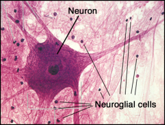
Name the 2 cells found in the nervous system: |
1. Neurons 2. Neuroglia (glial cells) |
|
|
Define Neurons |
-typically the largest cells -are the structural functional unit of the nervous system |
|
|
Define Neuroglia cells |
-aka glial cells -Cells provide support and protection for the neuron. |
|

|
|
|
|
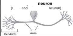
Typical Neuron Structure : 7 parts |
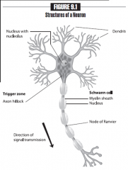
1. Cell Body 2. Dendrite 3. Axon hillock 4. Axon 5. Synaptic Knob 6 Nucleus 7 Myelin sheath |
|
|
Neuron Structure: Define Cell body |
contains nucleus and cellular organells |
|
|
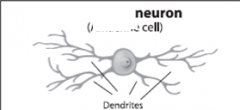
Neuron Structure: Define Dendrite: |
Multiple branching process that recieve impulses from other neurons
|
|
|
Neuron Structure: Define Axon hillock |
Region where axon leaves the cell body |
|
|
Neuron Structure: Define Axon: |
Single large process that sends impulses away from the cell body to another neuron or effector organ |
|
|
Neuron Structure: Synaptic knob |
Swelling at the end of an axon that forms a synapse with another neuron or effector organ. |
|
|
Neuron Structure: Define Myelin sheath |
Neuroglia cell that wraps itself around the axon in many circular layers -insulates the axon and provides a faster transmission of an impulse. |
|
|
Myelinated fibers VS Unmyelinated fibers |
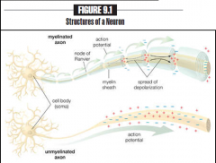
Myelinated Fibers -Axon wrapped by myelin sheath -insulates and provides a FASTER transmission of an impulse Unmyelinated Fibers -Axons w/out a myelin sheath -slower transmission of an impulse |
|
|
Neurons Classified two ways: |
1. by funtion 2.# of processes (dendrites and axons) they poses |
|
|
What are the 3 types of function of a neuron? |
1. Sensory (afferent) neurons 2. Interneurons (association) neurons 3. Motor (efferent) neurons |
|
|
Define Sensory (afferent) neurons: |
Conduct nerve impulses from sensory receptors located in the body to the central nervous system. |
|
|
Define Interneurons (association) neurons: |
-Act as a relay station for an impulse from one part of the brain or spinal cord to another. -Majority of neurons in the central nervous system are interneurons |
|
|
Define Motor (efferent) neurons: |
Conduct nerve impulses from the CNS to effector organs (muscles or glands) |
|
|
Question 1: How would you categorize a neuron that deliversa message from a sensory receptor to the brain or spinal cord? |
Sensory (afferent) neuron |
|
|
Question 2: How would you categorize a neuron that is sending messages back and forth from different area of the brain or spinal cord? |
Interneurons (association) neurons |
|
|
Question 3: How would you categorize a neuron that is sending a message to a muscle? |
Motor (efferent) neuron |
|
|
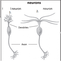
Classification of Neurons: # of processes... 4 types... |
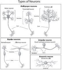
1. Multipolar neurons 2. Bipolar neuron 3. Unipolar neuron 4. Anaxonic |
|
|
Define Multipolar neuron: |
•Contain typically one axon and mutipledendrites -Make up 99% of nerve cells, including motor neurons and interneurons |
|
|
Define Bipolar neuron |
•Contain one axon and one dendrite. -Mainlyspecialized sensory neurons |
|
|
Define Unipolar neuron |
•Contain only one cell process thatdivides into two branches. -Typically make up some sensory neurons |
|
|
Define Anaxonic neuron |
Means “no axon”; contains only dendrites
|
|
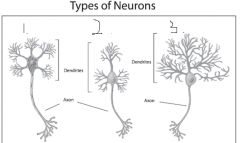
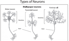
Type of Neuron? Names of each neuron? |
Type: Mulitipolar neuron (left to right) 1. Motor neuron 2. Pyramidal neuron 3. Purkinje cell |
|
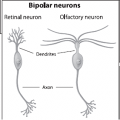
Type of Neuron? Name each neuron? |
Type: Bipolar Neuron (left to right) 1. Retinal neuron 2. Oflactory neuron |
|
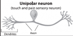
Type of neuron? Name of neuron |
Type: Unipolar neuron Name: Touch and pain sensory neuron |
|
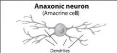
Type of neuron? Name of cell? |
Type: Anaxonic neuron Name: Amacrine cell |
|
|
Nervous tissue is classified into 2 areas on the gross anatomy level: |
White matter and gray matter |
|
|
White matter |
•consists mainly of myelinatedaxons
-Bundles of white matter: --- •Tracts – in the central nervous system(brain and spinal cord) --- •Nerves – in the peripheral nervous system |
|
|
Gray matter |
•consists mainly of neuron cell bodies,dendrites, and unmyelinatedaxons
-Bundles of gray matter: ----- •Nuclei – in the center nervous system ------•Ganglia – in the peripheral nervoussystem |
|
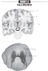
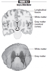
Name each part of the picture |
1. Longitudinal fissure 2. White matter 3. Grey matter (cerebral cortex) 4. White Matter 5. Gray Matter |
|
|
Organization of the Nervous System: (2) |
1. Central nervous system (CNS) 2. Peripheral nervous system (PNS) |
|
|
Central Nervous System (CNS) |
-Control center - Consists of the: BrainandSpinal cordSup - Sensory information is delivered to theCNS and interpreted - produces all motor impulses going to muscles or glands |
|
|
Peripheral Nervous System |
-Consists of Nerves thatconnect the brain and spinal cord to muscles, glands, and receptors - Cranialnerves: nerves that are connected to the brain -Spinalnerves: nerves that are connected to the brain Both cranial and spinal nerves can send impulses from receptors to CNS. From the CNS to muscles or glands |
|
|
PNS is diveded into two function, it is dived into what two subdivisions: |
1. Sensory Division 2. Motor division |
|
|
Sensory division: |
Consistof sensory neurons that send impulses from receptors in the body to the CNS tobe interpreted. |
|
|
3 different receptor of senory division |
-Somatic sensory receptors:Detect general sensations (touch, pressure, temperature, pain, and bodyposition) in the skin, skeletal muscles, and joints
-Visceral sensory receptors:Detect sensations in the internal organs -Special sensory receptors:Detect special sensations (smell, taste, vision, hearing, and equilibrium) |
|
|
Motor division |
Consistsof motor neurons that send impulses from the CNS to effector organs (musclesand glands).
|
|
|
Two parts of Motor Division |
-Somatic nervous system: Containsmotor neurons going form the CNS to skeletal muscle. This impulse pathwayproduces voluntary actions.
-Autonomic nervous system:Contains motor neurons going from the CNS to smooth muscle, cardiac muscle, andglands. This impulse pathway produces involuntary actions.Á |
|
|
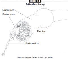
- Nerve: Epineurium - Nerve bundles (fascicles): Perineurium - Axon: Endoneurium |