![]()
![]()
![]()
Use LEFT and RIGHT arrow keys to navigate between flashcards;
Use UP and DOWN arrow keys to flip the card;
H to show hint;
A reads text to speech;
77 Cards in this Set
- Front
- Back
|
what is the neural retina |
typically refers to three layers of neural cells (photo receptor cells, bipolar cells, and ganglion cells) within the retina while the entire retina refers to these three layers plus a layer of pigmented epithelial cells |
|
|
what does the neural retina develop from |
developsas part of the neural tube nso is a part of the brain, the optic nerve hence isa central nerve. |
|
|
where does the neural retina end? |
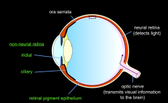
The neuralretina ends at the ora serrata (serrated edge), there is then (on theillustration) a black structure that goes over the ciliary bodies andback of the iris (ora serrata). |
|
|
what does the neural retina detect, and how does it send afferents to the brain |
The neural retina detects light and contains afferents,which go out and form the optic nerve, carrying signals back to the thalamus. |
|
|
what types of cells does the neural retina have to allow it to detect light |
havephotoreceptors here. Cones are for daylight vision, and rods are for ‘scotopic’(night time) vision |
|
|
how does the retina send afferents to the brain |
We have afferents with ganglion cells. Axons run across thesurface of the retina. The gap is bridged by inter-neurones in the inner nuclearlayer. |
|
|
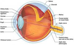
what is the fovea centralis |
a small depression in the retina of the eye where visual acuity is highest. The centre of the field of vision is focused in this region, where retinal cones are particularly concentrateq |
|
|
what is the visible which part of the eye made up of |
made up of collagen. Tough and flexible. |
|
|
what is the uveal tract of the eye and what does it contain |
The uveal tract is a layer of tissue located between the outer layer (cornea and sclera) and the inner layer (the retina) of the eye. The front portion (anterior) of the uveal tract contains the iris, and the back portion (posterior) of the uveal tract contains the choroid and the stroma of the ciliary body |
|
|
how is the cornea arranged |
collagen are v organized, and gluedtogether with proteoglycans. So have a transparent window through which thelight can pass |
|
|
the eye isnt made of solid structures. they are very flexible. so, how is the shape of they eye maintained |
shape of eye is maintained by thecontents. In posterior Segment of the eye you have the vitreous humour.In the anterior segment of the eye you have the aqueous humour. Shape of eye is maintained bypressure created by the aqueous humourbeing produced and drained. Allows eye to maintain its globular shape. |
|
learn |
. |
|
|
what are the characteristics of the choroid layer of eye, and why is it important |
Very vascularized layer that produceda very rapid flow of blood past the photoreceptors. It actually provides the o2and glucose and other demands for the outerpart of the retina. |
|
|
The cillary body of the eye contains two different compartements. what are these. |
1 – muscle that controls the shapeof the lens via the suspensory ligaments on either side. 2 – tissue thatproduces the aqeous humour. |
|
|
what is the optic nerve made up of |
Made up of the axons of retinal ganglioncells whichflow across surface ofretina. It is covered in the 3 layers of meninges (pia matter, arachnoid matterthen dura matter). |
|
|
inner layer of the eye has two main regions. what are these |
neural retina &Retinalpigementepithelium |
|
|
what is the retinal pigment layer |
The latter is a single layer of pigmentepithelial cells. |
|
|
how are the retina &Retinalpigementepithelium joint |
The two layers aren’t ever joinedtogether v strongly. They are parially glued together by proteoglycans. The pigment epitheliumpump fluid out from the gap between them creating some suction. It is thissuction and glue that holds the retina to the back of the eye . If that suction gives way, theretina may float free from the pigment. |
|
|
what does the neural retina do |
1 -The neuralretina detects light by use of rods and cones 2 - totransmit the information they detect to the brain by the afferents that then go out and form theoptic nerve. |
|
|
what are rod photoreceptors |
receptors that detect light in night vision,also called scotopic vision (work best in dim light). |
|
|
what are cone photoreceptors |
receptors that detect light. for daylightvision (work best in bright light) |
|
|
how does the neural retina transmit information it detects to the brain |
The axons of theganglion cells (in the inner layer of the retina) run over the surface of the retina and head to the optic nerve. |
|
|
what does the inner nucleur layer of the eye contain |
containsinterneurons which lay inbetween the photoreceptors to the ganglion cells. |
|
|
what is the importance of the bipolar interneuronem and where is is it situated |
in the innernuclear layer, thebipolar interneurone that is in the middle of the two cones. Theseconnect the photoreceptors to the ganglion cells. |
|
|
what is the ganglionic cell receptive feed |
Therays of the visual image that enter our eye that are detected by cones, and form the retinal receptive field for this ganglion cell |
|
|
why is it adaptive for the cones to converge their input into one single ganglion cell? |
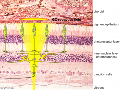
When lightenters the eye, it has to pass through the vitreous and the multiple layers ofthe retina before it reaches the cones. In fact it is the outer-portion of thecones which actually detect the change in illumination. light that hitsthe retina being detected is blurred because it has had to pass through theseouter layers first. These pool ofcones will converge input onto a single ganglion cell so make sense of multiple blurry images |
|
|
what is the peripheral and central retina |
|
|
|
why is peripheral vision poor |
the fact thatthe cone photoreceptors in the periphery are large and spaced widely apart totry gather as much of this scattered/blurred light cones in periphery have large receptivefields -> Thesecones gather this blurred light and converge it onto single ganglion cells andthus all of this info of this region of scattered light is brought to one cellto transmit the info |
|
|
within the central retina, we have the Macula Lucida (area of yellow pigment). what is the importance of this |
Within themacula lucida is the fovea centralis, this is a very specialised and small areaof the eye. The fovea has no overlying blood vessels or capillary bed. |
|
|
why is the fovea so important/ why does the fovea allow us to avoid blurry vision in the cenral retina |
The fovea isspecalised because it contains a small region, only 0.35mm across where theganglion cells and interneurons have been pushed back and halted to leave afoveal pit. This directlyexposes the photoreceptors and means that light that passes into the eye andvitreous does not have to pass through multiple layers first and thus not besubject to getting scattered. This is the onlypart of the eye where vision is very well focused/ resolution and it visualisesabout the size of your thumbnail at arms-length. |
|
|
how do the characteristics of cones in the fovea and the peripheral retina vary |
cones in thefoveal pit are very slender and thus a huge number are packed in together(150,000mm2). Eachphotoreceptor then synapses onto a single ganglion cell, so there is noconvergence. This means the fine image (because no scattering!) detected byeach single photoreceptor is signaled to a single ganglion cell so there is no“noise” added to the fine image and instead is signaled back to the braincompletely as the fine image.well focused |
|
|
how is the fovea specialised for high resolution |
-> Overlyinglayers and blood vessels are absent so the image is well focused/not scattered -> The foveaonly contains cone photoreceptors which are narrow and closely packed -> Thesignals from photoreceptors are kept separate |
|
|
what happens to light hitting they eye (externally) |
someof these rays will hit your cornea and pass through. Someof the rays that pass through will be blocked by the iris others will passthrough the pupil and these will be brought into focus/further refracted by thelensin |
|
|
what has the primary role in focusing light rays |
It is the corneathat refracts the light the most and hence is primary responsible for focusinglight rays. Howeverthe cornea is fixed in its refractory ability |
|
|
what structure of the eye accomodates focusing on near or far items |
the lens |
|
|
what is the punction of the iris |
The iris controlsthe amount of light that enters the eye via the pupil. |
|
|
why does the pupil dilate and constrict |
If the pupil wasa pinpoint, then the image would be clearly focused on the back but it would bevery dim as hardly any light would get through. The larger thepupil the more light enters in and will hit the outside of the lens which isnot as good at focusing the image and it leads to more blur. |
|
|
what muscles control the optics of the eye |
1 - The sphincter pupillae is a ring muscle and when it contracts it makes the pupil smaller 2 - ciliary muscle-a ring shaped muscle going around the front part of the eye. 3 - dilator pupillae,when the muscle contracts it pulls the pupil open |
|
|
what is the role of the sphincter pupillae muscle |
The sphincterpupillae is a ring muscle and when it contracts it makes the pupil smaller. Whensphincter pupillae is relaxed, the pupil is large and dilated |
|
|
how are the sphincter pupillae controlled |
Under thecontrol of the short ciliary nerves which are parasympathetic nerve fibres,they use Ach as their neurotransmitter. The shortciliary nerves come from the ciliary ganglion, a parasympathetic Thepre-ganglionic fibres going into this pathway of contracting sphincter pupillaeare actually primarily driven by the retina itself! This is thedirect/consensual light reflex! |
|
|
what nerve carriesparasympathetic pre-ganglionic fibres to the ciliary ganglion? |
CN III (oculomoter nerve) |
|
|
what is the light reflex |
Inthe retina we have the retinal ganglion cell which will be activated if thereis illumination of that retina
Signalsfrom that ganglion cell -> sent to pretectal nucleus -> sends axons to Edinger-Westphalnucleus -> sends preganglionic parasympathetic fibres to cilliary ganglion. ciliary ganglion axons synapse onto postgangionic short ciliary nerves -> release of Ach onto the sphincter pupillae so eye constricts |
|
|
why does illumination of one eye lead to constriction in both eyes |

pretectalnucleus neurones synapse onto both sides of the Edinger-Westphal nucleus, soboth parasympathetic pathways get activated! |
|
|
what is the function of the dilator pupillae. what does this muscle react to |
when the musclecontracts it pulls the pupil open This dilation isbrought about by the long ciliary nerves which innervate dilator pupillae. Thisis a sympathetic pathway. When the eyedilates it is NOT in response to a decrease in light rather it is due to strongemotional drive. For example, fear, love. |
|
|
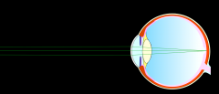
what is the characteristics of a normal eye |
thelength of their eye is perfectly matched to the strength of their optics |
|
|
how does a normal eye react to seeing something far away |
If the person islooking at something very far away, the light rays will enter into their eyenear enough parallel and only need a little extra refraction in the lens to getthe image onto the retina (so lens will be flattened). |
|
|
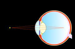
how does a normal eye react to seeing something close |
If an individualwants to look at an object close to them the light rays will be diverging morethus more refractive power is needed to bring the rays together. The lens willbe made fatter and this will refract the light nicely onto the retina and it iswell focused |
|
|
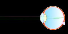
what is myopia |
theoptics of their eye are simply too strong, the refractive power is too much forthe length of their eyeball. so individuals are short-sighted. |
|
|
how does a myopic eye react to seeing something close |
The individualwith a myopic eye will look at something close and lens will bulge as lightrays are more divergent. However, the power of the optics is too strong andthus the light rays and image will be refracted so strongly it will focus infront of the retina. This is what amyopic eye is, when objects are focused in front of the retina. canhowever still focus on close objects by flattening out the lens for closeobjects and thus reducing the refractive power to get the image onto the retina. This flattened lens would usually only be for distant objects, so person isessentially using long distance vision to view close objects. |
|
|
how does a myopic eye react to seeing something far away |
However as themyopic eye has compensates by using long distance vision to look at closeobjects. With far away objects the lens cannot be made sufficiently more flatto get the image on the retina. The optics are still too strong. Hence theimage will focus in front of the retina |
|
|
how can you fix myopic lems |
The way to fixmyopia is with a concave (negative) lens. This will diverge the rays a bit moreand thus weakens the overall optics of the eye so the image will be able tofocus onto the retina.retina. |
|
|
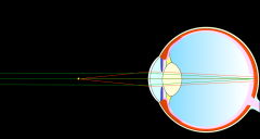
what are the characteristics of a hypermetropic |
the optics of apersons eye are too weak and the refractive ability of their eye is too weakfor the length of their eye. Then the eye is hypermetropic, this is where theimage focuses behind the retina as the optics are not powerful enough to focusit properly! being long-sited |
|
|
what is meant by the optics of the eye |
the basic optic function of the eye is to form an image of whatever object being looked at (fixated on) on the retina. This image formation is done by the combination of the cornea and the lens which behaves like a convex lens |
|
|
how does a hypermetropic eye respond to seeing things far away |
longdistance image with its rays very straight anyway, cannot be focused enough. lenscan compensate and adapt in order to focus far away objects by bulging out andincreasing the refractive power (as if the object were close!) This brings theobject into focus on the retina4 |
|
|
how does a hypermetropic eye respond to seeing things that are close |
The problem thenarises with near objects, here the light rays are much more divergent thusrequiring more refraction by the lens. However the lenshas already refracted strongly for distant objects and cannot get any more fatand has reached its maximum refractive power. This level it is not enough andthe image will be focused after the retina. |
|
|
how can hypermetropia be corrected for functional vision |
Hypermetropiacan be corrected with a convex (positive) lens that increases the refractivepower of the overall optics. This helps bring the rays together and gets themfocused onto the retina |
|
|
why does vision decrease with age |
the lens has avery little capacity to regenerate and thus can fail with age. |
|
|
why does the eye have such little ability to regenerate |
that in order tohave such a transparent structure it needs to be made up of cells that containvery few organelles. Due to this they have very little regenerative capacity |
|
|
why do many middle aged people require lenses for both short and long distances |
Inmiddle age, the lens becomes stiff and stops responding to changes in theciliary muscle. Because of this focus becomes fixed (presbyopia) and theindividual may need two pairs of glasses to see properly both long and shortdistance. |
|
|
when may an individual develop a cataract. how can this be resolved |
In old age or inpathological conditions things can get worse, the lens can become opaque, thisis a cataract. The only solution here is to take it out and replace it with anartificial lens. |
|
|
what is the primary visual pathway. what other pathways run alongside it |
pathwayis the pathway from the eye, to the thalamus to the primary visual cortex inthe occipital lobe Alongside thisprimary visual pathway we also have another pathway that is associated withreflexes and orientating responses. |
|
|
what is the role of the superiorcolliculi |
integrating sensory information (visual, auditory, somatosensory) into motor signals that help orient the head toward various stimul involved inreaching movements, turning the head and eyes towards a stimulus requiringattention and navigating in locomotion. |
|
|
where is the pathway for visual perception |
start of at the retinal ganglion cells -> project back to the lateralgeniculate nucleus (LGN) in thethalamus -> From here axons go back to the to calcarine sulcus (which is in the primary visual cortex in the occipital cortex) |
|
|
what is the optic radiation |
The optic radiation (also known as the geniculocalcarine tract, the geniculostriate pathway, and posterior thalamic radiation) are axons from the neurons in the lateral geniculate nucleus to the primary visual cortex. |
|
|
where does light coming from the left visual feild hit (in both eyes) |
Light comingfrom the left visual field will fall onto the right side of our retinas, inboth eyes. In the right eyeit will fall onto the temporal retina, in the left eye it will fall onto thenasal retina. |
|
|
where does light coming from the right visual feild hit (in both eyes) |
Light comingfrom the right visual field will fall onto the left side of our retinas, inboth eyes. In the left eyeit will fall onto the temporal retina, in the right eye it will fall onto thenasal retina. |
|
|
how is left visual field information processed |
As left visual field information will be processed in the right hemisphere: Right temporal fibre (from Right eye temporal retina) does not need to cross the optic chiasm as it is on the same side as the hemisphere the information will be processed. Left nasalganglionic fibres (from left eye nasal retina) will be carrying information from theleft visual field (red) and will be processed in the right hemisphere andtherefore need to cross over at the optic chiasm to get to the right hemisphere. |
|
|
how is right visual field information processed |
As right visual field information will be processed in the left hemisphere: left temporal fibre (from left eye temporal retina) does not need to cross the optic chiasm as it is on the same side as the hemisphere the information will be processed. right nasal ganglionic fibres (from the right eye nasal retina) will be carrying information from the right visual field and will be processed in the left hemisphere and therefore need to cross over at the optic chiasm to get to the left hemisphere. |
|
|
what happens of there is damage in front of the optic chiasm |

thedamage is independent of the two pathways. if an optic nerve is damaged, it affects only the visual field of that eye. So in otherwords there will be different deficits in the two eyes. |
|
|
what happens of there is damage in behind the optic chiasm |
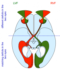
Behind the chiasm axons from a matching part of the 2retinae lie close together to any lesion will take out a matching part of thevisual field that is being projected back from the 2 eyes. If it matches inboth eye you know it is from behind the chiasm. |
|
|
Primaryvisual pathways have a pathway associated with reflexes. where are the nuclei involved in this reflex |
Allnuclei involved in this kind of movement of the eye are down in the brain stem,the biggest and most prominent is the superior colliculus |
|
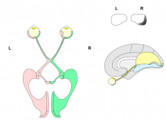
what defect can be seen here |
Inthis case, there has been loss of the left visual field in the right eye dueto a detached retina |
|
|
what is glaucoma |
a condition of increased pressure within the eyeball, causing gradual loss of sight. |
|
|
what may a lesion of one optic nerve cause |
A lesion of one optic nervecauses monocular blindness in the corresponding eye (as the nerve contains allthe fibres for the visual field of that eye) |
|
|
what may be the effect of a lesion of the optic chiasm |
- A lesion of the optic chiasmadestroys crossing nasal fibres (nasal fibres look at the temporal field on bothsides) this leads to a bitemporal hemianopia |
|
|
what is hemianopia |
blindness of half the field of vision |
|
|
what may a lesion of the optic tract cause |
A lesion of the optic tract(which is carrying axons for the right OR left visual field only) will lead toa contralateral homonymous hemianopia (contralateral as other side e.g. rightoptic tract will lead to left hemianopia, homonymous as in both eyes,hemianopia as in half visual field loss) |