![]()
![]()
![]()
Use LEFT and RIGHT arrow keys to navigate between flashcards;
Use UP and DOWN arrow keys to flip the card;
H to show hint;
A reads text to speech;
94 Cards in this Set
- Front
- Back
|
4 Major regions of the Brain |
• The Cerebral hemispheres include the frontal, parietal, temporal and occipital lobes and the basal nuclei (ganglia). • The diencephalon includes the thalamus and hypothalamus. • The brainstem includes the midbrain, pons and medulla oblongata. The cerebellum |
|
|
Brain Folds |
• Since the brain grows more rapidly than the skull, the brain forms folds, allowing it to fit inside the skull. . A raised region is called a gyrus •A wrinkle towards the interior of the brain is called a sulcus • A deep wrinkle is called a fissure. |
|
|
Grey Matter |
The grey matter of the CNS consists of, neuron cell bodies, while the white matter consists of myelinated/nonmyelinated axons. • CNS has a central cavity surrounded by grey matter, with a layer of white matter externally. • The brain stem has additional grey matter nuclei scattered within the white matter. • The cerebral hemispheres and cerebellum have an outer layer of grey matter, called the CorteX |
|
|
Cerebral White Matter |
Responsible for communication between cerebral areas and the cerebral cortex and lower CNS centres. |
|
|
3 types of fibres that make up cerebral white matter. |
1. AssoCiatiOn fibres 2. Commissural Fibres 3. Projection Fibres |
|
|
Association Fibres |
Are tracts of cerebral white matter that run horizontally, connecting different parts of the same hemisphere. |
|
|
Commissural Fibres |
Run horizontally and connect corresponding areas of grey matter in the two hemispheres, allowing the hemispheres to function together as a whole (includes the corpus callosum). |
|
|
Projection Fibres |
Run vertically, and connect the cerebral cortex to the lower brain or cord centres, tying together the rest of the nervous system to the body's receptors and effectors. |
|
|
Cerebral Hemispheres |
The cerebrum is the largest region of the brain, separated into right and left hemispheres • Each cerebral hemispheres has 3 regions: the superficial cortex of grey matter, internal white matter, and areas of grey matter deep within the white matter, the basal nuclei. • Each hemisphere is divided into 4 visible lobes: frontal, parietal, temporal and occipital. A 5th region, called the insula, forms part of the floor of the lateral sulcus and is covered by the lobes. |
|
|
Cerebral Cortex |
• The cerebral cortex is the location of the conscious mind, allowing us to communicate, remember, and understand. It has motor areas, sensory areas, and association areas. Each hemisphere has contralateral control over sensory and motor functions, meaning that each hemisphere controls the opposite side of the body. • The hemispheres exhibit lateralization of function, meaning that there is specialization of one side of the brain for certain functions. |
|
|
Motor Areas of the Cerebral Cortex |
• Motor areas of the cortex are in the posterior of the frontal lobes and control voluntary movement. • The primary motor cortex (located in the pre-central gyrus) allows conscious control of skilled voluntary movement of skeletal muscles. • The premotor cortex is the region controlling learned motor skills. • Broca's area is a motor area that controls muscles involved in making words when speaking or writing. |
|
|
Cortical Homonculus |
This is an artistic depiction of how the human body would look if the body parts were scaled proportionately to the size of the region of your cerebral cortex that processes sensory information from those body parts. |
|
|
Sensory Areas of Cerebral Cortex |
• The Primary somatosensory cortex processes sensory information from the body, such as touch and temperature, as well as the location of stimulation. It is located in the postcentral gyrus of the parietal lobe. The somatosensory association cortex integrates sensory information and produces an understanding of the stimulus being felt. • The primary visual cortex and visual association area in the occipital lobe receive and interpret visual stimuli. |
|
|
Sensory Areas of Cerebral Cortex 2 |
• The primary auditory cortex and auditory association area are in the temporal lobe. They allow detection and recognition of sound . • The vestibular cortex is in the parietal lab and the insula. It is responsible for awareness of balance • The olfactory cortex processes smell and the gustatory cortex perceives taste. They are found at the inferior frontal lobe and insula. • The visceral sensory areas in the insula are involved in conscious awareness of visceral sensation. |
|
|
Multimodal Association Areas |
The anterior association area, or prefrontal cortex, is involved with intellect, cognition, recall, and personality. It is closely linked to the limbic system. • The posterior association area aids in recognition of patterns and faces, as well as understanding of written and spoken language. This includes Wernicke's area, which links words and meanings. • The limbic association area deals with emotions surrounding situations and includes the cingulate gyrus, parahippocampal gyrus, and hippocampus. |
|
|
Late realization of Cortical functioning |
• Some medical studies have found varying degrees of lateralization of cortical functioning, in which each cerebral hemisphere has unique control over abilities not shared by the other half. • In many cases, the left hemisphere dominates language abilities, math and logic, while the right hemisphere dominates visual-spatial skills, ntuition, emotion, and artistic and musical skills. • Both sides of the brain are involved in virtually all skills and processes. |
|
|
Basal Nuclei |
• Basal nuclei (sometimes inaccurately called basal ganglia) consist of a group of subcortical nuclei that have overlapping motor control with the cerebellum that regulate cognition and emotion. They appear to be involved in deciding which action you should take and inhibiting other actions. • Nuclei are clusters of neuron bodies in the CNS. Ganglia are clusters of neuron bodies in the PNS. The brain is part of the CNS, so "basal ganglia" is a misnomer. |
|
|
Thalamus |
Mediates sensation, motor activities, cortical arousal, learning, and memory. It plays a role in the maintenance of consciousness. • It acts as a relay station to conduct Sensory impulses to the primary sensory areas of the cerebral cortex and transmits motor information from the cerebellum and basal nuclei to the primary motor area of the cerebral cortex. |
|
|
Hypothalamus |
The control centre of the body, regulating autonomic nervous system activity. • It initiates physical responses to emotions, and regulates body temperature, food intake, water balance, thirst, sleep-wake cycles, and endocrine function. It is the integration centre of the autonomic nervous system. • The hypothalamus also regulates the pituitary. gland and produces oxytocin and antidiuretic hormone. |
|
|
Higher Mental Functions |
• The interconnected structures of the brain allow higher mental functions. • The ability to both speak and understand language is produced through coordination of several brain areas, notably Broca's area and Wernicke's area. • Memory is the storage and retrieval of information and there are different kinds of memory: declarative (fact-based), procectural (skills), motor, and emotional. |
|
|
Declarative Memory |
Has 2 stages: short-term memory, and long-term memory. short-term memory, or working memory - allows the memorization of a few units of information for a short period of time. Long-term memory allows the memorization of potentially limitless amounts of information vor very long periods. |
|
|
More about declarative memory |
• Transfer of information from short-term to long-term memory can be affected by emotional state (best if alert, motivated, excited or surprised), rehearsal, association of new information with old, or the automatic formation of memory while concentrating on something else. • Damage to the hippocampus or surrounding temporal lobe structures on either side result in only slight memory loss. Bilateral destruction causes widespread amnesia. |
|
|
Consciousness |
Can be clinically defined on a continuum that measures behaviour in response to stimuli and ranges through several stages: alertness, drowsiness or lethargy, stupor, and coma. • Syncopy (fainting) is a brief loss of consciousness, most often due to inadequate cerebral blood flow caused by low blood pressure or sudden, severe emotional stress. • Coma is unconsciousness for an extended period. Oxygen consumption is lower than during sleep. |
|
|
Sleep |
• Sleep is a state of partial unconsciousness from which a person can be aroused and has two major types that cycle: non-rapid eye movement (NREM) and rapid eye movement (REM). NREM sleep is restorative REM sleep allows the brain to analyze events or eliminate meaningless information. Dreaming occurs & skeletal muscles are inhibited during REM sleep |
|
|
Protecting the CNS |
The meninges are 3 connective tissue membranes that cover & protect the CNS. • The Dura mater is the most durable, outermost covering that extends inward in certain areas to limit movement of the brain within the cranium. • The arachnoid mater is the middle meninx that forms a loose brain covering. • The Pia mater is the innermost layer that clings tightly to the brain. |
|
|
The blood brain barrier |
• The blood-brain barrier is a mechanism that helps maintain a protective environment for the brain. • It contains exceptionally impermeable tight junctions keep brain separated from many bloodborne substances. • The blood-brain barrier is made up of three layers: • Continuous endothelium of capillary walls. • Thick basal lamina around capillaries. • Feet of astrocytes surrounding neurons. |
|
|
Blood brain barrier 2 |
• Nutrients, essential amino acids, and some electrolytes are allowed to pass into cerebrospinal fluid, but metabolic wastes, proteins, toxins, and most drugs are excluded. • Lipid-soluble molecules, including alcohol, nicotine, and anaesthetics, can cross the blood-brain barrier. It is absent in some areas, such as the vomiting centre and the hypothalamus, because these brain areas need to monitor chemical composition and temperature of blood. |
|
|
Cerebrospinal Fluid (CSF) |
The fluid found w/in ventricles and surrounding the brain and spinal cord. CSF gives buoyancy to the brain, protects the brain and spinal cord from impact damage, and is a delivery medium for nutrients and chemical signals. • The four ventricles are lined with ependymal cells and are filled with cerebrospinal fluid. The ventricles of the brain are connected to one another and to the central canal of the spinal cord. |
|
|
CSF 2 |
• The 4 ventricles in the brain are paired lateral ventricles deep within each cerebral hemisphere, a third ventricle within the diencephalon, and a fourth ventricle at the cerebellum. Ependymal cells are responsible for keeping CS circulating. • Arachnoid granulation villi are pockets of the arachnoid membrane that extend into the dura mater sinuses, such as the superior sagittal sinus. This is where CSF can be reabsorbed into venous blood to re-join the circulatory system. |
|
|
Brain stem |
Midbrain, Pons, Medulla Oblongata |
|
|
Cerebral Peduncles (midbrain) |
Contain pyramidal motor tracts that descend toward spinal cord |
|
|
Corpora Quadrigemina (midbrain) |
Control visual & auditory startle behaviours |
|
|
Pons (brain stem) |
Contains fibre pathways between the brain and spinal cord, as well as giving rise to some cranial nerves, and contains some important nuclei, including one that helps control breathing. |
|
|
Medulla (brain stem) |
The location of the medullary pyramids, which act as crossover points for corticospinal motor tracts, resulting in the contralateral control of voluntary movements. It also houses neurons controlling vital functions such as cardiac and respiratory rate |
|
|
Cerebellum |
The second-largest area of the brain. It adjusts Motor output, ensuring coordination and balance. Processes inputs from several structures and coordinates skeletal muscle contraction to produce smooth movement. • The cerebellum may also play a role in thinking, Janguage, and emotion, |
|
|
Structure of Cerebellum |
Consists of 2 cerebellar hemispheres with 3 lobes each. Anterior and posterior lobes coordinate body movements, and the flocculonodular lobes adjust posture to maintain balance. Three paired fibre tracts, the cerebellar peduncles, communicate between the cerebellum and the brain stem. • Grey matter is located in outer cortex (folia) and in deeper nuclei. White matter tracts are called arbor vitae |
|
|
Limbic System |
Is involved with emotions and is extensively connected throughout the brain, allowing it to integrate and respond to a wide variety of stimuli. |
|
|
Reticular Activating System |
Extends through the brain stem and keeps the cortex alert while suppressing familiar, repetitive, or weak sensory inputs. 99% of sensory input is not relayed to consciousness. |
|
|
Spinal Cord |
2 main functions: 1. The spinal cord provides a COnduction pathway. Each column of white matter in the spinal cord contains bundles of axons (tracts). These tracts have a common origin (or destination) and carry similar information. Tracts are continuous with white matter tracts in the brain. 2. also serves as an integration centre for spinal reflexes. |
|
|
Structure |
Extends from the foramen magnum of the skull to the level of the first or second lumbar vertebra. It provides a two-way conduction pathway to and from the brain and serves as a reflex centre. • Fibrous extensions of the Pia mater anchor the spinal cord to the vertebral column and coccyx, preventing excessive movement of the cord. • The spinal cord has 31 pairs of nerves (part of the PNS) along its length that define the cord segments |
|
|
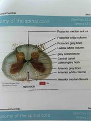
Anatomy of the Spinal Cord |
• There are cervical and lumbar enlargements for the nerves that serve the limbs and a collection of nerve roots at the inferior end of the vertebral canal (cauda equina) that travel through the vertebral column to their intervertebral foramina. The ventral (anterior) median fissure and dorsal (posterior) median sulcus divide the spinal cord into left and right halves. A CSF-filled central canal runs the length of the spinal cord at its centre. |
|
|
Anatomy of the Spinal Cord 2 |
• The grey matter resembles a butterfly or "H" shape n cross section: the dorsal horns contain interneurons; the ventral horns consist most of cell bodies of somatic motor neurons • In the thoracic and superior lumbar regions, there are also paired lateral horns that extend laterally between the dorsal and ventral horns, and contain cell bodies of autonomic motor neurons. |
|
|
Anatomy if the Spinal Cord 3 |
The grey commissure is a bridge of grey matter that connects masses of grey matter on either side. Ventral roots exit the spinal cord carrying axons of motor neurons, and join with dorSAl roots entering the spinal cord from peripheral receptors from outside of the spine to form spinal nerves. Dorsal root ganglia are the cell bodies of sensory neurons. The white matter (myelinated and nonmyelinated nerve fibres) of the spinal cord allows communication between the cord and brain |
|
|
Anatomy of Spinal Cord Diagram |
Back (Definition) |
|
|
Trauma to Spinal Cord |
Any localized damage to the spinal cord or its roots leads to paralysis (loss of motor function) × 50 or paresthesias (loss of sensory function). Severe damage to the ventral root or ventral horn results in flaccid paralysis, since nerve impulses are not transmitted to the skeletal muscles. When upper motor neurons of the primary motor cortex are damaged, spastic paralysis occurs, in which voluntary control over skeletal muscle is lost. |
|
|
Spinal Cord Trauma |
If damage to the spinal cord occurs between T1 (first thoracic vertebra) and L1 (first lumbar vertebra), lower limbs are affected, resulting in paraplesia. Further down If the damage occurs in the cervical region, all four limbs are affected, resulting in quadriplegia. Spinal shock is a transient loss of function caudal to the trauma. |
|
|
Neuronal Pathways |
Carry sensory and motor information to and from the brain. Major spinal tracts are part of paired multineuron pathways that mostly: • Cross from one side to the other (decussate) • Consist of a chain of 2 or 3 neurons •Exhibit somatotopy (a point-for-point relationship between an area of the body and a specific point in the central nervous system). |
|
|
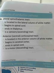
Examples of conduction pathways |
Back (Definition) |
|
|
Ascending Sensory Pathways |
Ascending pathways conduct sensory impuises upward through a chain of three neurons. 1. First-order neuron conducts the sensory info from the receptor to the spinal cord or brain stem. 2. Second -order neuron conducts the impulse to the thalamus. Third -order neuron conducts the impulse to the primary somatosensory cortex of the cerebrum. |
|
|
Ascending Sensory Pathways |
Nonspecific ascending pathways receive input from many different types of sensory receptors, and make multiple synapses in the brain. Specific ascending pathways mediate precise input from a single type of sensory receptor. •Spinocerebellar tracts convey information about muscle and tendon stretch to the cerebellum. They are ipsilateral (don't cross over) and only involve two neurons, not three. |
|
|
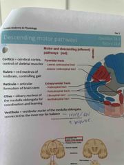
Descending Motor Pathways |
Descending pathways involve two neurons: upper motor neurons and lower motor neurons that innervate skeletal muscle. • In the direct or pyramidal, system, upper motor neurons from the cerebral cortex regulates fast, finely controlled or skilled movements. • In the indirect or extrapyramidal system, upper motor neurons from the brainstem regulate muscles for balance and coarse movements and head, neck, and eye movements for visual tracking. |
|
|
Descending Motor Pathways |
Back (Definition) |
|
|
Peripheral Nervous System |
Includes all neural structures outside the brain and spinal cord: sensory receptors, peripheral nerves and their associated ganglia, and efferent motor endings. |
|
|
PNS Sensory Receptors |
Sensory receptors are specialized'to respond to changes in their environment called stimuli. Activation of sensory receptors by a strong enough stimulus causes the production of graded potentials that trigger nerve impulses along afferent pathways to the central nervous system (CNS). |
|
|
Receptor Structure |
•Simple receptors are general senses and endings may be free (non-encapsulated) or encapsulated. • Non-encapsulated dendritic endings are free nerve endings that detect temperature, pain, itch, light touch, or are located at the base of hair follicles. Encapsulated dendritic endings consist of a dendrite enclosed in a connective tissue capsule and detect discriminatory touch, initial, continuous, and deep pressure, and stretch of muscles, tendons, and joint capsules. |
|
|
First Order Sensory Neurons |
• Graded receptor potentials can trigger action potentials that travel along the axon to the CNS |
|
|
Types of Stimulus |
Mechanoreceptors are stimulated by mechanical force, such as touch, pressure, vibration, and stretch. Thermoreceptors detect changes in temperature Photoreceptors detect light. Chemoreceptors are stimulated by chemicals, such as odours, tastes, or chemical components of body fluids. Nociceptors respond to painful stimuli. |
|
|
Location of Stimulus or Sensor |
Exteroceptors are located at or near the body surface and detect stimuli from outside the body, such as touch, pressure, pain, skin temperature receptors, and most of the special senses. • Interceptors, associated with internal organs and vessels, monitor chemical changes, stretch, or temperature. • Proprioreceptors are found within skeletal muscles, joints, and associated connective tissues and relay information about body movements. |
|
|
Sensation & Perception |
Sensation is the awareness of changes in the internal and external environment. Perception is the conscious interpretation of those stimuli. First-order sensory neurons carry impulses from receptors. Second-order neurons transmit to the cerebellum or third-order neurons, which carry impulses to the postfrontal gyrus. |
|
|
Pain |
The perception of pain protects the body from damage and is stimulated by extremes of pressure and temperature, as well as chemicals released from damaged tissues. • The pain threshold is the stimulus intensity at which we begin to perceive pain and is the same for most people. Pain tolerance is a genetically determined trait with learned aspects that varies from person to person. Like any other perception, perceived pain can be affected by the environment. |
|
|
Visceral Pain |
Visceral pain results from stimulation of receptors within internal organs from stimuli such as extreme stretch, ischemia (lack of blood flow), chemical irritation, and muscle spasms. Visceral pain travels along the same fibre tracts as somatic pain impulses, giving rise to reFerred pain that feels as though it is located in an area different from the affected body region. |
|
|
Peripheral Nerves |
• Peripheral nerves, either cranial or spinal, are classified according to the direction in which they transmit impulses. • Mixed nerves contain both sensory and motor fibres. Most nerves are mixed nerves. • Sensory, or afferent, nerves carry impulses only toward the CNS. • Motor, or efferent, nerves only carry impulses away from the CNS. |
|
|
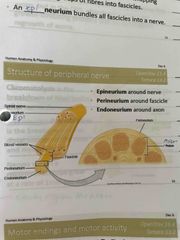
Nerve Structure |
A nerve is a cord-like organ consisting of parallel bundles of peripheral axons enclosed by connective tissue wrappings. Each axon within a nerve is surrounded by a thin layer of loose connective tissue, the endoneurium. A perineurium is a connective tissue wrapping that bundles groups of fibres into fascicles. An Epíneurium bundles all fascicles into a nerve. |
|
|
Structure of peripheral nerve diagram |
Back (Definition) |
|
|
Motor Endings & Motor Activity |
• All ventral branches except T2-T12 form interlacing networks called nerve plexuses. Within each plexus, fibres crisscross so that each branch contains fibres from several different spinal nerves. Fibres go to the body periphery via several routes, so that each limb muscle is innervated by more than one spinal nerve and damage to one does not cause paralysis Dermatomes are areas of skin innervated by cutaneous branches of a single spinal nerve. Most dermatomes overlap, so destruction of a single spinal nerve will not cause complete numbness. |
|
|
Motor Endings & Motor Activity |
The terminals of the somatic motor fibres that innervate voluntary muscles form neuromuscular junctions with their effector cells and they release the neurotransmitter acetylcholine. The junctions between autonomic motor endings and the visceral effectors form synapses en passant ("synapses in passing") involving varicosities and release either acetylcholine or epinephrine as their neurotransmitter. |
|
|
Varicosities |
• Autonomic nerve fibres innervate most smooth muscle. These muscle contractions tend to be a bit slower than the reactions of skeletal muscles. • Vesicles release neurotransmitters into a wide synaptic cleft (a diffuse junction). |
|
|
Peripheral Nerve Repair |
Ganglia are collections of neuron cell bodies associated with nerves in the PNS. Damaged CNS nerve fibres almost never regenerate, but if a PNS nerve fibre is cut or compressed, and the cell body remains intact, axons can regenerate. • Schwann cells participate in regenerating PNS axons, but in the CNS, oligodendrocytes have growth-inhibiting proteins that do not support regrowth of axons. |
|
|
Neuroregeneration |
Chromatolysis is the breakdown of Nissl bodies after nerve damage. Wallerian degeneration is the breakdown of the distal portion of the axon and myelin sheath. Nerves within a regeneration tube grow at a rate of 1mm per day |
|
|
Autonomic Nervous System |
Lecture 35 |
|
|
Reflexes |
• Reflexes are unlearned, rapid, predictable motor responses to a stimulus and occur over highly specific neural pathways called reflex arcs. The spinal cord is the integration centre for these arcs. • Inborn, or intrinsic, reflexes are unlearned, unpremeditated, and involuntary. Learned, or acquired, reflexes result from practice, or repetition. |
|
|
Reflex Arc |
• A reflex arc is a very specific neural path that controls reflexes and has 5 components: a receptor, a sensory neuron, an integration centre, a motor neuron, and an effector. • Somatic reflexes activate skeletal muscle. • Autonomic reflexes involve smooth muscle, cardiac muscle and/or glands. |
|
|
Stretch Reflex |
The stretch reflex causes contraction of a muscle that has been stretched. muscle spindle is stretched and excited by either an external stretch or an internal stretch. This stretch can trigger a reflex to protect the muscle by contracting to oppose the stretch. |
|
|
The Autonomic Nervous System |
The peripheral nervous system can be divided into a somatic nervous system, which innervates skeletal muscle, and an Autonomic nervous system (ANS). • The autonomic nervous system is also called the involuntary nervous system. It helps to maintain homeostasis by controlling smooth muscle, cardiac muscle and glands. • The ANS has parasympathetic and sympathetic divisions. Both divisions usually serve the same visceral organs, but cause opposite effects. |
|
|
Somatic VS Autonomic |
• In the somatic motor pathway, motor neuron bodies are in the CNS. Axons extend to skeletal muscles via cranial or spinal nerves. In the autonomic motor pathway, there are 2 neurons in sequence • The first neuron has its cell body in the CNS and is referred to as a preganalionic neuron. This neuron terminates in a ganglion (outside of the CNS) where it synapses with the postganglionic neuron. * the postganglionic neuron has an unmyelinated axon & Carries the signal to an effector organ (smooth muscle, cardiac muscle or glands). |
|
|
Sympathetic Division |
The "fight-or-flight" system, enables the body to cope with potential threats to homeostasis, by stimulating heart rate and force of contraction, increasing breathing, sweat production, pupil dilation, and glucose release from the liver, while inhibiting nonessential tasks, such as digestion. |
|
|
Parasympathetic Division |
rest-and-digest" system, keeps body energy use as low as possible, stimulates the gastrointestinal tract and directs digestion and waste elimination. |
|
|
Parasympathetic Division |
rest-and-digest" system, keeps body energy use as low as possible, stimulates the gastrointestinal tract and directs digestion and waste elimination. |
|
|
Autonomic Neurotransmitters |
• The neurotransmitter released by the somatic motor neurons is acetvlcholine, which always has an excitatory effect (contraction) on skeletal muscle. • The neurotransmitters released by the ANS are epinephrine and acetylcholine, and both can have either an excitatory or an inhibitory effect. Higher brain centres (specifically the hypothalamus) coordinate the somatic and autonomic nervous systems, so that there is cooperation between skeletal muscle and visceral organ functions. |
|
|
Differences in Neurotransmitters |
The preganglionic neurons of both divisions release acetylcholine (Ach) The postganglionic neurons differ, with most postganglionic sympathetic neurons releasing norepinephrine (NE) and most postganglionic parasympathetic neurons releasing Ach. Sampathetic division effects tends to be longer-lasting with body-wide effects because NE is inactivated more slowly than Ach and because hormones from the adrenal medulla can persist. |
|
|
Differences in Receptors Used |
• Autonomic neurons can be classified by the neurotransmitter they produce and release. • Cholinergic neurons release acetylcholine. There are 2 different types of cholinergic receptors. In addition to responding to acetylcholine, we have found drugs that can also activate these receptors, so they are described as either nicotinic (stimulated by nicotine, an anti-herbivore chemical) or muscarinic (stimulated by muscarine, a mushroom poison) receptors. |
|
|
Nicotinic Cholinergic Receptors |
Found on all postganglionic neurons, hormone-producing cells of the adrenal medulla, and skeletal muscle cells at the neuromuscular junction, and are always excitatory. |
|
|
Nicotinic Cholinergic Receptors |
Found on all postganglionic neurons, hormone-producing cells of the adrenal medulla, and skeletal muscle cells at the neuromuscular junction, and are always excitatory. |
|
|
Muscarinic cholinergic receptors |
Occur on all parasympathetic target organs and a few sympathetic targets, such as eccrine sweat glands, and may be excitatory or inhibitory. |
|
|
Adrenergic Receptors |
(adrenalin-related, responding to norepinephrine and epinephrine). Adrenergic a, and B, receptors cause excitation and a, and B, cause inhibition of the sympathetic division. |
|
|
Adrenergic Receptors |
(adrenalin-related, responding to norepinephrine and epinephrine). Adrenergic a, and B, receptors cause excitation and a, and B, cause inhibition of the sympathetic division. |
|
|
ANS Physiology |
• Sympathetic and parasympathetic tone are the result of having both divisions active. • Sympathetic tone throughout the vascular system allows the firing rate of sympathetic neurons to control the diameter of blood vessels, regulating systemic blood pressure. • Parasmpathetic, tone is usually dominant in the heart, digestive system and urinary tracts, maintaining normal levels of function unless overridden by the sympathetic system during stress, |
|
|
Differences in effects on organs |
, Most visceral organs receive dual innervation by both autonomic divisions, allowing for a dynamic antagonism to exist between the divisions and precise control of visceral activity (example: heartrate). • The sympathetic is dominant during vigorous activity while the parasympathetic is dominant during resting periods. The two divisions can also show a cooperative effect, such as in the external genitalia during sexual excitement and release. |