![]()
![]()
![]()
Use LEFT and RIGHT arrow keys to navigate between flashcards;
Use UP and DOWN arrow keys to flip the card;
H to show hint;
A reads text to speech;
47 Cards in this Set
- Front
- Back
|
describe the anatomy of the outer ear |

The outercartilaginous bit of the ear forms the outermost part of the outer ear, this isthe visible bit and forms a funnel shape called the pinna. This connectswith the ear canal which projects back to the where the tympanic membrane is(in the middle ear). |
|
|
describe the anatomy of the middle ear |
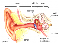
contains the tympanic membrane (otherwise known as the eardrum). Behind thetympanic membrane is an air filled cavity, which is bridged by three articulated bones collectively referred to as the ossicles. The head of thefirst bone (hammer) sits against the tympanic membrane while the head of thefar end bone (the stirrup) sits against the oval window (a membrane covering ahole in the bone of the skull). Behind this is the inner ear. |
|
|
describe the anatomy of the inner ear |

the inner ear is a set ofchannels and chambers carved out of the temporal bone, it represents a space inthe bone. Thecochlea is the auditory part of the inner ear |
|
|
what is the bony labyrinth of the inner ear |
The bonylabyrinth is the rigid bony outer wall of the inner ear in the temporal bone,it consists of three parts the vestibule, semicircular canals and the cochlea. These cavitiesare hollowed out of the substance of the bone. thebony labyrinth is filled with a sodium-rich extracelllar fluid called perilymph. |
|
|
what is the memranous labyrinth of the inner ear |
within the bonylabyrinth there is also a smaller membrane bound compartment that is filledwith endolymph, this is fluid low in sodium and high in potassium. This this is themembranous labyrinth. |
|
|
where is the cochlear |
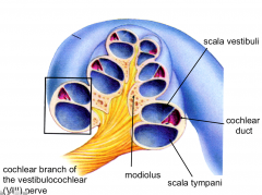
part of the bony labyrinth of the inner ear's bony labyrinth and is a spiral shaped structure containing 3 chambers separatedby membranes. Scala Vestibuli (upper chamber) & Scala Tympani (lower chamber) contain perilymph. Chochlear duct (3rd chamber) is separated from upper chamber by membranous labyrinth. contains endolymph. these structures are innervated by the axons of the vestibulocochlear nerve. |
|
|
what is structure of each turn of the cochlear |
There are twomembranes, the basilar membrane on the bottom and vestibular membrane on thetop, which separates the cochlear duct from the S. Vestibuli and S. Tympani. There is then abony compartment in which the nerve fibres enter, the afferents go back to thespiral ganglion. Sittingon the basilar membrane is the spiral organ |
|
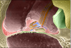
what is the internal structure of the cochlear duct |
hereare the auditory hair cells which face upwards into the endolymph. They haveprojections called stereocilia The green flapseen is the tectorial membrane, during life this covers over andattaches to the spiral organ. The sterocilia of the auditory hair cells join tothe tectorial membrane. There are innerhair cells and outer hair cells.The inner haircells lay closer to the origin of the tectorial membrane while the outer haircells lay further back |
|
|
what are sound waves |
Soundwaves are propagating waves of pressure |
|
|
how are sound waves transferred to the inner ear |
These soundwaves are captured by the pinna and channeled towards the tympanic membrane. This causes thetympanic membrane to vibrate, this vibration is carried through the ossiclesand the final bone the stirrup transfers these vibrations into the fluid filledcompartment of the inner ear via the oval window. |
|
|
how do we convert the vibrations of the ear into waves |
ossicles capture the vibrations in the air and allow them to beconverted to waves in the fluid. If they weren’t there, then the sound waveswould just bounce off the membranes as fluid is much denser. They are also usedto amplify the sound. |
|
|
how do the hair cells of the cochlear become deolarised |
pressure waves start at the S. vestibuli, and spiral (pass forward and back) thus passing into the other chambers. as the waves pass throughthese membranes they cause vibration of those membranes. It is vibration of thebasilar membrane that causes the hair cells to depolarise!much denser. |
|
|
what haoppens to the stereocilia of the hair cells as vibration passes through? |
The basilar membrane has the hair cells embedded, andthe stereocilia of the hair cells join to the tectorial membrane. Thebasilar membrane moves up and down, vibrating due to the soundwaves. As the membrane moves up and down the stereociliawill tilt from side to side (sterociliaarranged in height order with their tips linked to their neighbour) As thestereocilia is pulled away from its shorter neighbours it tugs against the tiplink! mechanically opens gated channels on adjacentsteriocilia membrane .'. K+ can enter the sterocilia down its electrical gradient This causesdepolarisation of the cell, resulting in an increased release of glutamate anda burst of action potentials fired. When thesterocilia tilts in the other direction, which it will do during the soundwaves, the tip links relax and the mechanical channels close.This stops any K+entering and the cell hyperpolarises which stops release of glutamate and theafferent stops firing briefly |
|
|
what is the basal state of the stereocilia |
In the neutralposition:(at rest) the sterocilia are straight up. cell = -40mV (partly depolarised) .'. releases tonic glutamate onto the afferent nerve whichwill fire streams of APs. The stereocilia project into endolymph (has very high K+ so is +ve charged) which as a potential difference of +80mV. |
|
|
how does the potential difference of the stereocilia change when there is a low frequency sound |
the afferentwill fire bursts of action potentials at the frequency of the sound (i.e. every“wave” of pressure will cause the sterocilia to tilt and then hyperpolarise andover again). |
|
|
how does the potential difference of the stereocilia change when there is a high frequency sound |
For highfrequency sounds, there will just be continuous depolarisation and firing ofthe afferent because the stereocilia is unable to depolarise and hyperpolariseso quickly |
|
|
from where is endolymph produced |
Endolymph iscreated by the stria vascularis, the endolymph needs to be continually replacedbut also excess fluid must be removed. |
|
|
why must excess endolymph be drained? |
too much fluidwould lead to increased pressure which would damage the inner ear.Hence productionand removal must be perfectly balanced in order to keep pressure normal. |
|
|
what happens if there is an increase in endolymph (eg in meniere's diease) |
It will leaddamage the cochlea, there will be damage to the hair cells and they willgradually lose their hearing. They also getringing in their ears and feel dizzy due to problems with the vestibular parts. |
|
|
where are the auditory hair cells and what are their projections called |
Sitting on the basilar membrane arethe auditory hair cells which face upwards into the endolymph. They haveprojections called stereocilia (type of microvillae). |
|
|
Thehair cells are very specialised and very delicate receptors, they are easilydamaged what happens if they are damaged |
they cannot be replaced. This is because there are no stem cells to replacethem with. |
|
|
there are two main things that can cause damage to the hair cells. what are these? |
1 - ototoxicity (thesecan damage the stereocilia, meaning the transduction process will not work. Ifthey are damaged enough the hair cell may just die) 2 - noise induced damage (Veryhigh noises can damage the stereocilia and cause the hair cells to die) |
|
|
what does Ototoxicity mean |
the property of being toxic to the ear (oto-), specifically the cochlea or auditory nerve and sometimes the vestibular system |
|
|
what may cause ototoxicity |
-> Aminoglycosides (e.g. kanamycin, gentamicin) -> Anti-cancerdrugs (e.g. cisplatin) -> EvenNSAIDs in some cases |
|
|
what happens if there is noise induced damage to the ear |
-> Very high noisescan damage the stereocilia and cause the hair cells to die -> Worse, ifthe cells are traumatised (rupture), it may cause release of loads of glutamatewhich can be toxic and damage the afferents |
|
|
how do outer hair cells amplify signals + what is the importance of this |
basilar mebrane vibrations = stereocilia sway -> outer hair cells depolarise -> outerhair cells shorten -> pulls down & amplifies the sensitivity of the system. The outer haircells are essential for the inner hair cells to do their job, because otherwisethe inner hair cells would get a very poor signal |
|
|
as sound increases how do the mechanics of hearing change? |
Largervibrations, which cause bigger receptor potentials (In the hair cells, due togreater tugging on tip links) and thus more action potentials |
|
|
how is the cochlea adapted to differentiate pitches |
spiralshaped cochlea is that it is the method to separate out sound frequencies. Themechanics of the cochlea means some hair cells will only be stimulated by lowfrequency sounds while others only by high frequency sound |
|
|
how does the auditory system determine pitch of a sound |
The structure of the basilar membrane +associated hair cells varies along the length of the cochlea so differentpoints along the membrane have a natural tendency to oscillate at differentfrequencies The braincan therefore determine the pitch of a sound from the location of the cellsthat respond to it |
|
|
where are the hairs cells that are activated in response to high frequency sounds |
close to the base of the cochlear |
|
|
where are the hairs cells that are activated in response to low frequency sounds |
close to the apex of the cochlear |
|
|
where to the afferents from excited hair cells go |
hair cells excite afferents that have their cellbodies in the spiral ganglion. these axons exit the bony labyrinth via theinternal auditory canal. They join the afferents of the vestibular system tocreated the 8th nerve |
|
|
where do the inner hair cell afferents go |
inner HC affent -> dorsal cochkea nuclei (in brainstem) -> fibres project to inferior coliculu -> fibres to medial geniculate nucleus (in thalamus) -> fibres terminate in the temporal lobe primaryauditory cortex(within lateral sucus) |
|
|
why do bilateral lesions to A1 of lateral sulcus not cause total deafness? |
it DOES damage discriminative hearing not total deafness bc. other areas still process sound. (othercortical areas also receive primary auditory input) |
|
|
how would a bilateral lesion of the lateral sulcus affect a patient> |
damage their discriminative hearing
So the individualwill be aware a sound has occurred but unable to discriminate between thefrequencies heard. They would beunable to understand a voice as they would be unable to hear the differentpitches. |
|
|
why is higher pitched hearing often lost first as we age? |
eachpitch will activate a specific set of hair cells in the cochlea The vulnerabilityhere is that high-pitched sound is more energetic and hence more damaging! |
|
|
what is fluent aphasia |
complex sound such as speech must be decoded in wernicke's area. |
|
|
where is speech decoded |
complex sound such as speech must be decoded in wernicke's area damage hereproduces a form of deafness where the individual can hear what is being saidbut cannot comprehend language. They can speakfluently back but their speech will be nonsensical as they have lost theability to comprehend and self monitor their speech |
|
|
what is presbyacusis |
is the loss of hearing that gradually occurs in most individuals as they grow older |
|
|
how can we discriminate sound origin |
The location ofa sound source is determined by comparing the sound detected by the two ears. This is done bythe superior olivary nuclei, which compare the noises coming into the two earsand determine the laterality of that noise. However thesound waves are so far apart that the medial superior olivary nuclei can detectthe arrival of the sound at one ear vs the other. (which ear it hits first!)They are verysensitive to differences in timing. |
|
|
how is information about origin of sound |
afferents sent -> ventral cochlear nuclei -> superior olives -> project up to inferior colliculus -> medial genticulate nucleus -> primary auditory cortex
|
|
|
what does the central auditory pathway comprise of |
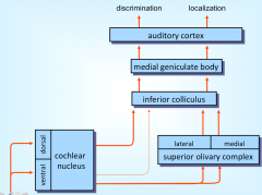
the pathways for dicrimination of sound and localisation put together |
|
|
why are afferents going to cochlear nuclei vulnerable to space occupying lesions |
Afferents goingto the cochlear nuclei have to run through the auditory canal, this is a narrowspace surrounded by thick bone. A narrow spacegives vulnerability to space-taking lesions which can damage the auditory(cochlear) nerve. |
|
|
what does the myelination of the vestibular nerve change along its length |
The vestibularnerve, for most of its length is myelinated by oligodendrocytes as it is acentral axon, but right at the end it turns into a peripheral nerve myelinatedby Schwann cells |
|
|
what is a neuroma |
a tumour on a nerve |
|
|
how can the vestibular nerve be prone to neuromas |
the Schwann cells can start to proliferate to form a benign tumour on the nerve. |
|
|
why is a vestibular neuroma also called an acoustic neuroma |
Asa neuroma forms on the vestibular nerve it will grow and press on the other nerves, thefirst thing the patient is likely to experience is ringing in the ears. |