![]()
![]()
![]()
Use LEFT and RIGHT arrow keys to navigate between flashcards;
Use UP and DOWN arrow keys to flip the card;
H to show hint;
A reads text to speech;
126 Cards in this Set
- Front
- Back
|
Hypervolemia |
a condition in which the liquid portion of the blood (plasma) is too high |
|
|
Hypovolemia |
a decreased volume of circulating blood in the body. A condition in which the liquid portion of the blood (plasma) is too low. |
|
|
Anemia |
condition of the blood in which the # of functional red blood cells or their hemoglobin content is below normal. |
|
|
Polycythemia |
Disorder characterized by an above-normal hematocrit (above 55%) in which hypertension, thrombosis, and hemorrhage can occur. |
|
|
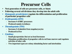
Erythropoietin |
A hormone released by the juxtaglomerular cells of the kidneys that stimulates red blood cell production. |
|
|
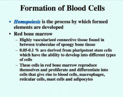
Hemopoiesis |
Blood cell production, which occurs in red bone marrow after birth.
|
|
|
Thrombopoietin |
Hormone produced by the liver that stimulates formation of platelets (thrombocytes) from megakaryocytes.
|
|
|
Carbon Monoxide Poisoning |
A potentially fatal condition caused by inhalation of carbon monoxide gas which competes favorably with oxygen for binding with hemoglobin and thus interferes with the transportation of oxygen and carbon dioxide by the blood.
|
|
|
Transferrin |
a protein of the beta globulin group that binds and transports iron in blood serum.
|
|
|
Ferritin |
a protein produced in mammalian metabolism that serves to store iron in the tissues.
|
|
|
Heme |
an iron-containing compound of the porphyrin class that forms the nonprotein part of hemoglobin and some other biological molecules.
|
|
|
Globin |
The globins are a family of globular proteins, which are thought to share a common ancestor. These proteins all incorporate the globin fold, a series of eight alpha helical segments. Two prominent members of this family Siclmyoglobin and hemoglobin, which both bind the heme (also haem) prosthetic group. |
|
|
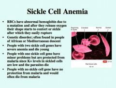
Sickle cell Anemia |
a severe hereditary form of anemia in which a mutated form of hemoglobin distorts the red blood cells into a crescent shape at low oxygen levels. It is most common among those of African descent.
|
|
|
Iron-deficiency anemia |
Too few healthy red blood cells due to too little iron in the body.
|
|
|
Pernicious anemia |
A decrease in red blood cells when the body can't absorb enough vitamin B-12 .
A deficiency in the production of red blood cells through a lack of vitamin B12. |
|
|
Aplastic anemia |
deficiency of all types of blood cells caused by failure of bone marrow development.
|
|
|
Hemolytic anemia |
hEA form of anemia due to hemolysis, the abnormal breakdown of red blood cells (RBCs), either in the blood vessels (intravascular hemolysis) or elsewhere in the human body (extravascular). |
|
|
Hemorrhagic anemia |
Acute posthemorrhagic anemia or acute blood loss anemia is a condition in which a person quickly loses a large volume of circulating hemoglobin. Acute blood loss is usually associated with an incident of trauma or a severe injury resulting in a large loss of blood.
|
|
|
Hypoxia |
Lack of adequate oxygen at the tissue level. |
|
|
MHC antigens |
The major histocompatibility complex (MHC) is a set of cell surface molecules encoded by a large gene family which controls a major part of the immune system in all vertebrates by determining histocompatibility.
Major histocompatibility complex (MHC), group of genes that code for proteins found on the surfaces of cells that help the immune systemrecognize foreign substances. MHC proteins are found in all higher vertebrates. In human beings the complex is also called the human leukocyte antigen (HLA) system. |
|
|
Chemotaxis |
A movement of a motile cell or organism, or part of one, in a direction corresponding to a gradient of increasing or decreasing concentration of a particular substance
|
|
|
Diapedesis |
The passage of blood cells through the intact walls of the capillaries, typically accompanying inflammation. |
|
|
Emigration |
Process whereby white blood cells (WBCs) leave the bloodstream by rolling along the endothelium, sticking to it, and squeezing between the endothelial cells.
|
|
|
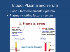
Serum |
Blood plasma minus its clotting proteins.
Liquid part of blood after coagulation, therefore devoid of clotting factors as fibronogen |
|
|
Plasma |
The colorless fluid part of blood, lymph, or milk, in which corpuscles or fat globules are suspended.
The liquid cell-free part of blood that has been treated with anti-coagulants. |
|
|
Clotting Factors |
Any of a number of substances in blood plasma that are involved in the clotting process, such as factor VIII.
Any of various plasma components involved inthe clotting of blood, including fibrinogen, prothrombin, thromboplastin, and calcium ionAlso called coagulation factor. |
|
|
Hemophilia |
A hereditary blood disorder where there is a deficient production of certain factors involved in blood clotting, resulting in excessive bleeding into joints, deep tissues, and elsewhere.
|
|
|
Thrombus |
A stationary clot formed in an unbroken blood vessel, usually a vein.
|
|
|
Thrombosis |
The formation of a clot in an unbroken blood vessel, usually a vein.
|
|
|
Embolus |
A blood clot, bubble of air or fat from broken bones, mass of bacteria, or other debris or foreign material transported by the blood
|
|
|
Deep Vein Thrombosis
|
The presence of a thrombus (blood clot) in a deep vein of the lower limbs. It may lead to (1) pulmonary embolism, if the thrombus dislodges and then lodges within the pulmonary arterial blood flow, and (2) postphlebitic syndrome, which consists of edema, pain, and skin changes due to destruction of venous valves.
|
|
|
Atherosclerosis |
A progressive disease characterized by the formation in the walls of large and medium-sized arteries of lesions called atherosclerotic plaques.
|
|
|
Pulmonary Embolism |
The presence of a blood clot or a foreign substance in a pulmonary arterial blood vessel that obstructs circulation to lung tissue.
|
|
|
Hemolytic disease of the newborn |
A hemolytic anemia of a newborn child that results from the destruction of the infant's erythrocytes (red blood cells) by antibodies produced by the mother; usually the antibodies are due to an Rh blood type incompatibility. Also called erythroblastosis fetalis (e-rith?-rō-blas-TŌ-sis fe-TAL-is). |
|
|
Rhogam |
Given to a pregnant woman whose blood type is Rh-negative to keep the baby's blood from interacting with the mother's. Also treats a blood cell disorder called idiopathic thrombocytopenic purpura (ITP).
|
|
|
Mediastinum |
The broad, median partition between the pleurae of the lungs that extends from the sternum to the vertebral column in the thoracic cavity.
|
|
|
Auricle |
a structure resembling an ear or earlobe.another term for atrium (of the heart).strictly, a small muscular appendage of each atrium.
|
|
|
Pectinate muscles |
Projecting muscle bundles of the anterior atrial walls and the lining of the auricles.
|
|
|
Interatrial septum |
The interatrial septum is the wall of tissue that separates the right and left atria of the heart
|
|
|
Trabeculae carnea |
Ridges and folds of the myocardium in the ventricles.
|
|
|
Chordae tendinae |
Tendonlike, fibrous cords that connect atrioventricular valves of the heart with papillary muscles.
|
|
|
Papillary muscles |
Muscles located in the ventricles of the heart. They attach to the cusps of the atrioventricular valves (also known as the mitral and tricuspid valves) via the chordae tendineae and contract to prevent inversion or prolapse of these valves on systole (or ventricular contraction).
|
|
|
Pulmonary Trunk |
The pulmonary trunk is a major vessel of the human heart that originates from the right ventricle. It branches into the right and left pulmonary arteries, which lead to the lungs.
|
|
|
Stenosis |
An abnormal narrowing of a heart valve, duct or opening.
When a heart valve does not open completely. |
|
|
Insufficiency/incompetence |
The inability of a heart valve to close completely. |
|
|
Prolapse |
Improper closure of the valve between the heart's upper and lower left chambers.
|
|
|
Rheumatic fever |
A noncontagious acute fever marked by inflammation and pain in the joints. It chiefly affects young people and is caused by a streptococcal infection.
A disease that can result from inadequately treated strep throat or scarlet fever. It can weaken entire heart wall; but most of the effect is seen bicuspid and aortic valves. |
|
|
Anastomoses |
Connections between arteries that provide alternate routes for blood to reach a particular organ or tissue.
Route 934 |
|
|
Ascultation |
Examination by listening to sounds in the body; specifically heart.
|
|
|
Systole |
In the cardiac cycle, the phase of contraction of the heart muscle, especially of the ventricles.
|
|
|
Diastole |
In the cardiac cycle, the phase of relaxation or dilation of the heart muscle, especially of the ventricles.
|
|
|
Coronary artery disease
|
A condition such as atherosclerosis that causes narrowing of coronary arteries so that blood flow to the heart is reduced.
Damage or disease in the heart's major blood vessels. |
|
|
Heart attack (myocardial infarction) |
Gross necrosis of myocardial tissue due to interrupted blood supply. Also called a heart attack.
Occurs when there is insufficient blood channeled to the heart muscle, and as a result the heart is damaged from lack of oxygen and suffers death (necrosis) of regions of the heart muscle. Blood flow is often blocked by plaque rupture and thrombus formation in the coronary artery. |
|
|
Sudden cardiac arrest |
The unexpected cessation of circulation and breathing due to an underlying heart disease such as ischemia, myocardial infarction, or a disturbance in cardiac rhythm.
|
|
|
Congestive heart failure |
A weakness of the heart that leads to a buildup of fluid in the lungs and surrounding body tissues.
occurs when the heart cannot pump vigorously enough to pump blood to all cells of the body-many causes |
|
|
Atrial fibrillation |
Asynchronous contraction of cardiac muscle fibers in the atria that results in the cessation of atrial pumping.
|
|
|
Angioplasty |
surgical repair or unblocking of a blood vessel, especially a coronary artery.
|
|
|
Defibrillation |
The stopping of fibrillation of the heart by administering a controlled electric shock in order to allow restoration of the normal rhythm.
|
|
|
Vasoconstriction |
A decrease in the size of the lumen of a blood vessel caused by contraction of the smooth muscle in the wall of the vessel.
|
|
|
Vasodilation |
An increase in the size of the lumen of a blood vessel caused by relaxation of the smooth muscle in the wall of the vessel.
|
|
|
Metarteriole |
A blood vessel that emerges from an arteriole, traverses a capillary network, and empties into a venule.
|
|
|
Thoroughfare channels |
Metarterioles exist in the mesenteric microcirculation, and the name was originally conceived only to define the "thoroughfarechannels " between arterioles and venules. In recent times the term has often been used instead to describe the smallest arterioles directly prior to the capillaries.
|
|
|
Vasomotion |
Change in caliber of blood vessels.
Vasomotion is the spontaneous oscillation in tone of blood vessels, independent of heart beat, innervation or respiration. Intermittent contraction and relaxation of blood vessels. |
|
|
Anastomoses |
Connections between arteries that provide alternate routes for blood to reach a particular organ or tissue
|
|
|
End Arteries |
An end artery (or terminal artery) is an artery that is the only supply of oxygenated blood to a portion of tissue. Examples of an end arteryinclude the splenic artery that supplies the spleen and the renal arterythat supplies the kidneys.
|
|
|
Collateral Circulation |
The alternate route taken by blood through an anastomosis.
|
|
|
Blood reservoirs |
Systemic veins and venules that contain large amounts of blood that can be moved quickly to parts of the body requiring the blood.
|
|
|
Venoconstriction |
Venoconstriction is the constriction of a vein and vasoconstriction of the blood vessels in general
|
|
|
Hemorrhage |
Bleeding; the escape of blood from blood vessels, especially when the loss is profuse.
|
|
|
Systemic Circulation |
The routes through which oxygenated blood flows from the left ventricle through the aorta to all the organs of the body and deoxygenated blood returns to the right atrium.
|
|
|
Hepatic Portal Circulation |
The flow of blood from the gastrointestinal organs to the liver before returning to the heart.
|
|
|
Pulmonary Circulation |
The flow of deoxygenated blood from the right ventricle to the lungs and the return of oxygenated blood from the lungs to the left atrium
|
|
|
Fetal Circulation |
The cardiovascular system of the fetus, including the placenta and special blood vessels involved in the exchange of materials between fetus and mother.
|
|
|
Resistance |
How cardiac output is distributed into circulatory routes depends on 2 things. *Pressure Difference-drives blood flow through tissue *Resistance-- to blood flow in specific blood vessels *blood flows from high pressure to low pressure *The greater the pressure difference, the great the blood flow *The higher the resistance the smaller the blood flow |
|
|
What is blood? |
Blood is liquid connective tissue and composed of plasma (liquid) and suspended cells. |
|
|
What is plasma? |
Watery, liquid matrix with dissolved substances |
|
|
What is interstitial fluid? |
Interstitial fluid is the fluid that bathes cells. |
|
|
What are the 3 functions of blood? |
*Transport-blood transports oxygen, CO2, nutrients, hormones, heat and wastes *Regulate- blood helps maintain homeostasis of body fluids through regulation of ph, body temperature and water content *Protect- blood loss is prevented via clotting mechanisms -antibodies and phagocytic WBC protect the body from infection |
|
|
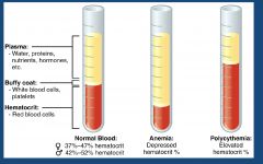
If blood is centrifuged what are the 3 layers? |
Plasma, buffy coat layer, and RBCs |
|
|
What are the characteristics of blood? |
*Blood is dense, slightly viscous, and sticky *Temp of blood is slightly higher than reg body temp (100.4) *Is slightly alkaline *Constitutes 20% of ECF; 8% total body mass |
|
|
Components of blood: Formed Elements composition |
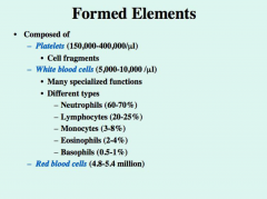
45% of blood *Cells and cell fragments *99% of formed elements are Red Blood Cells *1% is White Blood Cells and fragments |
|
|
Components of blood: Blood plasma |
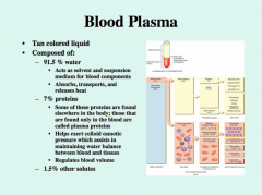
50% *Watery liquid matrix with dissolved substances |
|
|
Blood and Plasma |
|
|
|
Albumins |
*Produced by liver *Small,most numerous plasma proteins *Transports several steroid hormones and fatty accids |
|
|
Globulins |
*Produced by liver and plasma cells (develop from B cells) *Antibodies (immunoglobins) help attack viruses and bacteria -When stimulated by viral or bacterial invasion, specific antibodies are produced -Antibodies bind to specific antigens to form a complex that disables invading microbes -Alpha and beta globulins transport iron lipids, and fat soluble vitamins |
|
|
Fibrinogens |
*Produced by liver *Plays role in blood clotting |
|
|
WBCs/Leukocytes types and relative abundance |
Neutrophils-60-70% Lymphocytes-20-25% Monocytes- 3-8% Eosinophils-2-4% Basophils- .5-1% |
|
|
Hematocrit |
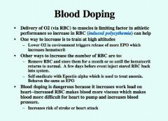
*Determination of %RBC in total blood volume -42 is average for femals -47 is average for males +Testosterone stimulates RBC production via erythopoietin (EPO) *Anemia describes low Hematocrit level *Polycythemia describes abnormally high level of RBC (65% or higher hematocrit) due to tissue hypoxia, abnormal RBC production, dehydration, and blood doping by athletes. |
|
|
Hematocrit examples |
|
|
|
Blood doping |
|
|
|
Complete blood count |
A complete blood count (CBC) gives important information about the kinds and numbers of cells in the blood, especially red blood cells ,white blood cells , and platelets. A CBC helps your doctor check any symptoms, such as weakness, fatigue, or bruising, you may have. A CBC also helps him or her diagnose conditions, such as anemia, infection, and many other disorders.
|
|
|
Differential White Blood Cell Count |
White blood cell types (WBC differential). The major types of white blood cells are neutrophils, lymphocytes, monocytes, eosinophils, and basophils. Immature neutrophils, called band neutrophils, are also part of this test. Each type of cell plays a different role in protecting the body. The numbers of each one of these types of white blood cells give important information about the immune system. Too many or too few of the different types of white blood cells can help find an infection, an allergic or toxic reaction to medicines or chemicals, and many conditions, such as leukemia
|
|
|
Reticulocyte count |
A reticulocyte count (also known as a retic count, reticulocyte index, or corrected reticulocyte) is a measurement and percentage of how many reticulocytes are in the blood. This count indicates whether enough red blood cells are being produced in the bone marrow.
|
|
|
Leukocytosis |
an increase in the number of white cells in the blood, especially during an infection.
|
|
|
Leucopenia |
a reduction in the number of white cells in the blood, typical of various diseases. |
|
|
Hemorrhage |
an escape of blood from a ruptured blood vessel, especially when profuse |
|
|
acute leukemia |
A type of cancer of the blood and bone marrow with excess immature white blood cells. |
|
|
Chronic leukemia |
Many types of chronic leukemia; some produce too many cells, some produce too few. It involves mature cells. |
|
|
Lymphoma |
*Malignant transformation of lymphocytes *Two types: Hodgkin's and Non-Hodgkin's *People with HIV, EB and HTLV infections have higher incidence |
|
|
Multiple myeloma |
*Malignant transformations of lymphocytes destined to be plasma cells (B lymphocytes) *Usually tumors in multiple sites; destroys bones *Most frequent in older people; median age is 71. Rarely found in someone under 50. |
|
|
Homeopoiesis |
The process by which formed elements are developed |
|
|
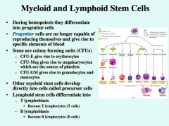
Slide on Homeopoiesis |
|
|
|
Slide on Homeopoiesis
|

|
|
|
Slide on Homeopoiesis
|
|
|
|
Slide on Homeopoiesis
|
|
|
|
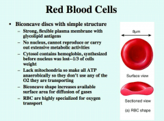
Structure of RBCs |
Hemoglobin: oxygen carrying protein that gives blood its red color |
|
|
Red Blood Cells |
|
|
|
Hemoglobin |
*Each RBC has about 280 million hemoglobin molecules; each can carry 4 molecules of oxygen *2 parts: Protein called globin made of 4 polypetide chains :4 pigment molecules (called hemes) which have a central iron molecule that can reversibly bind to oxygen : One heme binds to each polypetide chain *Hemoglobin transports oxygen from lungs to interstitial fluid and transports a portion of the CO2 back to lungs *Hemoglobin helps in regulation of blood flow and blood pressure |
|
|
Sickle Cell Anemia |
|
|
|
CO Poisoning |
*CO is colorless, odorless, tasteless gas produced by the incomplete combustion of fossil fuels *Hemoglobin binds to oxygen but if CO is present it will preferentially release the oxygen and bind CO *Always CO in the air but if CO levels get too high (800ppm) too much oxygen will be displaced and you'll start to get headaches, eventually have convulsions and die *Treatment is 100% oxygen-if delayed too long, patient could still die, or suffer heart damage, brain damage, or permanent memory loss. |
|
|
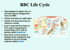
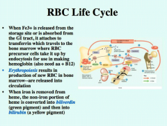
RBC Life Cycle |
*Live approximately 120 days *No nucleus or organelles, so can't replace damaged components * Damaged RBC are removed by fixed macrophages in the spleen and liver and the breakdown products are recycled |
|
|
RBC Life Cycle
|
|
|
|
RBC Life Cycle
|
|
|
|
RBC Life Cycle
|

|
|
|
Major Histocompatibility Complex |
*WBC and all other nucleated cells have unique proteins that protrude from the plasma membrane called MHC which serve as identity markers and help us recognize self from non-self.
|
|
|
ABO blood group antigens |
*Blood plasma has isoantibodies agglutinins that react with A and B antibodies if they are mixed *You have antibodies for antigens your blood cells lack *Type A has anti B; Type B has anti-A; Type AB has neither; Type O has both |
|
|
Compatible Donors |
A-A,O B-B,O AB- A, B, AB, O O-O O is universal donor; AB is universal recipient |
|
|
Emigration |
Process whereby white blood cells (WBCs) leave the bloodstream by rolling along the endothelium, sticking to it, and squeezing between the endothelial cells.
|
|
|
Selectins |
are displayed on endothelial cells near ares of inflammation and injury they stick to surface of neutrophils which causes them to slow down and roll along the surface. |
|
|
Integrins |
Adhesion molecules on neutrophils and other cells, called integrins tether neutrophils and assist their movement through the blood vessel wall and into the interstitial fluid |
|
|
Lysosomes and Defensins |
After neutrophil engulfs bacteria it releases several chemicals to destroy it including: lysozyme, strong oxidants such as hydrogen peroxide, hypochlorite ion and proteins called defensins that poke holes in the plasma membranes of the invading cells |
|
|
Chemotaxis |
Chemicals released by microbes attract phagocytic cells through the process of chemotaxis |
|
|
Platelet Characteristics |
Thrombopoietin is a hormone that causes myeloid stem cells to differentiate into megakaryocyte colony forming units which diff. into megakryoblasts and then megakaryocytes *Megakaryocytes are huge cells that splinter into 2000-3000 pieces *Each piece is surrounded by plasma membrane and = platelets *Platelets break off from megakaryocyte in the bone marrow and enter the bloodstream |
|
|
Platelet Char. Cont. |
* 150,000-400,000 plateltes/microliter in blood *Disc-shaped and have many vesicles but no nucleus *Help stop blood loss from damaged blood vessels by forming platelet plug *Platelet granules also contain chemicals that help promote clotting when they are released from the platelets * Live 5-9 days and are removed by fixed macrophages in the spleen and liver. |
|
|
Homeostasis |
* Sequences of responses that stops bleeding *Must be quick, localized to specific region, and carefully controlled *Helps control loss of blood from small vessels-loss of blood from bigger vessels require medical attention *Hemorrhage is loss of large amount of blood from blood vessels *Three Mechanisms: Vascular Spasm :Platelet plug formation :Blood clotting or coagulation |
|
|
Steps of Homeostasis
|
1) Vascular spasm: Smooth muscle contracts, causing vasoconstriction 2) Platelet plug formation: Injury to lining of vessel exposes collagen fibers; platelets adhere : Platelets release chemicals that make nearby platelets sticky; platelet plug forms 3) Coagulation: Fibrin forms a mesh that traps red blood cells and platelets, forming the clot |