Reading...
![]()
Play button
![]()
Play button
![]()
Use LEFT and RIGHT arrow keys to navigate between flashcards;
Use UP and DOWN arrow keys to flip the card;
H to show hint;
A reads text to speech;
66 Cards in this Set
- Front
- Back
|
What are the layers that make up the lining of the oral cavity?
|
1. Mucosa
2. Submucosa |
|
|
The mucosa of the oral cavity consists of a _______ epithelium and a ________ of ____ CT.
|
stratified squamous NONkeratinized epithelium
Lamina Propria of Loose CT |
|
|
Deep to the mucosa in the oral cavity is the ____. This consists of a layer of ___ that contains _____ glands.
|
Submucosa
Connective Tissue Muco-serous Glands (secrete mucous and serous) |
|
|
The outer surface of the lip is skin (epidermis and dermis). What is the core of the lip made up of? What is the inner surface?
|
Core- Orbicularis Oris (skeletal muscle)
Inner surface- Oral Mucosa |
|
|
What kind of glands are in the oral mucosa of the lip?
|
Labial Glands
(Situated between the mucosa and the orbicularis oris muscle.) |
|
|
What is the vermillion border and where is it found?
What is another name for it and why is it called this? |
This is a transition from skin to the mucosa of the oral cavity.
Also called the red margin because it is thin and the high dermal papillae here carry blood close to the surface giving it it's red color. |
|
|
What is the structure of the epithelial lining of the red margin?
How is the lamina propria of the red margin different than in other parts of the lip? |
The epithelial lining of the red margin is lightly keratinized.
The lamina propria in the red margin does not have glands. |
|
|
The epithelium of the mucosa of the tongue is what type?
|
Stratified squamous non-keratinized to keratinized.
|
|
|
What epithelial layer is mainly responsible for forming papillae on the dorsal surface of the tongue?
|
Stratum Corneum
|
|
|
If you burn your tongue, which layer of epithelium are you most likely damaging?
|
Stratum Corneum
|
|
|
Papillae have a small core of CT coming from what layer?
|
Lamina Propria of Mucosa (which makes sense because the lamina propria is made of loose CT)
|
|
|
The papillae contain receptors for what?
|
Taste (Taste Buds)
|
|
|
What are the four types of papillae?
|
1. Filiform Papillae
2. Fungiform Papillae 3. Foliate Papillae 4. Circumvallate Papillae |
|
|
What do Filiform Papillae look like?
|
Elongated and partially keratinized. They look little flames and are very abundant.
|
|
|
Where are Filiform Papillae located?
|
They cover the dorsal surface of the anterior two-thirds of the tongue.
|
|
|
Do the Filiform Papillae contain taste buds?
|
No.
|
|
|
What do Fungiform Papillae look like? Where are they found on the tongue?
|
Mushroom Shaped.
Scattered over the dorsal surface of the tongue. |
|
|
Do Fungiform Papillae contain taste buds?
|
Yes
|
|
|
Which type of Papillae is uncommon in adults and seen more often in children?
|
Foliate Papillae
|
|
|
Where are Foliate Papillae located?
|
On the posterolateral margin of the tongue.
|
|
|
Do the Foliate Papillae contain taste buds?
|
Yes
|
|
|
What do Circumvallate Papillae look like?
|
They are very large (about 4 times as large as the other papillae) and tooth shaped. They are surrounded by a groove or moat.
|
|
|
Where are Circumvallate Papillae located?
|
These lie along the sulcus terminalis which is like a line that divides the anterior two-thirds of the tongue from the posterior one-third.
|
|
|
About how many Circumvallate Papillae are on a tongue?
|
Only about 7-12.
|
|
|
Where are the Glands of Von Ebner located?
|
In the Lamina Propria (which is the loose CT part of the mucosa) deep to Circumvallate Papillae.
|
|
|
What do the Glands of Von Ebner do?
|
They are serous secreting glands that secrete into the moats surrounding Circumvallate Papillae. They wash away tastes so that you dont taste what you ate a long time ago.
|
|
|
What might cause food to become lodged in the moats surrounding Circumvallate Papillae?
|
If the Glands of Von Ebner are not secreting properly, they wont be able to wash food out of the moats.
|
|
|
Do the Circumvallate Papillae have taste buds?
|
Yes
|
|
|
Which types of Papillae have taste buds? Which don't?
|
DO have tastebuds:
Fungiform Papillae Foliate Papillae Circumvallate Papillae DON'T have tastebuds: Filiform Papillae |
|
|
What is the structure of a taste bud?
|
Taste-buds are barrel shaped structures that extend through the full thickness of the epithelium.
|
|
|
What is the opening into the taste bud called?
|
Taste Pore
|
|
|
What are the cell types found inside the taste bud?
|
1. Neuroepithelial taste cells
2. Basal Cells 3. Sustentacular Cells |
|
|
What is the function of Neuroepithelial Taste Cells in the Taste Buds? How do they transmit information to the CNS?
|
Act as chemoreceptors.
Transmit info to the CNS via the Facial Nerve (12) and the Glossopharyngeal Nerve (9). |
|
|
What is the function of the Basal cells in the taste bud? With this in mind, what layer are they usually located in?
|
Basal cells can differentiate into the taste cells. They are usually found at the base of the taste bud in the Stratum Basale.
|
|
|
How are the muscles of the tongue structured?
|
The intrinsic muscles of the tongue are bundles of skeletal muscle that run in longitudinal, transverse, and vertical directions so that the shape of the tongue can be altered in many directions.
|
|
|
Small amounts of _____ are found amoung the muscle fiber groups of the tongue as well as minor glands that are predominately _____ secreting.
|
Adipose tissue
Serous secreting |
|
|
These are rounded masses of lymphatic tissue that cover the posterior third of the tongue.
|
Lingual Tonsils
|
|
|
What is the part of the tooth that projects above the gingiva?
|
Crown
|
|
|
What is the name of the mucous membrane that is attached to the alveolar periosteum and surrounds the neck of the tooth?
|
Gingiva
|
|
|
What is the part of the tooth that projects below the gingiva?
|
Root
|
|
|
What is the name for the junction between the crown and the root of a tooth?
|
Neck
|
|
|
The roots of the teeth are embedded in the _____ ____ of the ____ and ____.
|
Alveolar Bone
Mandible Maxilla |
|
|
The pulp makes up the ___ of the tooth. It is made of ____. Where is it found?
|
The pulp makes up the core of the tooth. It is made of loose CT and is found in the Pulp Cavity.
|
|
|
What is contained in the pulp of the tooth? How do these structures enter the pulp cavity?
|
The pulp contains nerves (for pain) and blood vessels.
These enter through the apical foramen which is the opening into the pulp cavity (located at the base of the root). |
|
|
What covers the pulp of the tooth?
|
A calcified tissue- Dentin
|
|
|
____ is the second hardest substance in the body.
|
Dentin
|
|
|
What covers the dentin at the crown of the tooth?
At the root? |
Enamel at the crown
Cementum at the root |
|
|
What type of cells line the pulp cavity? What do these cells do?
|
Odonotoblasts
Make dentin. |
|
|
What happens to the pulp cavity with age?
|
The pulp cavity gets progressively smaller with age. (Dentin formation continues throughout life but decreases with age.)
|
|
|
This part of the tooth is sensitive to touch, cold, and acid.
|
Dentin
|
|
|
Can dentin persist without odonotoblasts?
|
Yes
|
|
|
What is the hardest substance in the body?
|
Tooth enamel
|
|
|
What do ameloblasts do and where are they located?
|
Make enamel.
Ameloblasts cover the surface of the developing tooth. |
|
|
When do you have ameloblasts?
|
Only on the surface of the developing tooth. They die once the tooth erupts from the gingiva.
|
|
|
Cementum is similar in structure and composition to what?
|
Primary Bone
|
|
|
What cells make cementum and where are they located?
|
Cementoblasts
Located against the periodontal ligament (which is what attaches the root to the alveolar bone). |
|
|
What are the cells within the cementum called?
|
cemetocytes
|
|
|
What kind of fibers make up the Periodontal Ligament? What does this ligament do?
|
Collagen Fibers
These attach the cementum to the adjacent bone of the jaw. |
|
|
When your baby teeth are hanging by a string, what is the string?
|
Periodontal Ligaments
|
|
What is this structure?
|
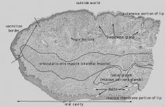
Lip
|
|
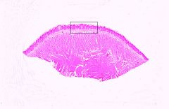
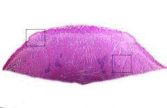
What types of Papillae can be seen here?
|
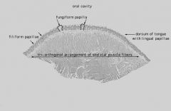
Fungiform Papillae and Filiform Papillae
|
|
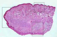
What is the overall structure here?
What sub-structures are in the boxes? |
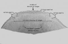
Circumvallate Papillae
Von Ebner's Glands (serous only) |
|
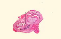
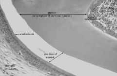
What structure is shown here?
|
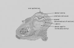
Developing Tooth
|
|

What are the structures numbered?
|

1. Enamel
2. Ameloblasts 3. Dentin 4. Odontoblasts 5. Developing Pulp Cavity |
|
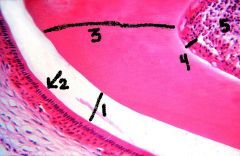
What are the structures numbered?
|
1. Enamel
2. Ameloblasts 3. Dentin 4. Odontoblasts 5. Developing Pulp Cavity |
|

What are the structures within the box?
|
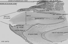
Taggable:
Enamel Dentin Pulp |