Reading...
![]()
Play button
![]()
Play button
![]()
Use LEFT and RIGHT arrow keys to navigate between flashcards;
Use UP and DOWN arrow keys to flip the card;
H to show hint;
A reads text to speech;
65 Cards in this Set
- Front
- Back
|
What is the equivalent of rugae in the small intestine?
|
Plicae Circulares
|
|
|
What are Plicae Surculares and what is their function?
|
They are like rugae but in the small intestine. They help increase surface area for absorption.
|
|
|
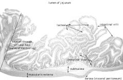
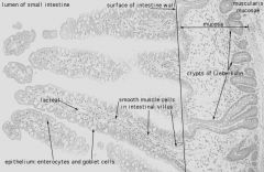
What are the 3 morphological adaptations in the gut tube that increase surface area?
|
Plicae Circulares
Villi Microvilli |
|
|
What makes up the core tissue of the Plicae Circulares?
|
Submucosa
|
|
|
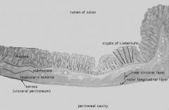
What are the main layers that make up the small intestine?
|
Serosa
Muscularis Externa Submucosa Mucosa |
|
|
What are the folds in the mucosa of the small intestine called?
|
Villi
|
|
|
Villi in the small intestine are finger-like projections containing a core of ____ and covered with ____.
|
Lamina Propria
Epithelium |
|
|
What structures make up the core of a villus?
|
Connective Tissue
Smooth Muscle Capillary Network Lacteal (Lymphatic) |
|
|
What epithelium covers the surface of the small intestine?
|
Simple Columnar
|
|
|
What type of simple columnar epithelium cells make up the lining of the small intestine?
|
1. Enterocytes
2. Goblet Cells 3. Antigen Presenting M-cells |
|
|
What is an enterocyte and what structure do these have?
|
Absorptive cells of the lining of the small intestine.
These have a microvilli brush border. |
|
|
What is the function of Goblet cells in the small intestine?
|
Secrete mucous that helps protect the lining of the intestine from pancreatic enzymes and bacteria increase.
|
|
|
What is the function of antigen presenting M Cells? Where would you find these?
|
They take antigens from the lumen by phagocytosis and present them to lymphocytes scattered throughout the mucosa.
Found scattered throughout the epithelium of the small intestine. |
|
|
Where would you find a Crypt of Lieberkuhn?
|
Small and Large Intestines.
Between the villi. |
|
|
What are the cell types in the Crypts of Lieberkuhn?
|
1. Undifferentiated stem cells
2. Enteroendocrine cells 3. Paneth cells |
|
|
Crypt of Lieberkuhn:
Where are the Undifferentiated stem cells found? What do they give rise to? |
Found in the lower half of the crypt.
Give rise to absorptive cells (enterocytes), goblet cells, and enteroendocrine cells. |
|
|
What are the products of the enteroendocrine cells located in the Crypts of Lieberkuhn?
|
1. Secretin
2. Cholecystokinin 3. Gastric Inhibitory Peptide 4. Motilin |
|
|
What is the function of secretin? What kind of cells secrete it in the small intestine?
|
Stimulates growth and secretion of the exocrine pancreas.
Enteroendocrine cells of the Crypts of Lieberkuhn. |
|
|
What is the function of Cholecystokinin (CCK)?
Where in the small intestine is it secreted? |
Stimulates growth and secretion of the exocrine pancreas AND contraction of the smooth muscle of the gall bladder. Also INHIBITS gastric emptying.
Secreted from the enteroendocrine glands of the Crypts of Lieberkuhn. |
|
|
What is the function of Gastric Inhibitory Peptide (GIP)?
Where in the small intestine is GIP secreted? |
Inhibits HCl secretion by the parietal cells of the stomach.
Secreted by the enteroendocrine cells of the Crypts of Lieberkuhn. |
|
|
What do paneth cells do?
Where are they found? |
Secrete the antibacterial enzyme lysozyme.
Found in the base of the Crypts of Lieberkuhn. |
|
|
What is the structure of the muscularis mucosa of the small intestine?
What does this layer help the small intestine to do? |
Usual inner circular and outer longitudinal smooth muscle.
Strands of smooth muscle from this layer extend up into the villi, causing their movement and aiding the circulation of lymph through the lymphatic vessels. |
|
|
What special structures are found in the Submucosa of the duodenum? the ileum? throughout the small intestine?
|
Brunner's Glands
Peyer's Patches Meissner's Plexus |
|
|
What do Brunner's Glands do?
Where are they found? |
Secrete alkaline mucus that protects the mucosa from acid and raises the pH for pancreatic enzyme activity.
Found in the Submucosa of the Duodenum. |
|
|
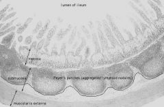
What are Peyer's Patches?
|
Patches of lymphoid tissue found in the submucosa of the ileum.
|
|
|
What are the 3 sections of the small intestine from top to bottom?
|
duodenum
jejunum ileum |
|
|
What is the function of the Muscularis Externa in the small intestine?
|
Peristalsis
|
|
|
What plexus is found between the two layers of the Muscularis Externa?
|
Auerbach's Plexus
|
|
|
What are the parts of the large intestine from start to finish?
|
Cecum
Vermiform Appendix Ascending Colon Transverse Colon Descending Colon Rectum Anal Canal |
|
|
What are the main functions of the colon and rectum?
|
Absorption of water and electrolytes.
Compaction and elimination of undigested food. |
|
|
What valve is located between the small and large intestine?
|
Iliocecal Valve
|
|
|
What is the structure of the Iliocecal Valve?
|
It consists of two horizontal folds of mucous membrane that project around the orifice of the ileum.
|
|
|
What controls the flow of contents from the ileum into the colon?
|
The circular muscle (Iliocecal Sphincter) of the lower end of the ileum controls the flow of contents.
|
|
|
In what way is the Mucosa of the Colon similar to that of the small intestine? (What cell types do they share?)
|
Both have Simple Columnar Epithelium cells, Goblet Cells and M cells.
Lamina Propria with Crypts of Lieberkuhn and lymphatic tissue. Muscularis Mucosa. |
|
|
In what ways does the Mucosa of the Colon differ from that of the small intestine?
|
No Villi!
No Paneth Cells! |
|
|
The mucosa of the colon is ____ than the mucosa of the small intestine.
It also has more ____ cells. |
Thicker
Goblet |
|
|
In what layer of the colon would you find the Meissner's Plexus?
|
Submucosa
|
|
|
What is the location of the hemorrhoidal plexus of veins in the colon?
|
Lower rectum
Located in the Submucosa and extending into the lamina propria. (These veins have no valves. There may also by lymphatic nodules present in this area.) |
|
|
What is the arrangement of the Muscularis Externa in the Colon?
|
The outer longitudinal layer is arranged into 3 thick bands called the Teniae Coli.
|
|
|
What are Teniae Coli?
|
3 thick bands of outer longitudinal muscle in the colon.
|
|
|
How do the Teniae Coli work?
|
They are shorter than the colon so they gather its wall into sacculations called haustra.
|
|
|
What plexus is located between the layers of the Muscularis Externa in the Colon?
|
Auerbach's Plexus
|
|
|
Hirschsprung's Disease (Congenital Megacolon)
What causes it? What happens? |
Caused by an absence of ganglion cells in the Meissner's and Auerback's Plexuses of the rectum.
Feces remain in the colon causing it to enlarge. |
|
|
How can you tell the Vermiform Appendix from the Colon?
|
It is similar in structure but it has shorter Crypts of Lieberkuhn and NO TENIAE COLI.
|
|
|
The Lamina Propria and Submucosa of the Vermiform Appendix contain large amounts of _____ tissue.
|
Lymphoid Tissue
|
|
|
Anal Canal:
The epithelium is ____. The lamina propria contains ____ crypts. |
Simple Columnar
Short Crypts |
|
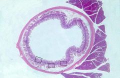
What is this structure?
What glands can be seen? |

Duodenum
Brunner's Glands (secrete alkaline mucous) |
|
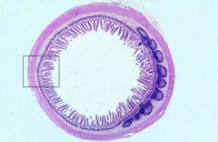
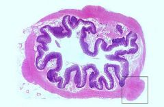
What is this structure?
|
Jejunum
(Looks a lot like ileum BUT- longer, more slender villi, and no Peyer's Patches) |
|

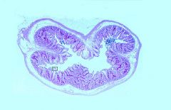
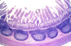
What is this structure?
What are the basophilic blobs? |

Ileum
Peyer's Patches |
|
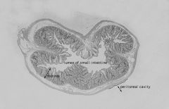
What is this structure?
What are the basophilic blobs? |

Ileum
Peyer's Patches |
|
What are the basophilic structures?
Where are these found? |
Peyer's Patches
Ileum |
|

What cell type is shown?
Where are these found? |
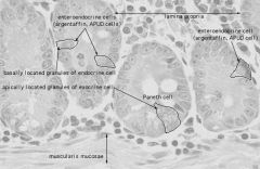
Paneth Cells
Located at the base of Crypts of Lieberkuhn |
|
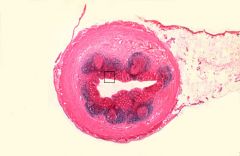
What is this structure?
|

Colon (Notice Teniae Coli)
|
|
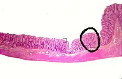
What are the circled structures?
What layer are these in? |
Crypts of Lieberkuhn (in colon here)
Lamina Propria |
|
What is this structure?
|
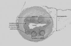
Appendix
|
|
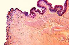
What is this?
(What other structures are taggable here?) |
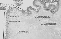
Anal Canal
(Hair and Apocrine Glands) |
|
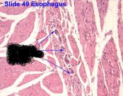
What are the arrows pointing to?
|
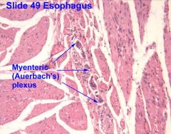
Auerbach's Plexus
(Between layers of Muscularis Externa) |
|

What structure is circled?
What layer is this? |
Meissner's Plexus
Submucosal Layer |
|
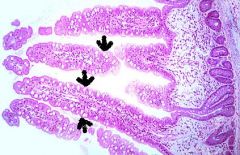
What cells are the arrows pointing to?
|
Goblet Cells
|
|
|
What are the main layers of the anal canal?
|
Mucosa
Submucosa Muscularis Externa ADVENTITIA |
|
|
What kind of epithelium makes up the mucosa of the upper and lower halves of the anal canal?
|
Upper half= Simple Columnar
Lower half= Stratified squamous nonkeratinized |
|
|
Does the lamina propria in the anal canal have Crypts of Lieberkuhn?
|
No.
|
|
|
The submucosa of the anal canal contains _____ glands associated with hair and ____ sweat glands.
|
Sebaceous
Apocrine |
|
|
What muscle layer makes up the involuntary internal anal sphincter?
|
Made of the inner circular layer of smooth muscle of the Muscularis Externa
(In the upper 3/4 of the canal) |
|
|
What muscle forms the voluntary external anal sphincter?
|
Skeletal muscle from the pelvic diaphragm (the muscular floor holding important organs in place.)
|