![]()
![]()
![]()
Use LEFT and RIGHT arrow keys to navigate between flashcards;
Use UP and DOWN arrow keys to flip the card;
H to show hint;
A reads text to speech;
280 Cards in this Set
- Front
- Back
|
Lymph system function
|
-Carry proteins and fluids (that have leaked out of capillaries) back to the veins
-Absorb fats from the small intestine and bring them back to the blood stream -Defend the body against foreign organisms: -Lymphocytes produce antibodies -Monocytes attack foreign cells and bacteria |
|
|
What organs make up the Lymph system
|
-organs, lymph nodes, lymph ducts, lymph vessels move lymph to veins
-Includes tonsils, adenoids, spleen, thymus -Major part of body's immune system |
|
|
Lymph fluid
|
-flows out of capillary walls to bathe the body tissues
-carries oxygen to cells, carries waste away (CO2) -contains white blood cells -lymph vessels carry lymph away to the lymph duct to be drained so lymph nodes aren't swollen |
|
|
Lymph facts
|
-Lymph is the fluid that filters out of capillaries into the spaces between the cells
-fluid that surrounds body cells is called interstitial fluid -lymph is similar to blood (leukocytes, water, sugar, metabolic wastes but doesn't contain erythrocytes, platelets, and has less protein than blood |
|
|
Lymph vessels
|
-have thicker walls than capillaries
-contain valves so lymph flows in one direction toward duct (thoracic) -all vessels empty into left thoracic duct and right lymphatic duct--ducts carry lymph into large veins in the neck where lymph enters bloodstream |
|
|
Lymph node
|
-mass of lymph cells and vessels surrounded by a fibrous connective tissue capsule
-produce lymphocytes, filter lymph, and trap substances from infections and inflammations -makes immune cells to help fight off infections -when bacteria is detected in lymph, lymph nodes make more lymphocytes--causing them to swell -remove foreign material like cancer cells and bacteria |
|
|
Chyle
|
fluid from intestines--contains proteins and fats
|
|
|
B Cells,
T Cells |
B Cells- produce antibodies
T Cells- attack bacteria and foreign cells |
|
|
Lymph node locations
|
-inguinal, cervical, axillary, mediastinal
|
|
|
Spleen
|
-aging erythrocytes are destroyed here by macrophages
-Bacteria and foreign materials are filtered here from blood -B cells are activated by antigens to produce antibodies and T cells are activated to attach to foreign materials |
|
|
Thymus
|
important in developing an effective immune system in childhood
|
|
|
Immune System
|
-defends body against foreign substances (toxins, bacteria, foreign RBCs)
-Includes WBCs (neutrophils, monocytes, macrophages) that are phagocytes found in blood and tissue -Lymphoid organs: spleen, thymus, lymph nodes and their products (antibodies and lymphocytes) |
|
|
Antigen
Pathogen |
substance that body recognizes as foreign
-any virus/bacteria/substance that causes disease |
|
|
Immunity
|
protection against diseases--natural and acquired immunity
|
|
|
Natural Immunity
|
-innate
-genetic predisposition -phagocytes -macrophages -NK cells |
|
|
Acquired Immunity
|
-development of antibodies against antigens Active: vaccination, transfer of immune cells from a donor, having an infectionPassive: maternal antibodies, immunoglobulin, antitoxins |
|
|
Phagocytosis
|
cellular eating and destroying substances--usually by neutrophils and macrophages
|
|
|
Lymphaden/o
|
lymph node (gland)
|
|
|
Immun/o
|
immune, protection
|
|
|
tox/o
|
poison
|
|
|
Ana-
|
again, anew
|
|
|
-edema
|
swelling
|
|
|
-phylaxis
|
protection
|
|
|
Immunodeficiency (Pathology) |
-severe combined immunodeficiency disease -group of rare, sometimes congenital disorders characterized by little or no immune response -defect in B and T-lymphocytes -bubble boy disease -susceptible to infections -Treatments: bone marrow, stem cell transplant |
|
|
Acquired Immunodeficiency Disease (Pathology) |
-caused by the human immunodeficiency virus (HIV) -symptoms and signs that indicate a suppressed immune system -AIDS is characterized as infections, secondary neoplasms, and neurologic problems -Ex: Kaposi sarcoma and lymphoma -NO CURE: but medications can slow it down *Sexually transmitted infection -protease inhibitors are used to treat AIDS |
|
|
Hypersensitivity (Allergy) |
-abnormal hypersensitivity acquired by exposure to an antigen
-Sensitized person: person who was recently exposed to allergen reacts to a subsequent exposure -mild to severe (anaphylaxis) |
|
|
Lymphoma |
-malignant tumor of lymph nodes and tissue -Hodgkin lymphoma (formerly called disease)--malig tumor of the spleen's lymphoid tissue and lymph nodes -lymphadenopathy, splenomegaly, weakness, loss of appetite -Diagnosis made when Reed-Sternberg cells are found in the lymph nodes -high cure rate (chem, radiation, stem cell) *Caused when there's a DNA mutation in B cells--they live past their prime and stay in lymph node. they overcrowd the area and cause signs/symptoms |
|
|
Stage 1 Lymphoma |
Cancer found in one or more lymph nodes in one lymph node group (thymus, spleen) -Waldeyer's ring |
|
|
Stage 2 Lymphoma |
cancer found in two or more lymph nodes below OR above the diaphragm |
|
|
Stage 3 Lymphoma |
cancer found in one or more lymph nodes below AND above diaphragm |
|
|
Stage 4 Lymphoma |
Cancer found outside the lymph nodes throughout one or more organs -in lungs, bone marrow, or spread to lymph nodes far away from that organ |
|
|
Lymphoma (Non Hodgkin) |
malignant tumor originates in the lymphocytes -more common than Hodgkin lymphoma -Ex: follicular lymphoma (collection of lymphocytes in a follicle or nodule) -Ex: large cell lymphoma: large lymphocytes that infiltrate nodes and tissues -Medicines used for treatment can suppress immune system |
|
|
Malignant Myeloma |
-Malignant tumor of bone marrow cells (composed of plasma cells) -high levels of one of the specific immunoglobulins (IgG) -Symptoms: hypercalcemia (causes excessive thirst, nausea, constipation, confusion) -kidney failure -fatigue -bone damage and fractures (osteolytic/lytic)--appears as punched out spots on X-rays (back, pelvis, skull) -myeloma cells replace oxygen carrying cells in bone marrow -treatment: chemo, bone marrow transplant, radiation |
|
|
Thyoma |
-malignant tumor of the thymus gland -grows slowly -rarely spreads to areas beyond thymus -patients with thyoma usually have autoimmune disorder (myasthenia gravis, lupus, rheumatoid arthritis) |
|
|
Edema |
-accummulation of excess fluid in intercellular space--can be caused by lymph vessel blockage |
|
|
Pitting Edema |
-edema -when you press on skin, an indent remains |
|
|
Elephantitis |
-edema of lower extremities due to blockage of lymph vessels -caused by filarial worms (filariae) -filariae: small parasitic worms that travel through mosquitoes. they invade tissues as embryos and block lymph vessels when they grow |
|
|
Vaccination |
-administration of weakened or killed pathogen, or protein of pathogen, so body can produce antibodies against it for future protection |
|
|
Immunosuppression |
-usually prescribed for autoimmune disorders -use of chemotherapy or immunosuppressants to interfere with immune system |
|
|
Tolerance |
recognizing and accepting body's own antigens as self -ability for body to fight off infections/antigens before getting sick |
|
|
Autoimmunity |
when one's immune system attacks its own tissues and cells |
|
|
Hypersensitive |
exaggerated response to a stimulus |
|
|
rejection |
immunologic response of incompatibility to a transplanted organ or tissue |
|
|
Systemic |
body as a whole |
|
|
Virulent |
extremely toxic pathogen |
|
|
Immunoelectrophoresis |
-test that measures immunoglobulins in the blood -immunoglobulins are separated by charge-to-mass rations - |
|
|
ELISA (enzyme linked immunosorbent assay) |
-lab test -a screening that tests for HIV antibodies in blood |
|
|
Western blot |
-lab test that confirms for HIV antibodies |
|
|
CD4+ Cell Count |
-measures the CD4 T cells (T helper cell) count in the blood of patients with AIDS -HIV uses CD4 receptor to latch on to T cell and infect it |
|
|
Blood System functions |
-transport nutrients, gases, and wastes to and from the cells in body -transport hormones from secretion sites to other areas -Protection: transport WBCs and platelets |
|
|
Blood Composition |
-45% formed elements (cells)--erythrocytes RBCs to carry oxygen, leukocytes (WBCs) for immunity, platelets/thrombocytes (clotting--hemostasis), 55% is liquid/plasma--water, dissolved proteins, sugars, wastes, salts, hormones -45% formed elements, 55% plasma -plasma is 91% water, proteins 7%, other solutes 2% |
|
|
Blood Component Count |
-Platelets: 250-400 thousand WBC: 5-9 thousand RBC: 4.2-6.2 million |
|
|
Blood cell formation |
-all blood cells originate in bone marrow -stem cells undergo process of differentiation |
|
|
Hematopoiesis |
-formation of blood cells -hemocytoblast->blast->cyte |
|
|
Erythrocytes |
-during maturation, loses nucleus and forms bioconcave shape -contains hemoglobin (heme: iron containing pigment, globin: protein)--enables erythrocyte to carry oxygen -oxygen + hemoglobin=red color |
|
|
Erythropoietin |
-secreted by the kidney -stimulates the production of erythrocytes -life span of blood cell: 120 days -two to ten mill blood cells are destroyed every second -macrophages break them down |
|
|
Breakdown of hemoglobin |
-broken down by macrophages -Heme->bilirubin, iron-> excreted by liver in bile, reused or stored in liver, spleen, bone marrow -Globin-> protein |
|
|
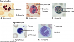
Leukocytes |
-white blood cells -5 types of mature leukocytes: -Granulocytes: Basophils, Eosinophils, neutrophils -Mononuclear cells: Monocytes, Lymphocytes |
|
|
Basophils |
contains histamine--is released during allergic reactions -contains heparin (prevents clotting) -0-1% |
|
|
Eosinophils |
increase in allergic reactions and engulf substances that trigger the allergic reaction -1-4% -phagocytic |
|
|
Neutrophils |
are phagocytes--accumulate at sites of infection and engulf bacteria -50-70% |
|
|
Monocytes |
phagocytic cell that becomes a macrophage and fights off disease -3-8% |
|
|
Lymphocytes |
make antibodies and destroy foreign antigens 20-40% |
|
|
efgb |
|
|
Thrombocytes or Platelets |
-formed from giant cells in bone marrow called megakaryocytes -tiny fragments break off to form platelets -help blood clot |
|
|
Coagulation |
blood clotting -complicated enzymatic cascade that ends in formation of fibrin clot -platelets begin process -anticogulation substances inhibit clotting when finished |
|
|
Clotting factors |
any of the plasma components involved in clotting |
|
|
Coagulation |
clotting-liquid to solid |
|
|
Fibrin |
-produced by fibrinogen -elastic fibrous protein -needed for clotting |
|
|
Clotting pathway |
Prothrombin->thrombin->fibrinogen->fibrin |
|
|
Injury pathway |
injury -platelet aggregation -tissue, clotting factors, calcium, prothrombin, thrombin -fibrinogen to fibrin clot |
|
|
bas/o |
base, opposite of acid |
|
|
chrom/o, chromat/o |
color |
|
|
cyt/o |
cell |
|
|
granul/o |
granules |
|
|
morph/o |
shape |
|
|
myel/o |
bone marrow, spinal cord |
|
|
path/o |
disease |
|
|
phag/o |
eat, swallow |
|
|
phleb/o |
vein |
|
|
plas/o |
formation, development |
|
|
thromb/o |
clot |
|
|
pro- |
before, forward |
|
|
-apheresis |
removal |
|
|
-blast |
immature cell |
|
|
-cytosis |
sight increase in numbers -condition of cells |
|
|
-emia |
blood condition |
|
|
-globin, -globulin |
protein |
|
|
-oid |
derived from, resembling |
|
|
-penia |
deficiency |
|
|
-phage |
eat, swallow |
|
|
-philia |
attraction for |
|
|
-poiesis |
formation |
|
|
-rrhage |
bursting forth (blood) |
|
|
-sis |
condition, state |
|
|
-stasis |
to stop, control |
|
|
-y |
condition, process |
|
|
Hemolytic |
-pertaining to the rupture or destruction of RBCs |
|
|
Anemia |
deficiency in erythrocytes or hemoglobin |
|
|
Aplastic anemia |
bone marrow doesn't produce enough RBCs -cause is usually unknown |
|
|
Pancytopenia deficiency |
when stem cells don't produce enough WBCs, RBCs, platelets |
|
|
Pernicious Anemia |
-caused by inability to absorb Vitamin B12 -when number of RBCs does down -causes enlargement of individual cells (macrocytes) |
|
|
Iron deficiency Anemia |
-most common type -hemoglobin is unable to transport O2 due to lack of iron |
|
|
Sickle Cell Anemia |
Hereditary -prevalent in black people -Sickle shaped erythrocytes -caused by abnormal hemoglobin (Hgb S) -oddly shaped -can't fit in blood vessel->infarction -Hemolysis -painful |
|
|
Polycythemia |
-increase amount of RBCs -thick/viscous blood -bone marrow is hyperplastic -treatment: plebotomy, myelotoxic drugs to suppress blood cell count |
|
|
Hemophilia |
-inability of blood to clot due to deficiency of a clotting factor -treatment: administer deficient factor |
|
|
Thrombocytopenia |
low levels of platelets in blood -normal platelet count (140-400,000) |
|
|
Autoimmune Thrombocytopenia Purpura |
-body makes antibodies that destroys their platelets -results in bruising and bleeding from mucous membranes |
|
|
Petechiae |
tiny hemorrhages |
|
|
Ecchymoses |
large scale bruise |
|
|
Leukemia |
-increase in number of cancerous WBCs -malignant cells fill bone marrow and blood stream -acute: primarily immature leukocytes -chronic: "" mature "" |
|
|
Composition of blood |
45% formed elements (erythro, leuko, platelets) 55% liquid or plasma (water, salts, hormones, etc) |
|
|
Formation of blood |
-blood cells form in bone marrow -stem cells undergo a process of differentiation |
|
|
-apheresis |
removal |
|
|
-cytosis |
condition of the cell; increase in numbers |
|
|
-oid |
derived from, resembling |
|
|
-poiesis |
formation |
|
|
Pernicious Anemia |
inability of B12 to be absorbed |
|
|
Aplastic Anemia |
bone marrow doesn't produce enough RBCs |
|
|
-penia |
deficiency |
|
|
Pancytopenia |
deficiency in all blood cells (erythro, leuko, platelets) |
|
|
Sickle Cell Anemia |
-oddly shaped -prevalent amongst blanks -caused by abnormal hemoglobin -thrombosis and infarction -hemolysis |
|
|
polycythemia vera |
-too many RBCs -thick blood -treatment: plebotomy, myelotoxic drugs to reduce cell production |
|
|
Hemophilia |
deficiency of clotting factor |
|
|
Thrombocytopenia |
low levels of platelets |
|
|
Autoimmune Thrombocytopenic Purpura |
body makes antibodies that destroy platelets -results in bruising and bleeding from mucous membranes |
|
|
Petechiae |
small bruises |
|
|
Ecchymoses |
large scale bruise |
|
|
Leukemia |
malignant cancerous white blood cells -malig leukocytes fill bone marrow and blood stream -acute: primarily immature leukocytes -chronic: prim mature leuk treatment: chemo then blood transfusion |
|
|
Mononucleosis |
infectious disease -increased numbers of leukocytes and enlarged cervical nodes -caused by Epstein Barr Virus -symtoms: pharyngitis, lymphadenitis, hepatomegaly splenomegaly -mode of transmission: saliva |
|
|
complete blood count |
automated count of all blood cells |
|
|
Erythrocyte sedimentation rate |
measuring the rate at which RBCS settle at bottom of test tube -increased rate with infection/inflammation |
|
|
Hematocrit |
% of RBCs in a volume of blood |
|
|
RBC count |
used to test number of RBCs to diagnose anemia |
|
|
WBC count |
to diagnose disorders, infections, monitoring treatment |
|
|
Platelet count |
diagnose bleeding disorders |
|
|
Cross-matching |
blood typing test to see compatibility between donor and recipient |
|
|
Prothrombin |
test of the ability for blood to clot |
|
|
WBC differential count |
% of total WBC count made up by different types of leukocytes |
|
|
Apheresis |
separation of blood into component and parts and removal of a selected part (WBCs, platelets, etc) |
|
|
Blood component therapy |
transfusion of specific blood components (plasma, RBCs, platelets) |
|
|
Blood type A |
antigen A -anti-B |
|
|
Blood type B |
antigen B -anti A |
|
|
Blood type AB |
antigen A and B neither anti A or anti B |
|
|
blood type O |
no antigens for A or B -anti A and anti B |
|
|
Rh positive |
antigens D -no anti-D |
|
|
Rh negative |
No D antigen -anti D |
|
|
Autologous blood |
blood donated by the same patient for the future -usually pre-surgical |
|
|
Homologous blood |
blood donated by same species so a compatible recipient can use |
|
|
Bone marrow aspiration |
removal of a small amount of fluid and cells from inside the bone with a needle |
|
|
bone marrow transplant |
transfer of bone marrow from one person to another |
|
|
cardiovascular system |
heart and blood vessels |
|
|
cardiovascular structure |
heart has four chambers and valves -heart wall has three tissue layers heart muscle and tissues are specialized -capillaries allow for gas exchange -veins return blood back to the heart |
|
|
capillaries |
carry blood from arteries/arterioles to body cells |
|
|
-arteries have endothlial cells to reduce blood clotting and promote growth of blood vessels |
efg |
|
|
capillarie walls are only endothelial cells in thickness waste products pass from body cells to caps to venules |
hb |
|
|
superior vena cava |
-brings de-oxygenated blood from body to heart -blood comes from upper body (head and limbs) |
|
|
inferior vena cava |
de-oxygenated blood from lower side of body |
|
|
circulation of blood |
1. inferior/superior vena cava 2. right atrium 3. right ventricle 4. pulmonary artery (de-oxy)->lung caps 5. pulmonary veins (oxy) 6. left artria 7. left ventricle 8. AORTA 9. rest of body |
|
|
Aorta structure |
Ascending aorta is divided into separate arteries (subclavian, brachiocephalic, common carotid) |
|
|
Carotid arteries |
carry oxy-gen blood to head and neck |
|
|
Atrioventricular valves |
between atrium and ventricle -tricuspid (right) -bicuspid (left)p |
|
|
pulmonary valve vs. aortic valve |
jnf |
|
|
Septa |
partitions that separate the four chambers of the heart -interatrial septum: separates two upper chambers -interventricular septum: separates two lower chambers |
|
|
Three layers of the heart |
-endocardium lines interior of the heart and heart valves -myocardium: the middle layer (thickest layer) -pericadium: sac that surrounds the heart |
|
|
Diastole |
relaxation period -ventricle walls relax -blood flows from the vena cava and pulmonary veins "lubb" -closure of tricuspid and bicuspid valves |
|
|
Systole |
contraction -ventricles contract -atria relax |
|
|
angi/o, vas/o, vascul/o |
vessel |
|
|
ather/o |
yellowish plaque |
|
|
sphygm/o |
pulse |
|
|
steth/o |
chest |
|
|
thromb/o |
clot |
|
|
-ectasia |
dilation, widening |
|
|
-graphy |
process of recording |
|
|
-ium |
tissue, structure |
|
|
-stenosis |
narrowing, tightening |
|
|
patent |
open or exposed |
|
|
sphygmic |
pertaining to pulse |
|
|
precordial |
pertaining to anterior left chest |
|
|
dysrhythmia |
abnormal heart rhythm |
|
|
Partial heart block |
failure of conduction occurs occasionally |
|
|
complete heart block |
no impulses reach the AV node |
|
|
Flutter |
rapid contractions of the atria -symptomatic of heart disease |
|
|
Fibrillation |
-fast, irregular contractions of the heart |
|
|
Congenital heart disease |
small holes in walls between atria and ventricles -septa are affected -some holes close on their own while others require open heart surgery |
|
|
nitroglycerin |
a vasodilator that increases coronary blood flow and lower blood pressure |
|
|
aspirin |
used to prevent clumping of platelets |
|
|
beta blockers |
to lower blood pressure and lower spped and force of heart beat |
|
|
artherosclerosis |
buildup of plaque on the inside of artery walls |
|
|
Aortic Stenosis |
narrowing of the aortic valve opening |
|
|
Cardiac tamponade |
increase in fluid in the pericardium->compression of heart |
|
|
Coarctation of the aorta |
narrowing of the aorta causing hypertension, ventricular strain, and ischemia |
|
|
Congestive heart failure |
inefficiency of cardiac circulation causing edema and pulmonary congestion |
|
|
Embolus |
made up of a thrombus -vascular blockage |
|
|
Peripheral arterial disease |
any disorder of arteries outside of the heart |
|
|
Raynaud disease |
vasoconstriction of hands and feet causing cyanosis. caused by cold temps or emotional things |
|
|
Rheumatic heart disease |
rheumatic fever causes valvular disease |
|
|
Aneurysm |
widening of an arterial wall -usually caused by artherosclerosis and hypertension or congenital weakness in a vessel wall -common in aorta |
|
|
varicose veins |
abnormally swollen and twisted veins -usually in leg -caused by damaged valves that don't prevent backflow of blood -blood collects -thrombosis may occur |
|
|
creatine kinase |
enzyme that is released if heart has been damaged by a heart attack - |
|
|
troponin test |
proteins released when heart muscle has been damaged |
|
|
C-reactive protein |
a blood test used to measure the level of inflammation in the body--may indicate conditions that lead to cardiovascular disease |
|
|
electrolyte panel |
a blood test used to measure the ion levels -can diagnose acid-base or pH imbalance that may cause arrhythmias, muscle damage, or death |
|
|
lipid panel, lipid profile |
can measure high/low blood pressure -high density lipoprotein/or low |
|
|
Arteriography |
recording an artery after injection of a dye |
|
|
Angioscopy |
insertion of a catheter with an attached camera to visualize a structure or vessel |
|
|
Doppler sonography |
using sound waves to record velocity of blood flow |
|
|
Echocardiography |
recording the structure and function of the heart at rest and with exercise v |
|
|
vascular sonography |
placing ultrasound transducer at tip of a catheter within a blood vessel to assess blood flo |
|
|
Holter monitoring |
portable ECG that is used for 24 hrs to detect cardiac arrhythmias |
|
|
Cardioversion |
defibrillation -using electricity to restore normal heart beat |
|
|
Endarterectomy |
removal of plaque from artery |
|
|
coronary artery bypass graft |
veins are grafted from leg, arm, or breast and put on the heart to supply heart with oxygenated blood |
|
|
Extracorporeal circulation |
blood circulates through heart-lug machine while heart is being repaired |
|
|
Mitral valve prosthesis |
valve replacement |
|
|
-ectasis |
dilation |
|
|
Larynx |
voice box -air passageway between pharynx and trachea -epiglottis covers upper region of larynx during swallowing -glottis (vocal cords)--vibrate to produce sound |
|
|
trachea |
air passes from larynx to thorax |
|
|
Alveolus |
lined with epithelial cells to allow for gas exchange between alveolus and capillary -erythrocytes carry oxygen to the body and CO2 to the lungs to be exhaled |
|
|
pleura |
double folded membrane that surrounds the lungs and lines the pleural cavity |
|
|
Thorax |
area between neck to diaphragm -formed by sternum, thoracic vertebrae, and ribs |
|
|
Mediastinum |
area of thoracic cavity between lungs -contains aorta, heart, esophagus, thymus, trachea |
|
|
external respiration |
inspiration and exhalation |
|
|
Inspiration |
chest expands -diaphragm contracts |
|
|
expiration |
chest contracts -diaphragm relaxes |
|
|
internal respiration |
exchange of gases between blood and cells |
|
|
ventilation |
distribution of gas into and out of lungs |
|
|
apnea |
temporary absence of breathingd |
|
|
dyspnea |
labored or difficulty in breathing |
|
|
orthopnea |
difficulty breathing when lying down |
|
|
eupnea |
normal respirations |
|
|
hyperpnea |
breathing that is deeper than normal |
|
|
hypoxia |
less than normal oxygen |
|
|
anoxia |
absence of oxygen |
|
|
capn/o |
carbon dioxide |
|
|
cost/o |
rib |
|
|
lob/o |
lobe of lung |
|
|
ox/o |
oxygen |
|
|
phon/o |
voice |
|
|
phren/o |
diaphragm |
|
|
pneum/o |
lung/air |
|
|
spir/o |
breathing |
|
|
tel/o |
complete |
|
|
pan- |
all |
|
|
-osmia |
smell |
|
|
-pnea |
breathing |
|
|
-ptysis |
spitting |
|
|
-sphyxia |
pulse |
|
|
Dysphonia |
pertaining to disorders of the voice |
|
|
Pleurodynia |
pertaining to pain in pleura |
|
|
percussion |
tapping on a surface to determine difference in density below the surface |
|
|
rales (crackles) |
popping or clicking--means that there's fluid in alveoli |
|
|
rhonchi |
rumbling sounds -bronchi is obstructed by sputum (material expelled from bronchi, lungs, resp tract |
|
|
pleural rub |
friction sounds -when pleural surfaces rub against each other |
|
|
stridor |
whistling sound heard during inspiration -obstruction in pharynx or larynx |
|
|
wheeze |
narrowed airway (asthma) |
|
|
Croup |
cough and stridor (whistling when inspirating) -acute viral infection in infants -obstruction of larynx |
|
|
Pertussis (whooping cough) |
-caused by bacterium Bordtella pertussis -acute infectious inflammation of larynx, trachea, bronchi -highly contagious -vaccine preventable -coughing that ends in loud whooping inspiration |
|
|
Asthma |
chronic bronchial inflammation disorder with airway obstruction due to edema and excess mucous -Symptoms: dyspnea, wheezing, coughing |
|
|
Cystic fibrosis |
inherited disorder of exocrine glands -blood test to detect gene -affects cells that produce mucous, sweat, and digestive juices -instead of acting like lubricant, it's a plug |
|
|
Emphysema |
also known as chronic obstructive pulmonary disease -alveoli are progressively destroyed -gradually have loss of breath -leads to increase in pulmonary artery pressure -smoking is leading cause of emphysema -right side of heart works harder to pump blood--right ventricular hypertrophy and heart failure |
|
|
Pneumonia |
acute inflammation and infection of the alveoli, which fill with pus or products of the inflammatory reaction -alveoli gas exchange is inhibited by exudate (fluid, etc) -INFILTRATE: fluid filled area wtihin the lungs that can be visualized on a chest X ray or CT scan |
|
|
Pneumococcal pneumonia |
form of pneumonia caused by bacterial species Streptococcus pneumoniae |
|
|
Pulmonary abcess |
collection of pus in the lungs -bacterial infection |
|
|
Pulmonary edema |
air sacs and bronchioles fill with fluid |
|
|
Atelectasis |
Collapsed lung -causes include blockage (tumor, secretions) of bronchus or smaller bronchial tube |
|
|
Mesothelioma |
malignant tumor of the pleura -caused by asbestos exposure |
|
|
Pneumothorax |
air in the pleural space -may follow trauma |
|
|
acid fast bacilli (ABF) smear |
test to see if bacterium for TB is present |
|
|
Ventilation-perfusion scan |
test used to access distribution of blood flow and ventilation through both lungs |
|
|
Peak flow monitoring |
monitors how well your lungs are working -measures rate of air flow through airways -helps manage asthma symptoms |
|
|
Polysomnography |
monitoring and recording normal and abnormal activity during sleep |
|
|
pulse oximetry |
measurement of oxygen saturation in the blood |
|
|
Spirometry |
measures air flow and volume of air inspired and expired by the lungs using a spirometer |
|
|
Thoracoscopy |
endoscopic examination of the thorax done through a small opening in the chest wall |
|
|
septoplasty |
surgical repair of the sinus |
|
|
-tomy |
incision |
|
|
-stomy |
opening |
|
|
Endotracheal intubation |
tube inserted in the larynx and trachea to establish an airway for breathing purposes |
|
|
Hyperbaric Medicine |
medicinal use of high barometric pressure to increase O2 content of blood and tissues |