![]()
![]()
![]()
Use LEFT and RIGHT arrow keys to navigate between flashcards;
Use UP and DOWN arrow keys to flip the card;
H to show hint;
A reads text to speech;
73 Cards in this Set
- Front
- Back
- 3rd side (hint)
|
3 layers of the meninges (deepest to most superficial) and it’s functions |
Pia matar: deepest layer, thin and close to the contours of the brain and spinal cord Arachnoid matar: deep to the dura mater, spider web like connective tissue Dura matar: outermost layer, tough white fibrous connective tissue, layers separate to form dural venous sinuses |
P.A.D |
|
|
2 examples of skill memory & where is it stored? |
Riding a bike Writing In cerebellum |
Physical things |
|
|
2 examples of declarative memory & where is it stored? |
Factual things (studying) Math equations In hippocampus |
Mentally |
|
|
How does this protect the CNS? Skull and vertebral column |
Physical protection Boney structures
|
|
|
|
How does this protect the CNS? Meninges |
Physical protection Shock absorbance
|
|
|
|
How does this protect the CNS? Blood brain barrier (BBB) |
Chemical barrier Regulates content of CSF |
|
|
|
What lobe contains the primary motor cortex? |
Frontal lobe |
|
|
|
What lobe contains the primary somatosensory cortex? |
Parietal lobe |
|
|
|
What lobe contains the auditory association area? |
Temporal lobe |
|
|
|
What lobe processes visual information? |
Occipital lobe |
|
|
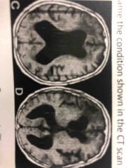
What condition is shown above? |
Hydrocephalus (CFS flow is obstructed) |
|
|
|
What happens to the ciliary muscle when looking at objects to achieve visual accommodation? |
Nearby objects- contract Far away objects- relax |
|
|
|
What happens to the suspensory ligaments when looking at objects to achieve visual accommodation? |
Nearby objects: loose Far away objects: tight |
|
|
|
What happens to the lens when looking at an object to achieve visual accommodation? |
Nearby objects- thick Far away objects- thin |
|
|
|
How does this protect the CNS? Cerebrospinal Fluid (CSF) |
Bouyance Prevents concussion Washes away metabolic waste |
|
|
|
What are the signs of meningitis and how do you get tested? |
Fever, stiff neck, lead to paralysis possible death Spinal tap (collects CFS), CT scan Inflammation of the meninges |
|
|
|
Where is white matter found? |
Inside brain-corpus cullosum & tracts (myelinated axon) Outside of spinal cord |
|
|
|
Where is grey matter found? |
Outside brain- cerebral cortex Thalamus Reticular formation Spinal cord-butterfly |
|
|
|
2 examples of spinal reflex |
Knee jerk reflex Babinski reflex |
|
|
|
2 examples of cranial reflex |
Coughing Swallowing Vomiting |
|
|
|
What lobe contains Wernicke’s area? |
Temporal lobe |
|
|
|
What lobe contains Broca’s area? |
Frontal lobe |
|
|
|
What is the composition and function of white matter? |
Myelinated axons that run together in bundles called tracts (ascending and descending) Enables communication between hemispheres |
|
|
|
What is the composition and function of Gray Matter |
Cell bodies and short nonmyelinated fibers Relays and filters sensory info (except smell) Forms sides and roofs of 3rd ventricle |
|
|
|
CNS |
Central Nervous system Brain and spinal cord composed of gray and white matter |
|
|
|
CSF (function and flow) |
Cerebrospinal Fluid Formed by glial cells (ependymal cells) found in choroid plexus (vascular tissue) Clear tissue that forms a protective layer around and within the CNS Creates buoyance which allows the brain to float in the skull Regulated by the blood brain barrier (BBB) |
|
|
|
Spinal Cord (structure and organization) |
OUTTER PERIPHERY: white matter (contains ascending and descending tracts) A- take sensory info to the brain (posterior) D- takes motor info from the brain (anterior) CENTER OF SPINAL CORD- gray matter (containing sensory neurons, motor neurons and interneurons) |
|
|
|
Cerebrum (Limbic system) |
Largest portion of brain, receives sensory input, carries out integration then commanding voluntary motor responses Higher thought process (for learning, memory, language) 2 halves (L/R cerebral hemispheres-connected by bridge of white matter-corpus callosum) |
|
|
|
Cerebral cortex (Limbic system) |
Thin layer of gray matter that covers the cerebral hemispheres Sensation, thought process and voluntary movements |
|
|
|
Primary motor area |
Frontal lobe Voluntary commands to skeletal muscle begins here Right primary motor area controls left side of body (vice versa) |
|
|
|
Primary somatosensory area |
Parietal lobe Sensory info from skin and skeletal muscle arrives here |
|
|
|
Association Area |
Integration occurs and memory stored Visual- associates new visual info with precious memories of visual info Auditory- same thing but with sounds |
V.A |
|
|
Broca’s Area |
Left frontal lobe Motor speech area (production of speech in a clear and fluent manner) Damaged—> Broca’s aphasia |
|
|
|
Wernicke’s Area |
Left hemisphere (temporal lobe) Damaged—> hinders ability to interpret written and spoken language (Wernicke’s aphasia) Cooperate with Broca’s area for human communication |
|
|
|
Medulla Oblongata |
90% of tracts cross the midline Reflexes (coughing sneezing swallowing vomiting Respiratory centre |
|
|
|
Thalamus |
Consists of 2 masses of gray matter, located at roof and sides of 3rd ventricle Receiving end of sensory input (except smell) (visual auditory and somatosensory info) Sends it to appropriate portions of the cerebrum |
|
|
|
Hypothalamus |
Forms the floor of third ventricle Regulates various processes related to homeostasis (hunger, sleep, thirst, body temp) Produces hormones secreted by posterior pituitary gland |
|
|
|
Pons |
Reflex center for stimulus involving visual/auditory and head movements 4 pairs of cranial nerves emerge here |
|
|
|
Cerebellum |
Maintain posture and balance Coordinate and communicate with cerebrum to execute smooth seamless muscle movement Stores skill memory |
|
|
|
Limbic System |
Reward system (survival and instinctive behaviours) Consists of: Amygdala (emotions), Thalamus (process memory), Hypothalamus (autonomic responses and hormone secretion), hippocampus (stores memory) |
|
|
|
Skill memory |
Repetition of motor tasks Involve cerebellum Highly persistent |
|
|
|
Declarative Memory |
Facts and impressions Temporal lobe |
|
|
|
Mechanoreceptors (sensory receptor) |
Stimulated by changes in pressure or body movement Inner ear—> body movement |
|
|
|
Retina |
Contains photoreceptors Rod cells: night and peripheral vision Cone cells: distinguish colour (fovea centralis where cones are densily packed) |
|
|
|
Optic nerve |
Carries info from retina to brain |
|
|
|
Thermoreceptors (sensory receptor) |
Stimulated by changes in external or internal temperature Located in skin and organs |
|
|
|
Nociceptors (pain receptor) |
Somatic- found in skin and skeletal muscle, responds to mechanical electrical or chemical damage to tissue (burning a finger) Visceral- react to excessive stretching of organs (oxygen deprivation, stomach is full) |
|
|
|
Referred pain |
Internal pain sometimes felt as pain from the skin Because somatic pain running through same spinal cord pathway as visceral pain |
|
|
|
Chemoreceptors |
Stimulated by changes in chemical concentration of substances (taste buds, smell receptors) |
|
|
|
Photoreceptors |
Located in the eye Stimulated by light |
|
|
|
Sclera (part of eye) |
Most superficial layer White and fibrous Eye muscles attached, cornea most anterior portion |
|
|
|
Choroid (part of eye) |
Contains blood vessels (nourish eye) Opening at front called pupil surrounded by muscular layer called iris Change in pupil size—>focusing on objects at different distances |
|
|
|
Lens (part of eye) |
Located behind iris and pupil Shape of lens controlled by suspensory ligament and ciliary body To maintain focus—> lens must change shape = visual accommodation |
|
|
|
Compartments of lens |
Anterior-aqueous humor Posterior- vitreous humor Important to maintain shape of eye ball |
|
|
|
Optic Chiasma |
X shaped formed by crossover of nerves Fibers: right half go to right optic tract, left half go to left optic tract Info: right field of view goes to left side of brain, left field of view goes to right side of brain |
|
|
|
Outter ear |
Pinna- visible parts of the ear Auditory canal- lined with fine hairs and sweet canals (secrete oily ear wax) |
P.A |
|
|
Middle ear |
Tympanic membrane- converts sound into mechanical movement (separates outer and inner ear) OSSICLES (amplifies sound x20) Malleus (hammer)- attaches to tympanic membrane Incus (Anvil) Stapes- attaches to oval window Auditory tube- equalizes pressure of the head (extends from middle ear to nasopharynx) |
T.O(MIS).A |
|
|
Inner ear |
Cochlea- spiral shaped passage needed for hearing Semicircular canals and Vestibule- needed for equilibrium and balance |
C.S.V |
|
|
Digestive system |
Ingest food Breakdown food Absorb nutrients Eliminate waste |
|
|
|
Layers of the GI tract |
Mucosa- layer of epithelium supported by connected tissue, secrete digestive enzymes and goblet cells (secrete mucus) Submucosa- loose connective tissue contains blood vessels, lymphatic vessels and nerves Muscularis- two layers of smooth muscle Serosa- very thin outer most layer, secretes fluid that keeps the surface of the intestines moist |
MSMS |
|
|
3 salivary glands |
Parotid gland Submandibular gland Sublingual gland |
|
|
|
Stomach |
Has sphincters: doesn’t allow food to re enter esophagus Cardiac sphincters keeps chyme in the stomach and regulates the flow into the small intestines |
|
|
|
3 layers of the stomach |
Oblique Circular Longitudinal |
|
|
|
Gastric glands |
Gastric glands (bottom) in gastric pits (top) 3 types of cells: Chief cells Parietal cells Mucous cells |
|
|
|
Carbohydrates |
Breakdown in mouth through amylase Amylose—>glucose |
|
|
|
Protein |
Break down in stomach through pepsin Proteins—> amino acids Chief cells: secrete pepsinogen which becomes pepsin when exposed to HCl |
|
|
|
Lipids |
Break down in duodenum through lipase Lipids—> glycerol + fatty acids |
|
|
|
Parietal cells |
Produce HCl, causes gastric juice in stomach to have a high pH |
|
|
|
Small Intestines |
Duodenum (digestion) Jejunum (absorption) Ileum (absorption) Circular folds, villi and microvilli help to increase surface area |
|
|
|
Bile |
Stored in gallbladder then secretes into duodenum through bile duct Liver makes this |
|
|
|
Liver |
Accessory organ Helps digestion of bile |
|
|
|
Pancreas |
Secretes pancreatic juices into duodenum Contain NaHCO3 and digestive enzymes (lactase, analase, lipase, pepsin) |
|
|
|
Large Intestines |
Material enters from ileum, absorbs water salts and some vitamins then eventually eliminated through the anus Cecum Colon Rectum |
|