Reading...
![]()
Play button
![]()
Play button
![]()
Use LEFT and RIGHT arrow keys to navigate between flashcards;
Use UP and DOWN arrow keys to flip the card;
H to show hint;
A reads text to speech;
3 Cards in this Set
- Front
- Back
- 3rd side (hint)
|
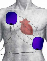
Defibrillation
|
Defibrillation is a common treatment for life-threatening cardiac dysrhythmias, ventricular fibrillation and pulseless ventricular tachycardia. Defibrillation consists of delivering a therapeutic dose of electrical energy to the heart with a device called a defibrillator. This depolarizes a critical mass of the heart muscle, terminates the dysrhythmia and allows normal sinus rhythm to be reestablished by the body's natural pacemaker, in the sinoatrial node of the heart. Defibrillators can be external, transvenous, or implanted (implantable cardioverter-defibrillator), depending on the type of device used or needed. Some external units, known as automated external defibrillators (AEDs), automate the diagnosis of treatable rhythms, meaning that lay responders or bystanders are able to use them successfully with little or no training at all.
|
|
|
|
AED
|
An automated external defibrillator (AED) is a portable electronic device that automatically diagnoses the life-threatening cardiac arrhythmias of ventricular fibrillation and ventricular tachycardia in a patient,[1] and is able to treat them through defibrillation, the application of electrical therapy which stops the arrhythmia, allowing the heart to reestablish an effective rhythm.
With simple audio and visual commands, AEDs are designed to be simple to use for the layperson, and the use of AEDs is taught in many first aid, certified first responder, and basic life support (BLS) level cardiopulmonary resuscitation (CPR) classes.[2] |
|
|
|
AF
|
Atrial fibrillation (AF or A-fib) is the most common abnormal heart rhythm. It may cause no symptoms, but is often associated with palpitations, fainting, chest pain, or congestive heart failure. The cause of an individual's AF may not be identified.
AF may be identified clinically when taking a pulse, and its presence can be confirmed with an electrocardiogram (ECG) that demonstrates the absence of P waves and an irregular ventricular rate.[1] |

|