Reading...
![]()
Play button
![]()
Play button
![]()
Use LEFT and RIGHT arrow keys to navigate between flashcards;
Use UP and DOWN arrow keys to flip the card;
H to show hint;
A reads text to speech;
10 Cards in this Set
- Front
- Back

This slide is an artery and vein (#22). What type of cells are on the epithelium?
|
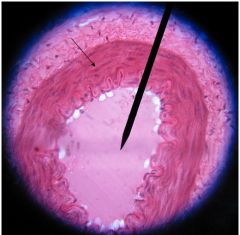
The simple squamous epithelium at the luminar surface of the artery and vein.
|
|
What type of epithelium is this?
|

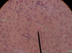
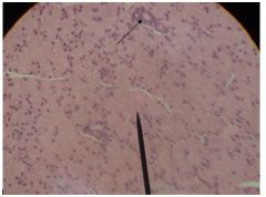
This slide is a thyroid gland #58. The epithelium is simple cuboidal epithelium of the thyroid follicle. These are a single layer of cuboidal cells, with the nucleus located in the center.
|
|
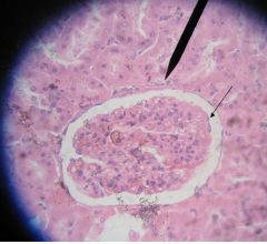
What is the glomerular epithelium?
|

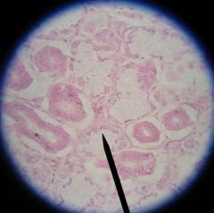
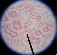
This is the kidney slide #45.
The layer that follows closely to the contour of the glomerulus is simple squamous epithelium. * Numerous tubules adjacent to the glomerulus are lined by simple cuboidal cells. |
|

What is the epithelium on this slide?
|
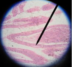
This is duodenum #39.
Simple columnar epithelium. Tall columnar cells are arranged in a single row. The ovoid nuclei are located in the basal region. Goblet cells are interspersed between the cells. |
|
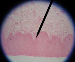
What is this epithelium?
|

This is the esophagus #34.
This is stratified squamous epithelium. Epithelium is composed of numerous layers of cells. The cells in the basal layer are columnar with oval nucleus, whereas in the upper region are several rows of squamous cells. Cells and nuclei become progressively flatter toward the free surface. |
|
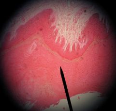
What type of epithelium is this?
|

This is a digital pad #69.
Stratified squamous epithelium. Observe the non-nucleated keratinized cells in the uppermost layer stratum corneum, which results from wear and tear. *Also see cornea for non-keratinized stratified squamous epithelium. |
|
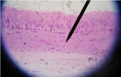
What type of epithelium is this?
|

This is a trachea slide #43.
Pseudostratified epithelium The cells appear to be in several layers because their nuclei are situated at different height but all cells are in contact with the basement membrane. * Observe the motile cilia on the surface of the cells |
|

What type of epithelium is this?
|
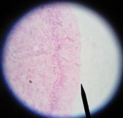
This is a bladder #47.
Transitional epithelium, composed of several layers of generally similar cells with rounded nuclei. A thin basement membrane is present. The epithelium has the ability to rearrange the number of layers according to whether it is in contracted or expanded condition. When contracted the cells are generally rounded. When distended number of cell layers are reduced, the cells are generally flatted and appear typical squamous. |
|
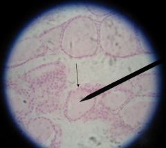
Where are the serous alveoli?
|
This is a parotid gland #31.
Observe the serous alveoli (arrow). Each alveolus is formed by pyramidal cells arranged around a small lumen. The small and rounded nucleus located in the basal zone of deeply basophilic cytoplasm and apical cytoplasm is lighter. |
|
Where are the serous and the mucous alveoli?
|
This is a sublingual gland #33.
Observe the serous alveoli (blue arrow) and the mucous alveoli (red arrow). The mucous alveoli are larger, more columnar, and utmost colourless staining reaction. The oval or flattened nuclei at the base of the cell larger lumen, Observe the mixed alveoli where mucous alveoli are surrounded by groups of serous cells. Also observe the ducts and identify their epithelium. |