![]()
![]()
![]()
Use LEFT and RIGHT arrow keys to navigate between flashcards;
Use UP and DOWN arrow keys to flip the card;
H to show hint;
A reads text to speech;
32 Cards in this Set
- Front
- Back
|
Name the attachments (anterior and posterior) of the choroid and the retina |
Choroid = Scleral spur (anteriorly), exit foramina vortex veins (posteriorly) |
|
|
Colour Doppler can be used to help differentiate melanoma from subretinal haemorrhages and haematomas because ......................
|
Subretinal haemorrhages and haematomas are avascular and melanoma's contain vascularity.
|
|
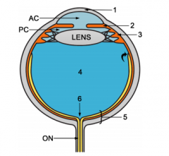
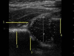
Label the diagram |
1 = Cornea 2 = Iris 3 = Ciliary Body 4 = Vitreous Chamber 5 = Posterior occular wall (formed by retina) 6 = Optic Disc |
|
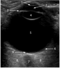
Label the diagram |
1 = Cornea 2 = Anterior Chamber 3 = Ciliary Body 4 = Lens 5 = Vitreous Chamber 6 =Retina 7 = Optic Nerve 8 = Echogenic Retrobulbar Fat 9 = Optic Disc |
|
|
What are the indications for an eye ultrasound? |
Corneal diseases, glaucoma, tumours, trauma, lens implant, foreign body localisation, prior to vitreoretinal surgery, elevated ICP, lens dislocation, vitreous haemorrhage.
|
|
|
What do you understand by developmental dysplasia of the hip [DDH]? List any 4 risk factors for DDH.
|
An abnormal growth and development of the hip which includes the osseous structures (acetabulum, proximal femur) as well as the labrum, capsule, and other soft tissues.
Risk factors: breech position, family history, oligohydramnios, first born child, females (increased oetrogen). |
|
|
Draw and name the structures seen on a standard transverse hip ultrasound image? |
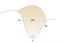
FM = Femoral metaphysis CFE = Capital Femoral Epiphysis ISC = Ischium P = Pubis |
|
|
Draw and name the structures seen on a standard coronal hip ultrasound image? |
|
|
|
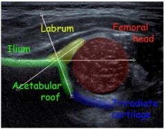
What is the Alpha angle and it's normal value? |
The Alpha angle is formed by the acetabular roof to the vertical cortex of the ilium. The normal value is greater than or equal to 60 degrees
|
|
|
What is the Beta angle and it's normal value? |
The Beta angle is formed by the vertical cortex of the ilium and the triangular labral fibrocartilage. The normal value is less than 77 degrees but is only useful in assessing immature hips when combined with the alpha angle
|
|
|
What is the Barlow Test when assessing for DDH? |
Barlow Test = examiner adducts the hip while applying a posterior force on the knee to promote dislocation
|
|
|
What is the Ortalani Test when assessing for DDH? |
Ortolani Test = examiner abducts the hip while applying an anterior force on the femur to reduce the hip joint
|
|
|
Currently the two widely accepted methods of ultrasound examination are the static method of ........... and the dynamic method of ..............
|
Static = Grafe Dynamic = Harcke |
|
|
A hip of an infant with a graf angle less than 49 degrees, and a beta angle of greater than 77 degrees will have a ......................
|
Dislocated or subluxed hip |
|
|
Treatment for DDH is best undertaken when? |
In the first 3 months |
|
|
What is a 'baseline' in relation to DDH? Which landmark is used to draw the baseline in assessing for DDH? |
The ilium is used to draw the baseline which is a line drawn parallel to the ossified lateral wall of the ilium using Graf’s method of assessing neonatal hip stability and is used to create the alpha and beta angles.”
|
|
Name structures 1-4 |
1 = Labrum 2 = Ilium 3 = Acetabular roof 4 = Femoral metaphysis |
|
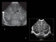
Which is the immature paediatric brain? |
Image A. In premature infants less than 24 weeks of age, the cerebral cortex has a smooth appearance with formation of only the Sylvian fissures. Specific sulci become visible as the infants mature with the parieto-occipital fissure visible at 24 weeks, the callosomarginal and cingulated sulci at 28 weeks, and branches arising from the cingulate sulcus at 30 weeks. As infants mature from 30 to 40 weeks, sulci gradually branch, becoming more complex. |
|
|
What is a subdural collection and how does it occur? |
Subdural collections may occur spontaneously or as a result of trauma causing a tearing of the dural folds or a rupture of the medullary veins allowing blood to collect around the periphery of the brain.
|
|
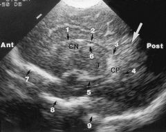
Label these structures of the paediatric brain? |
1. Frontal horn of lateral ventricle 2. Body 3. Atrium 4. Occipital Horn 5. Temporal Horn 6. Caudothalamic groove 7. Anterior cranial fossa 8. Middle cranial fossa 9. Posterior cranial fossa
|
|
|
What sonographic appearances are consistent with a severe case of periventricular leukomalacia? What would be the sonographic appearance of the brain in a few months with resolution? |
1.The presence of many cystic lesions within the brain parenchyma.
2. In a few months this resolves with brain atrophy and ventriculomegaly. Infants usually are afflicted with long term neurologic deficits. |
|
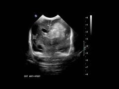
What grade is this intracranial haemorrhage and why? |
A grade 4 haemorrhage extends into the brain parenchyma as you can see in this image. Also note there is some midline shift due to the mass effect of the haemorrhage and associated venticulomegaly.
|
|
|
Name the three meningeal layers outermost to innermost |
Outermost layer - dura mater
Middle layer - arachnoid mater Inner layer - pia mater |
|
|
What is the difference between a fissure and a sulcus and give an example? |
Fissure - infolding of the brain tissue (longitudinal fissure)
Sulcus - depression separating gyri (central sulcus) |
|
|
Name the lobes of the brain, their locations and their role. |
Occipital lobe = located at the posterior aspect of the brain, houses the primary visual cortex
Temporal lobe = stretches from the temple posteriorly toward the occipital lobe, and is a processing center for language and memory. Parietal lobe = superior to the temporal lobe and adjacent to the occipital lobe, houses the somato-sensory cortex and touch and spatial navigation Frontal lobe = extends from the forehead posteriorly to the parietal lobe, and is the seat of executive function, reasoning, decision-making, integration of sensory information, and the planning and execution of movement |
|
|
What is a dorsal dermal sinus? |
- Dorsal dermal sinuses are characterized by an epithelialized fistulous tract extending inward from the skin surface.
- Have variable penetration, but up to two thirds extend into the spinal column. - Dermal sinuses arise from a focal area of non disjunction of cutaneous ectoderm from neural ectoderm during the phase of neurulation. |
|
|
What are the clinical symptoms of dorsal dermal sinus and what is the risk? What does this look like on US? |
- Patient usually present with a small midline dimple with or without discharge, and most commonly in the lumbosacral region.
- On ultrasound the sinus presents as an anechoic elongated structure extending superiorly from the skin surface into the spinal canal. -Early diagnosis is important to reduce the risk of meningeal infection |
|
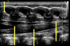
Label the structures 1 - 4 |
1. spinous process
2. filum terminale 3. nerve roots of cauda equine 4. conus medullaris |
|
|
What is the significance of a thickened filum terminale? |
A thickened filum terminale is a sign of a tethered cord. |
|
|
Describe the conus medullaris? |
- Cone shaped lower bulging of the spinal cord (caudal end)
- in healthy newborns tip of conus medullaris is located between L1 and L2 - Tip should NOT be positioned below L2-L3 |
|
|
Describe the Filum Terminale? |
- Fibrovascular tag that is 20cm long,
- Supports and protects the spinal cord. - continuation of pia mater of spine beyond the conus medullaris - stretches from end of spinal cord at level of L1 verterbra to attach to the coccyx |
|
|
Describe the cauda equina |
- Lies within thecal sac - Spinal nerves lying beyond the conus medullaris forming a bundle of nerves - arises from conus medullaris - visible as hyperechoic pulsatile streaks within intrathecal sac - Sensory, motor and parasympathetic functions |