![]()
![]()
![]()
Use LEFT and RIGHT arrow keys to navigate between flashcards;
Use UP and DOWN arrow keys to flip the card;
H to show hint;
A reads text to speech;
23 Cards in this Set
- Front
- Back
|
What are the layers of the pericardium? |
Superficial to deep: -fibrous pericardium-outermost layer, adjacent to pleural cavity -serous pericardium -parietal -pericardial cavit -visceral -myocardium -endocardium (lining of ventricles/atrium) |
|
|
What are the layers of the serous pericardium? |
Pareital Layer-on surface of fibrous pericardium vsiceral layer-directly on heart (similar to the way visceral pleura directly on lung) between the two-pericardial cavity containing a little bit of serous fluid |
|
|
What is the purpose of the pericardial cavity? |
It contains a little bit of serous fluid that allows for the movement of the heart. |
|
|
What is the mesocardium? |
Membrane connecting heart to the posterior body wall, dividing the left and right side of heart in embryo. 2 types: 1.dorsal mesocardium-connects all veins-->becomes transverse sinus 2. visceral mesocardium-connects the aorta and pulmonary trunk |
|
|
What are the two types of pericardial sinuses? |
Oblique-fold between the pulmonary veins at the back of the heart. Clinical:fluid not flow well there so has potential for bacterial infection there. transverse pericardial sinus-between two mesocardia, posterior to the pulmonary trunk and aorta but anterior to the pulmonary veins, SVC Clinical:Good place to pinch off blood flow to both veins and arteries in surgery. |
|
|
What is pericardiocentesis? |
When you remove serous fluid from the pericardial cavity either at the cardiac notch (5th/6th intercostal left to sternum) or the subxiphoid entrance. |
|
|
Where are all four of the valves in the heart situated? |
Aortic valve-At R4 level, between L ventricle and aorta Tricuspid valve-between R5-6 level, between R. atrium and R. ventricle. Pulmonary Valve-At R4 level, between R. ventrile and pulmonary A. Mitral/bicuspid valve-Between R4-5, between L. ventricle and L. atrium Bi/tricuspid valves=AV valves. associated with tendon chordinae and papillary muscles. aortic valve, pulmonary valve=semilunar valves. Passive systems-no contractile muscles involved. |
|
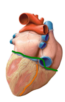
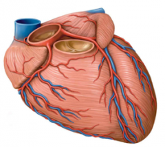
What sulci are associated with the heart? draw them on this view of the heart. |
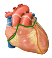
Anterior view. Green=coronary orange-interventricular Coronary sulcus-divides atria and ventricles interventricular sulcus-divide left and right ventricles. |
|
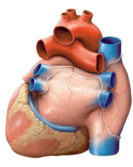
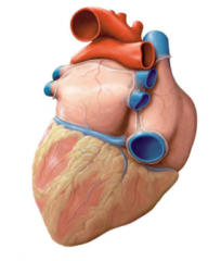
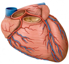
Draw the sulci of the heart on this view of the heart |
Green=coronary sulcus. you cant see interventricular sulci from posterior view. |
|
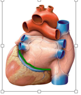
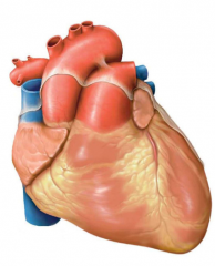
Draw the sulci on this view of the heart. |
Inferior view. Green=coronary sulcus orange=interventricular sulcus |
|
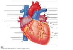
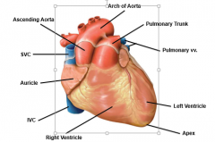
Label all the parts of this heart |
|
|
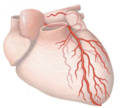
Label the Right coronary A. and its branches |
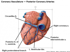
R. and L coronary arteries are first branches of the ascending aorta Anterior interventricular A aka LAD=left anterior descending artery |
|
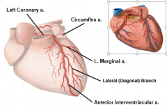
Label the Left coronary A. and its branches |
|
|
What are the posterior coronary A.? Label them. |
|
|
|
How is the L. dominant pattern of coronary vasculature different from the R. dominant? |
Fairly common variation.
R. coronary A. is small and circumflex is large so the posterior interventricular space is supplied by circumflex not coronary. |
|
|
Coronary Artery Bypass Graft (CABG) |
When you have a blocked artery, you takke another A. and attach it to the aorta and put the distal end right by the blcokage, effectively working AROUND blockage. Thoracic A. usually good subs, sometimes radial A. too. |
|
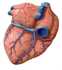
Label the relevant V. on this view of the heart |
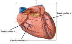
Anterior view. Great Cardiac. V follows LAD. Small Cardiac V. looks like it follows R. coronary A. |
|
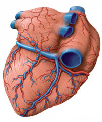
Label the relevant v. on this view of the heart. |
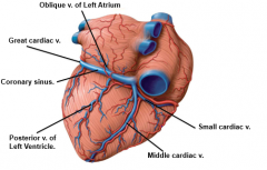
Coronary Sinus in coronary sulcus. Small Cardiac V. wraps around from the front. |
|
LABEL ALL THE THINGS |
use netters to check |
|
|
Pericardium innervation? |
Vagun N., sympathetic trunks, phrenic nn. phrenic nn=somatosensory so pericardial pain felt in supraclavicular region (Referred pain) |
|
|
Where do all the veins in the heart drain to? |
Coronary sinus which drains to R. atrium. |
|
|
What is the pattern of blood flow in the human heart (one sided pattern) |
I/S vena cava-->R. atrium--> tricuspid valve-->R. Ventricle-->pulmonic valve-->pulmonary artery-->lungs-->pulmonary vein (4)-->left atrium-->mitral valve-->left ventricle-->aortic valve-->aorta-->body |
|
|
Heart circulation (both sided pattern) |
1. Atria fill with blood 2. ventricles relax, AV valves open-->80% blood goes to ventricle 3. Atria pump-->left over 20% blood-->ventricles 4.ventricles contract-->pressure inc-->AV valves close, semilunar valves open 5. blood leaves-->ventricles relax-->semilunar valves close, AV valves open |