![]()
![]()
![]()
Use LEFT and RIGHT arrow keys to navigate between flashcards;
Use UP and DOWN arrow keys to flip the card;
H to show hint;
A reads text to speech;
19 Cards in this Set
- Front
- Back
|
what are the 4 different types of junctions between epithelial cells |
1. Zonula occludens (tightjunction) 2. Zonula adherens 3. Macula adherens (demosome, spotjunction) 4. Gap junction (nexus) |
|
|
What are tight junctions |
(zonula=band) Is the highest junction,which completely encircles the cell. It provides an impermeable seal betweenthe cells, seals cells together so that there is no intercellular space betweenthem at that part of the cell. functions by fusing the outer leaflets of the adjacent plasma membranes. |
|
|
what are zona adherens |
encircles the cell,providing some adhesion. Has a specific glycoprotein (A-CAM) in the cleftbetween the cells – functions by actin filaments inserting into dense plaques(myosin, tropomyosin etc) on the cytoplasmic surface |
|
|
what are desmosomes |
plaque likethickenings on membranes of adjacent cells, which form a thin dense line in themiddle of the intercellular space. Cytokeratin intermediate filaments meet together on plaque. Sites of attachment between cells and for the cytoskeleton. |
|
|
What are gap junctions |
close the parallelmembranes of adjacent cells but does not fuse them. Consists of intramembranous particles which consist of 6 sub-units arrangedaround a central pore – these particles attach to the particles on the adjacentcell (connected particles = connexons) This connection provides electrical coupling via ion flow between cells found in smooth cardiac muscles. They furthermore have a role of communication between cells as they providechannels for movement between them |
|
|
What are the 2 types of connective tissue and their subtypes and their properties |
Looseconnective tissue is flexible, and has a networkof fibers in between which many open spaces may exist. Examples are adiposetissue and areolar tissue underneaththe dermis. Denseconnective tissue has a far higher fiber content,and may be extremely hard due to mineralization and compaction. Denseirregular – resists tension from all directions -dermis of skin. Denseregular – parallel bundles of fiber resistsforce in one direction – tendons, ligaments. |
|
|
what are proteoglycans |
Part of the ground substance, they fill thespace between cells and fibres. Have a bottle brush shape with a core of protein (core down middle) andglycosaminoglycans (sugars coming off sides)Carbohydrates constitute approximately 80-90% of the mass Have a strong net negative charge due to phosphate and sulphate groups and asthus bind to many cations, usually Na+ and furthermore are intensely hydrated;proteglycans therefore hold water and allow movement of cells etc through themedium. Can sometimes aggregate and form a massive molecule. |
|
|
what is bone made of |
type 1collagen calcium hydroxynatite crystals (CaPO4) |
|
|
what are the three different bone formations |
woven compact/lamellar bone Cancellous/spongy bone |
|
|
what is woven bone |
disorganisedcollagenous arrangement, present in foetal bone, healing bone, anddiseased bone. |
|
|
what is lamellar/compact bone, what is it composed of and its constitutes functions |
it is strong bone that contains concentric rings of lamellae. It contains: Osteoblastsdeposit matrix in the form of lamellae (thin sheets). Collagen fibres withineach lamellae run parallel to one another and at oblique angles to each otherin different Haversian systems. Most individual lamellae form highly organised, concentric layers ofmineralised matrix around longitudinal canals within bone tissue (longtubes in the centre of the Haversian system = Haversian canal – containcapillaries and nerve fibres). These parallel cylindrical structures, which feature prominently, are Haversian systems/osteons, which are concentriclamellae (8-15) of mineralisedmatrix and run parallel to the long axis of the bone. Entombed in the lamellae are osteocyteswhich occupy spaces called lacunae andextend cytoplasmic processes/channels called canaliculi to communicated via gapjunctions with other osteocytes in other layers (is also a means ofdistributing nutrients, ions, metabolites, and oxygen to and from the Haversian canal) The Haversian Canal is lined by endosteum, is a neurovascular canal which passes centrally through the cavitywithin the concentric lamellae, running parallel to the long axis of the boneand serving to provide nutrient supply to the tissue. Volkmann’s canals run perpendicularto the Haversian canals, interconnecting osteons with each other and theperiosteum. The first few layers of lamellae immediately deep to the periosteum (adjacentto it) are the circumferential lamellae. Osteons are not permanent, but are continuallybeing reabsorbed whilst new bone is deposited. |
|
|
what is cancellous bone and what is its main purpose |
Branchingspicules or beams of bone that contain osteocytes in the lacunae. The bone lamellae are generallyarranged in parallel layers and not haversian systems. The bone matrix is mineralisedas in compact bone however has larger spacing between the bone – which isoccupied in turn by bone marrow. |
|
|
what are the 4 types of bone cells and their functions |
· Osteoprogenitor cells – flattened, mesenchymalcells located in the inner layer of the periosteum and endosteum that differentiate into osteoblastswhen new bone is being formed. · Osteoblasts - derived from osteoprogenitor cells. Synthesiscollagen (abundant RER). They lay down osteoid(unmineralised organic matrix) and then regulate (produce, initiate andcontrol) mineralization as a means of forming new bone, then becoming trapped in new bone as it lays the matrix around itself. · Osteocytes – modified osteoblasts which regulatenutrient/waste exchange as well as the composition of bone matrix. · Osteoclasts – Large, multinucleatedphagocytic cells, that are formed via the fusion of a monocyte and macrophage. They serve to reabsorb bone by releasing enzymes that breakdown collagen fibres of matrix and dissolve calcium matrices with acid. Found Howship’s lacunae (resorptioncavities) which remain in place after bone is degraded. |
|
|
What bones is intramembranous ossification used and what is its process |
Head bones, clavicle and thickening of some bones 1. At 7 weeks of gestation,primary ossification centres forms within condensations of mesenchymal tissue(undifferentiated loose connective tissue derived primarily from mesoderm)
2. Osteoblasts differentiated frommesenchyme make woven bone matrix (osteoid make of collagen fibres) which is subsequently mineralizedinto bone. 3. As osteoblasts are entombedby matrix, they then transform into osteocytes. Osteons form and woven boneis gradually transformed to lamellar bone. 4. Islands of developing boneare known as spicules – numerous in an ossification centre (cartilage betweenspicules contain growing blood vessels and haemangioblasts) 5. Bony spicules fuse. |
|
|
How does endochrondral ossification occur |
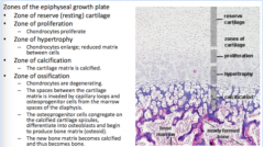
1. At 6 weeks, mesenchymal cells differentiate into chondroblasts, whichproduce a hyaline cartilage model, resembling adult bone shape. 2. Inner perichondrial cells of the shaft form a thin layer ofperiosteum which contains osteoprogenitor cells that mature intoosteoblasts (only form bone for appositional growth - width)Simultaneously Chondrocytes in the shaft undergo hypertrophy. 3. The region of hypertrophiedchondrocytes becomes calcified and the chondrocytes die. 4. Blood vessels invade the shaft centrally, bringing osteoprogenitorcells and restricting the proliferating chondrocytic cells to the endsof the cavity (epiphyses). (Invasion of periosteal bud) 5. Osteoblasts from invasion of the periosteal bud secrete osteoidalong the calcified matrix to produce cancellous bone. 6. Blood vessels and mesenchyme enter the upper epiphyseal cartilage andthe epiphyseal (secondary) ossification centre begins. (upper and lower butupper first) 7. The lower epiphyseal growthplate disappears. The upper disappears also - cartilage now onlypresent on the articular surfaces |
|
|
where are periosteal cells located and what are their function |
just beneath the periosteum they function in bone widening and fracture repair |
|
|
what are venous valves |
folds of endothelium with cusps |
|
|
where are venous valves important |
in perforating veins - veins which penetrate inwards into muscle fascia to connect the superficial veins to the deep veins - directly blood flow in one direction towards the heart |
|
|
what are venae comitantes and why are they important |
they are smaller veins that wrap around arteries they are important as they aid venous return upon the artery pulsations they also enable counter-current heat exchange between warm arterial blood and cooler veins |