![]()
![]()
![]()
Use LEFT and RIGHT arrow keys to navigate between flashcards;
Use UP and DOWN arrow keys to flip the card;
H to show hint;
A reads text to speech;
111 Cards in this Set
- Front
- Back
|
Components of tissue fluid |
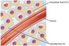
mostly water nutrients, dissolved gasses, and salts blood plasma without the plasma |
|
|
Function of tissue fluid |
bathes all tissues and moves freely through capillaries |
|
|
If tissue fluid is found in the lymphatic system it is called |
lymph |
|
|
Components of plasma |
1. dissolved proteins (albumins, globulins, fibrinogens), provides osmotic counterforce 2. glucose, electrolytes, dissolved gasses 3. hormones, neurotrasmitters, fats 4. clotting factors and waste 55% of blood volume |
|
|
What is serum |
blood plasma without clotting factors |
|
|
Where is blood found? What is blood made of? |
intravascular- confined to vessels and the heart plasma plus formed elements (blood cells) |
|
|
Define viscosity and relate the viscosity of blood to that of water. Define osmolarity |
Resistance to flow whole blood 5x more viscous than water. total molarity of dissolved particles |
|
|
Plasma is the liquid portion of blood. What are the three major categories of plasma proteins. Which is most abundant? What is their function |
1. albumins- contribute to viscocity and osmolarity, influence blood pressure, flow and fluid balance MOST ABUNDANT
2. globulins (antibodies) alpha, beta, globulins 3. fibrinogen - active form. precursor of fibrin threads that help from blood clots |
|
|
Kwashiorkor is an example of not enough of which blood protein |
albumins
|
|
|
Which of the plasma proteins are formed by the liver? |
albumins fibrinogen (not globulins, produced by B cells) |
|
|
How are blood types determined? |
glycoproteins and glycolipids
, |
|
|
Life span of RBC |
4 month life span |
|
|
Where are RBC produced |
stem cells in red marrow |
|
|
What is the major function of RBC? |
GAS TRANSPORT increased surface area/volume ratio 33% of cytoplasm of an RBC is Hemoglobin |
|
|
Erythrocytes produce what chemical? What it's function. |
Carbonic anhydrase (CAH) important role in gas transport and pH balance |
|
|
Describe the structure of Hg |
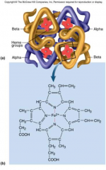
2A 2 B chains
4O2 molecules bind |
|
|
Define Hematocrit |
packed cell volume |
|
|
Normal Hematocrit for a male and a female |
42-52% male 37-48% female |
|
|
If a clinician wanted to measure how much oxygen a patients blood was able to carry, which lab values would be the most useful? |
Red blood cell count and HB |
|
|
What are the norm values for a male and females hemoglobin concentration |
men 13-18g/dL women 12-16 g/dL |
|
|
What are the norm values for a male and female Red Blood count |
men: 4.6-6.2 million/uL women: 4.2 - 5.4 million/ul |
|
|
What are the two reason that HCT, HB, and RBC count are lower in women? |
1. andgrogens stimluate RBC production (men have more) 2. women have periodic menstrual losses |
|
|
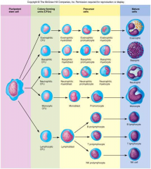
Function of hemopoietic tissues |
produce blood cells |
|
|
What is produced from each of the following hemopoietic tissues? Yolk Sac Spleen Red bone marrow |
Yolk sac: colonizes fetal bone, liver, spleen, and thumus Spleen: involved in WBC production Red bone marrow: -pluripotentstem cells -myeloidhemopoiesisproduces RBCs, WBCs and platelets |
|
|
What are the major areas where lymphoid hemopoiesis is distributed? |
thymus tonsils lymph nodes spleen peyers patches in intestine |
|
|
Myeloid pleuripotent stem cells give rise to which types of cells. |
RBC
WBC Platelets |
|
|
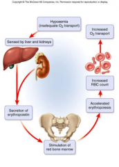
EPO Where is EPO produced. What is it's function? When is it released? |
Kidneys -stimulates production of RBC and stimulates angio genesis -released under hypoxic condition, increased exercise, loss of lung tissue (emphysema) |
|
|
Detail the negative feedback look for erythrocyte homeostasis. |
Negative feedback control. Drop in RBC count causes kidney hypoxemia. EPO stimulates bone marrow. RBC count up in 3-4d |
|
|
What is the key nutritional components needed for Erythropoiesis? |
Iron (has a low absorption of 10%. need 5-20mg/day) B12 and Folic Acid Vitamin C and Cu |
|
|
Where are erythrocytes recycled and disposed? What cells are of chief importance in this area? |
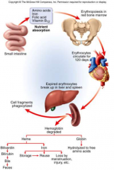
RBC lyse in narrow channels in the spleen Macrophages in the spleen digest RBC |
|
|
Describe how the RBC is recycled in the spleen. |
1. digested into membrane bits 2. heme and globin molecules are separated. 3. globins --> amino acids 4. Heme --> bilieverdin --> bilirubin (yellow) --> released into blood plasma of the kidney. Liver secretes bilirubin into bile. Concentrated via the gall bladder and released into small intestine. Urobilinogen makes poop brown. |
|
|
Color biliverdin |
green |
|
|
color bilirubin |
yellow |
|
|
color urobilinogen |
brown |
|
|
disorder resulting in excess RBC |
polycythemia |
|
|
Which type of polycythemia is a result of cancer of the erythropoetic cell line in bone marrow? |
Primary polycythemia Increases viscosity b/c too many RBC RBC: 11million/uL HCT 80% |
|
|
Which type of polycythemia is a result of dehydration, emphysema, high altitude or physical conditioning? |
Secondary RBC: 8 million/UL |
|
|
Describe the dangers of polycythemia |
increased blood volume pressure viscocity CAN lead to: embolism, stroke, heart failure |
|
|
Disorder characterized by the reduced oxygen capacity of blood? |
Anemia |
|
|
Below is a list of parameters that are affected by anemia. Describe each one. HCT HG Fe |
-Low HCT: hypoplasia: not enough RBC -Low HG: normal number of cells but each cell does NOT carry enough HG -Low Iron levels -can be a result hemorrhage -hemolysis- too much lysis or poison |
|
|
A microcytic anemia is likely due to? Describe microcytic |
iron deficiency small RBC |
|
|
A macrocytic anemia is likely due to? 2 Describe macrocytic |
alcoholic and COPD large RBC |
|
|
Describe three reasons that a pt may have inadequate erythropoiesis or hg synthesis which results in anemia. |
- low B12 or intrinsic factor (pernicious anemia) -iron-deficiency -kidney failure and insufficient EPO -aplastic anemia- deficiency in RBC- production stops |
|
|
The three major causes of Anemia. |
1. inadequate erythropoiesis or hg synthesis 2. hemorrhagic anemia 3. hemolytic anemias (poison) |
|
|
The three major effects of anemia are
|
-Hypoxia and necrosis (SOB and lethargy) -low blood osmolarity (tissue edema) -low blood viscosity (heart races and pressure drops) |
|
|
What are the conditions in which a person with sickle cell anemia will have sickling? |
hypoxia |
|
|
A person with sickle cell has chronic hypoxemia that reactivates hemopoietic tissues. The net result of this will be. |
1. spleen enlargement 2. bones of cranium enlarged |
|
|
An autosomal recessive disorder that produces defective hemoglobin, microcytic anemia, low Hg, low HCT and is found in mediterranean populations |
Thalasemia |
|
|
The two disorders that confer resistance to malaria |
sickle cell thalasemia |
|
|
Unique molecules on the surface of a cell |
Antigens |
|
|
_______ are secreted by plasma (B) cells and act as part of immune response to foreign matter |
antibodies |
|
|
The antibody and antigen molecule binding together and clumping |
agglutination |
|
|
At what age do antibodies appear in a child's blood? |
2-8 months after birth maximum concentration at 10yr * you do not form antibodies against your own antigens *antigen/antibody agglutination is responsible for mismatched transfusion reactions |
|
|
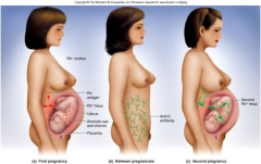
A disorder that occurs if mother has formed antibodies and is pregnant with 2nd Rh+ child. How is this prevented in a Rh- mom that is on her second Rh+ kid? |
Hemolytic Disease of the Newborn give RhoGAM |
|
|
Another term for WBC Nucleus? fx? how long live? |
Leukocytes conspicous nucleus protect against pathogens long lived amoebic movement |
|
|
Identify each of the following as either Granulocyte or agranulocyte neutrophil lymphocytes basophil monocytes eosinophils |
Granulocytes: neutrophils (60-70%) eosinophils basophils agranulocytes lymphocytes monocytes |
|
|
Function of neutrophils when are they increased? |
phagocytosis of BACTERIA release antimicrobial chemicals increased in bacterial infections |
|
|
Function of eosinophils when are they increased? |
phagocytosis of antigen/AB complexes & allergen & inflammatory chemicals release enzymes to destroy parasites increased during parasitic infection or allergies! |
|
|
Function of basophils when are they increased? |
1. secrete histamines (vasodilator) 2. secrete heparin (anticoagulant) increased during chicken pox, sinusitis, diabetes |
|
|
Function of lymphocyte when are they increased |
1. destroy cells (cancer, foreign, and virally infected cells-- SPECIFIC immune response 2. "present" antigens to activate other immune cells 3. coordinate actions of other immune cells 4. secrete antibodies and provide immune memory |
|
|
Function of monocyte when are they increased |
1. differentiate into macrophages 2. phagocytize pathogens and antigens 3. present antigens to activate other immune cells increased during viral infection and inflammation |
|
|
Three types of lymphocytes and function. What is their half life? |
T-cells - identify foreign antigens, present antigens to B cells, produced by thymus gland. Killer T-cells: destroy foreign cells directly B-cells: produce custom antibodies, become memory B cells 30 year half life |
|
|
Monocytes are circulating precursors to macrophages. Macrophages can be found in 4 distant localities, which are |
1. dendritic cells ( epidermis, oral mucosa, esophagus, vagina, lymphatic organs) 2. migroglia (CNS) 3. alveolar macrophages (lungs) 4. hepatic macrophages (liver) |
|
|
Name the 6 components of a CBC |
1. hematocrit 2. hemoglobin concentration 3. total count of RBC, reticulocytes, WBC, platelets 4. differential WBC count 5. RBC size (microcytic or microcytic) 6. hemoglobin concentration per RBC |
|
|
Each of the following pluripotent stem cells form leukocytes. Indicate what they form. myeloblasts (3) monoblasts (1) lymphoblasts (3) |
myeloblasts - Nuetrophils, basophils, eosinophils monoblasts- monocytes lymphoblasts- B and T lymphocytes, NK cells |
|
|
Where do T lymphocytes complete development? |
thymus |
|
|
What does red bone marrow store and release? |
granulocytes monocytes |
|
|
How long to circulation WBC stay in the blood stream? |
1. granulocytes leave in 8 hr live 5 days (N,B,E) 2. monocytes leave in 20 hr --> macrophages --> several years 3. WBCs provide long term immunity for decades |
|
|
A disorder with low WBC count that can be caused by radiation, poison (chemo), infectious disease. |
leukopenia |
|
|
A disorder with high WBC count that is caused by infection, allergy, and disease. |
Leukocytosis *differential count will tell you type of each leukocyte |
|
|
A cancer of the hemopoietic tissue. Both myeloid and Lymph which leads to uncontrolled WBC production. |
Leukemia can lead to normal cell % change impaired clotting |
|
|
Small fragment of megakaryocyte cytoplasm |
platelets |
|
|
The four functions of platelets are |
1. secrete clotting factors and growth factors for vessel repair
2. initiate formation of clot-dissolving enzyme 3. phagocytize bacteria 4. chemically attract neutrophils and monocytes to sites of inflammation |
|
|
Thrombopoiesis is the |
formation of platelets |
|
|
The stem cells that has repeatedly replicated DNA without diving cytoplasm that eventually forms a gigantic cell which remains in the bone marrow and is involved in thrombopoiesis |
Megakaryoblasts
BIast- BIG |
|
|
The infolding of cytoplasm splits off cell fragments that enter bloodstream as platelets which can also be stored in spleen |
megakarycocyte |
|
|
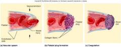
Three pathways of hemostasis |
1. Vascular Spasm 2. platelet plug formation 3. coagulation |
|
|
Which hemostasis pathway is activated by pain receptors that can directly innervate constrictors and only lasts for a short time. |
Vascular spasm. |
|
|
Which hemostasis pathway can be activated by smooth muscle injury which causes platelets to release serotonin which causes vasoconstriction. |
Vascular spasm |
|
|
The vascular spasm provides time for the other two clotting pathways which are? |
platelet plug formation and coagulation |
|
|
Describe the platelet plug formation cascade. |
|
|
|
The most effective defense against bleeding is |
clotting |
|
|
Describe the coagulation cascade |
1. plasma proteins --> fibrinogen --> insolube fibrin threads --> clot formation Clotting factors are in the plasma. Activate one to turn on cascade |
|
|
Differentiate between the intrinsic and extrinsic coagulation pathway. |
1. extrinsic: factors released by damaged tissue begin cascade 2. extrinsic: factors in blood begin cascade (platelet degranulation) |
|
|
Outline the cascade that leads to the completion of coagulation |
activation of factor X (liver production) --> production of prothrombin activator Prothrombin ----PTA------> thrombin Fibrinogen ------T------> fibrin Thrombin speed up formation of PTA Positive feedback loop. |
|
|
How long does it take a clot to retract? |
30 minutes |
|
|
Platelet derived growth factor is secreted by platelets and endothelial cells. What does PdGF do? |
it is a mitotic stimulant for fibroblasts and smooth muscle to multiply and repair damaged vessel |
|
|
Dissolution of a clot is called |
fibrinolysis |
|
|
The enzyme that is produced by the liver and is converted into plasmin, a fibrin-dissolving enzyme (clot buster). |
Plasminogen tPA = tissue plasminogen activator |
|
|
A genetic disorder described as lacking of a clotting factor that is sex-linked recessive |
Hemophilia |
|
|
Hemophilia A is missing what clotting factor? |
8 MC 83% |
|
|
Hemophilia B is missing what clotting factor? |
9 MC 15% |
|
|
Treatment for hemophilia |
transfusion of plasma or purified clotting factors or factor 8 by transgenic bacteria |
|
|
A coagulation disorder when a clot is caught in a vessel. |
Embolism |
|
|
A coagulation disorder with an abnormal clot in unbroken vessel most likely caused by a DVT. Can turn in to a PE |
Thrombosis
|
|
|
If a clot blocks blood supply to an organ it is called |
Infarction |
|
|
A disorder that results in delimitation of arteries. |
arterial dissection can be idiopathic, spontaneous, traumatic |
|
|
Most common vessels for an arterial dissection |
Carotids, Vertebrals, Aorta |
|
|
What is the function of a thrombylosis? |
actively dissolve a clot tPA by activating Plasmin --> plasminogen = no more clot |
|
|
Two examples of anticoagulants |
heparin and coumadin |
|
|
The disorder where there is a pathological activation of clotting mechanisms that results in exhaustion of clotting factors. |
DIC patient BLEEDS out DEATH IS COMING caused by vascular damage, sepsis, cytokines |
|
|
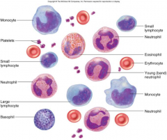
The formed elements of blood |
|
|
|
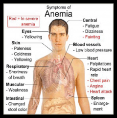
Symptoms of Anemia |
|
|
|
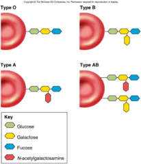
Blood Types |
|
|
|
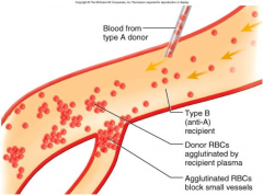
Transfusion Reaction |
|
|
|
Rh factor and Pregnancy |
|
|
|
Leukopoeisis |
|
|
|
Types of Hemostatis |
|
|
|
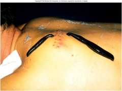
Medicinal Leeches |
|
|
|
Tissue fluid is |
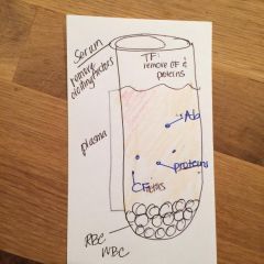
serum without plasma proteins ** Plasma has CF, proteins, SB **serum take out CF **tissue fluid take out plasma proteins |
|
|
Explain the relationship of osmolarity to BP and Edema. |
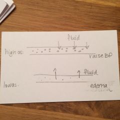
cause fluid absorption into blood RAISE BP |