Reading...
![]()
Play button
![]()
Play button
![]()
Use LEFT and RIGHT arrow keys to navigate between flashcards;
Use UP and DOWN arrow keys to flip the card;
H to show hint;
A reads text to speech;
38 Cards in this Set
- Front
- Back
|
The Spinal Cord fxn
|
• Spinal column protects the spinal cord
|
|
|
• Intervertebral disk between vertebral bodies for cushioning lead can be the basis of what injury
|
o Herniation of intervertebral disks is common
o Lateral disk prolapsed (C3) compressing root Can cuase disesthesia if on the dorsal root or muscle weakness if on the ventral root |
|
|
Cauda Equina:
|
is a bundle of dorsal & vental roots occupying the spinal canal below L2
|
|
|
Filum Terminale
What nerves go through the filum terminale?? |
is a delicate strand of fibrous tissue ~20cm long from the apex of the conus medullaris to the 1st coccygeal certebra (Co1) **NOT A NERVE!!!!!!
NO NERVES |
|
|
lumbar puncture
|
• Lateral decubitus or siing position
o Between L3 and L4 o Spinal cord ENDS at L2 o Use sacral anesthesia o Use the level of the ilac crests as a guide to find the correct spot for the puncture Cannot puncture just nerves-->like spaghetti |
|
|
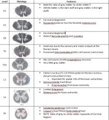
o The spinal cord is divided into 31 segments that are numbered according to their location with respect to vertebral level
|
o Cervical Nerves (1-8)
Innervate neck, shoulders, upper extremities Cervical Enlargement due to innervations of the upper extremities o Thoracic Nerves (1-12) Innervate truck and arms Enlarged intermediolateral horn o Lumbar nerves (1-5) Innervate hip region, lower back, feet and all lower limbs Lumbosacral enlargement for lower extremities Can get Sciatica from here o Sacral Nerves (1-5) Innervate posterior surface of the lower limbs |
|
|
• Spinal Cord Development
|
o Neural groove and neural plate begin invagination
o Neural crest cells form from ectoderm near the invagination o Neural crest cells form the DRG, skin begins growing over the neural tube that has completed formation o Alar plate(BMPs) -sulcus limitans(boundary b/w sensory and motor) - basal plate(SHH) differentiate (eventually separates the sensory (alar) and the motor (basal) fibers o Marginal layer (future white patter) forms on the outside of the spinal cord, - central canal is in the middle -the mantle layer (future gray matter, cell bodies) is at the bottom |
|
|
o General organization of Gray Matter
|
Rexed’s Laminae: I-IX & Area X
Gray matter is divided into 9 different layers (1-8 and X) X has to do with pain transmission 2,3 is substantia gelatinosa: pain, temp and touch 9 are motor neurons (DON’T FIT LAMINAR DISTRIBUTION) • Divided into those that innervate the appendicular skeleton |
|
|
Gray matter remanant of
|
-neural tube
|
|
|
o Somatotopic Organization of White Matter
|
o Sensory cortex is somatotopically oriented
o Dorsal columns are divided into two parts: MEDIAL: Gracile Fasciculus (Sacral, Lumbarmedial) DISTAL: Cuneate fasciculus (Thoracic, Cervicallateral) o Spinothalamic Tract: somatotpic distribution (OPPOSITE the dorsal column) o Lateral Corticospinal Tract: sends axons from the motor neurons, arranged in the somatotopic organization |
|
|
|
|
|
• General Organization of the Somatic Sensory System
|
o Pseudounipolar neurons in the DRG transmit information from peripheral receptors to the spinal cord
DRG=SENSORY -Peripheral process extends into periphery -Central process projects from DRG neurons into the spinal cord Proprioception -Ascend via dorsal column, synpase in cuneate -Fast Pain and temperature -slower than red -Synapses -CROSSES THE SPINAL CORD -ASCENDS UP THE SPINOTHALAMAC TRACT. |
|
|
• Dermatomes
-Receptive Field |
The cutaneous area supplied by a single dorsal root and its ganglion
o The border zones of successive dermatomes overla o Receptive Field: region of receptive surface that causes a sensory nerve cell to respond |
|
|
• Mechanoreptors of the SKIN (All of these except for the free nerve endings are in the MEDIAL division of the dorsal root)
|
o Pacinian Corpuscle: Touch and Vibration
o Meissner Corpuscle: Light touch o Ruffini’s Corpuscle: Stretch, signaling for position of fingers o Merkel’s Disks: Pressure o Free Nerve Endings: Pain |
|
|
• Spinal Sensory Systems
Posterior (Dorsal) Column System |
This pathway subserves FINE TOUCH and KINESTHESIS (position of limbs in space)
Axons arise uncrossed in the DRG and synapse in the nucleus cuneatus (upper body) or nucleus gracilis (lower body of the medulla) Axons of nucleus cuneatus and nucleus gracilis form the medial lemnicus (descussate in the medulla) and ascend to the thalamus (VPL) Thalamic projections synapse in the post central gyrus of the cortex |
|
|
• Spinal Sensory Systems
o Spinothalamic Tract (ALS |
Subserves TEMPERATURE, GROSS TOUCH & PAIN
Axons cross at or near the level they enter the spinal cord Ascend in the Lateral and Ventral (anterior ) funiculi |
|
|
• Spinal Reflexes:
o Descending motor input to the spinal cord: |
Pyramidal System: Corticospinal Tract
• Regulates voluntary movements Extrapyramidal System: • Rubrospinal Tracts: from basal ganglia, cerebellum and red nucleus • Tectospinal Tract: Coordinates head and eye movements • Reticulospinal Tract: Regulates muscle tone, particularly in antigravity muscle • Vestibulospinal Tract: regulates head position and posture |
|
|
• Spinal Reflexes
o Upper Motoneuron |
A motoneuron with the soma and axon entirely within the CNS
Directly or indirectly innervates LOWER motoneurons |
|
|
• Spinal Reflexes
o Lower Motoneuron: |
Typically an alpha-motoneuron whose axon innervates skeletal muscle
Soma in CNS, but axon leaves CNS and enters PNS |
|
|
• Spinal Reflexes
o Spinal Control of Movement: |
Local circuit neurons (Reflex coordination)Motor neuron pools (LOWER motor neurons)SKELETAL MUSCLES
|
|
|
• Spinal Reflexes
o The Motor Unit -innervation? size? fiber types? recruit more motor units? different motor units? |
= Muscle + the ONE nerve that it is innervated by
All muscle fibers innervated by at least one α-motoneuron • Most muscles are innervated by ONE nerve with fibers scattered all over the motor unit Motor units vary in size Muscle fiber type in a motor unit is constant • Slow motor units • Fast-fatigue resistant • Fast fatigable (Major FORCE generating fibers) With more effort, you recruit more motor units • The units become bigger with more effort because if the units were all the same size then the curve would be linear. • Small Motor Units: Fine delicate movements o Fewer muscle fibers per unit o Muscles of the hand, extraocular muscles, tongue muscles • Large Motor Units: Coarse movements o Many muscle fibers per unit o Biceps, gluteus, quadriceps |
|
|
Spinal Reflexes
are__ and reletively__ composed of? -graduation reflex = |
o Reflexes are nonvolitional, relatively fixed (potentially graded) biological responses evoked by specific stimuli
Composed of Afferent (SENSORY) INPUT and Efferent (MOTOR) RESPONSE Gradation of reflex refers to intensity of reflexive expression related to the magnitude of the stimulus or neural mechanisms outside the reflex |
|
|
Spinal Reflexes:
spinal cord is ___ of the ___ and final ___ motor coordination links? Basic form for motor coordination is ? which is defined by? occur where? actions? |
o The spinal cord is the most basic neural component of the motor system and the final decision point for controlling motor activity
Motor Coordination links the contractions of independent muscles so they function together • The most basic form of motor coordination is the reflex o Stereotyped response to a specific sensory stimulus of a body part o Simple reflexes have their neural circuitry WITHIN the spinal cord Such reflex responses can either promote movements (phasic muscle contractions) or maintain postures (tonic muscle contractions) |
|
|
o Receptors involved in movement:
|
Joint Afferents: located in the JOINTS and are most sensitive to extremes in the joint ankle (POSITION)
Muscle Spindles: located in the MUSCLE, very impt AFFERENT for POSITION and movement (VELOCITY) Golgi Tendon Organs (GTO): located in the MUSCLE TENDON, detect TENSION (FORCE) Tactile Afferents: touch receptors in the MSUCLE and OVERLYING SKIN |
|
|
o STRETCH REFLEX SEQUENCE (AKA Myotatic reflex, Jaw Closing Reflex, Knee Joint Reflex):
- simplest reflex? -receptor? -afferent limb? effector neuron? -Efferent limb? |
Simplest reflex is the STRETCH, MYOTATIC or MONOSYNAPTIC
• Receptor: records the stimulus and translates it into action potentials (Muscle Spindle, GTO) • Afferent Limb: conducts action potentials to the spinal cord via DRG, synapses in gray matter o Effector neuron: receives afferent signals, may adjust the impact of the signal depending on other inputs impinging on the neuron, integrates these messages and determines the appropriate response • Efferent Limb: axons conducting the output from the effector neuron to the target cell via the VRG o The effector is a target cell that mediates the response to the sensory stimulus (The muscle cell) |
|
|
The Muscle Spindle:
initiates? arranged? activating causes? maintians? |
• This SENSORY RECEPTOR initiates the stretch (deep tendon) reflex
• Arranged in PARALLEL with the muscle fibers • Activating the muscle spindle (STRETCH) increases firing in the Iα-afferents o Iα afferents increase activity of α-MN’s that innervate the same muscle (homonymous) o Iα afferents can inhibit antagonistic muscles • Maintains MUSCLE TONE, detects the stretch on the muscle fibers |
|
|
How to maintain spindle input?
o Gamma (γ)-loop reflex (Spindle + γ MNs) -maintain and innervate what? -y-MNs innervate and regulate what? -allows for what? Stretch reflexes also initate? which is assoicated with? Clinical Correlation : fasicular twtiches at rest--? -similar sx present with? |
o Gamma (γ)-loop reflex (Spindle + γ MNs)
Maintain sensitivity of spindle during contraction of muscle, innervates muscle fibers within the muscle spindle γ-MN’s innervate intrafusal muscle fibers (33% of VENTRAL root axons are from γ-MNs) γ-MN’s regulate the gain of the stretch reflex by adjusting the level of tension in intrafusal muscle fibers of the muscle spindle Allows for gradation of a muscle response o Stretch reflex also initiates RECIPROCAL INHIBITION Homonynous and synergistic muscles stimulated, BUT Antagonistic muscles inhibited (Interneuron) o Clinical Correlation (Twitches, Spasms, Cramps) Fascicular twitches at rest, if pronounced and combined with muscle weakness or atrophy, usually signify MN disease (ALS, progressive bulbar palsy) Similar symptoms present with tumor, ventral root injuries (herniated disk), syringomyelia (progressive degenerative spinal disease) |
|
|
o Golgi Tendon Organ (GTO)
are? detect? encode? respond to? Receptor is reponsible for? -in ___ with ___ Have what type of endings and where? Activating GTO activates what kind of fibers these fibers act by? only respond to? |
o Golgi Tendon Organ (GTO)
GTO are STRETCH RECEPTORS located within the TENDONS • Detecting the amount of stretch exerted by the muscles on the bones to which they are attached • Encode degree of stretch by the rate of firing • Don’t respond to length, but to how HARD THE MUSCLE IS PULLING (Responds to Contraction) Receptor is responsible for Tension Reflex • GTO are in SERIES with the EXTRAFUSAL MUSCLE FIBERS (outside of the motor unit) Encapsulated endings at the juncture of the muscle and tendon Activating the GTO (shortening) of muscle activates Iβ Afferent fibers • Iβ fibers INHIBIT α-MN to homonymous and synergistic muscles • Iβ can ACTIVATE ANATGONIST muscles • Doesn’t respond to stress, only Contraction |
|
|
Comparison of stretch and tension reflex:
Inverse myostatic reflex: Myotatic Reflex: Need Both? |
• Inverse Myotatic reflex: organized and regulated by the GTO, involves an inhibitory motor neuron
o SHUTS OFF THE SIGNAL TO CONTRACT • Myotatic Reflex: single synapse that causes the muscle to contract • NEED BOTH TYPES FOR A GRADED RESPONSE!!! o These are strictly at the level of the spinal cord |
|
|
Negative Feedback Regulationg of Muscle Tension by GTO
IB afferents synapse: IB inhibitory interneurons: |
• Iβ afferents synapse on INHIBITORY interneurons
o DECREASE activity in α-MN’s innervating the same muscle • Iβ inhibitory interneurons receive input from other sensory fibers & descending pathways o ****PREVENTS MUSCLES FROM GENERATING EXCESSIVE TENSION |
|
|
Withdrawal or FLEXOR REFLEX
-involves? -graded? -reflex modified? -local sign? -is used for? which uses? |
Withdrawal or FLEXOR REFLEX
• Involves SEVERAL Spinal Segments • Is exquisitely graded • Reflex is modified as appropriate • LOCAL SIGN: Modification of reflex such that it reflects the area being stimulated • This is a PROTECTIVE reflex that uses MULTISYNAPTIC RELAYS to maintain pressure • Reciprocal Innervation: Pain activates nociceptors o On the side IPSILATERAL to the stimulus Flexors Excited Extensors inhibited • Double Reciproval Innervation: o Produces the OPPOSITE pattern of muscle activation on the CONTRALATERAL SIDE Flexors inhibited Extensors excited |
|
|
Spasticity:
-composed of? -damage to? characterized by? -caused by? suggests? |
o One of a number sequelae of the UPPER MOTONEURON SYNDROME
Damage to DESCENDING motor pathways Characterized by: • Increased muscle tone, Hyperactive stretch reflex • Clonus: an oscillatory motor response to muscle stretching Caused by: REMOVAL of inhibitory descending influences exerted by the cortex and postural centers (Vestibular nuclei & reticular formation) Improved by lesions to vestibular nuclei (animal models) and deafferentation (REMOVAL OF DRG) in animal and human subjects • Suggests that spasticity represents an abnormal increase in afferent gain |
|
|
• ParesisParalysis
sequelae of? dammage to? characterized by? |
• ParesisParalysis
o Sequelae of the LOWER MOTONEURON SYNDROME Damage to VENTRAL HORN or Cranial MN’s Characterized by: • Paralysis (loss of movement) • Paresis (weakness) • Areflexia (loss of reflexes) • Loss of muscle tone & atrophy of muscle mass • Fibrillations and Fasciculations (spontaneous twitches) o Altered excitability of muscle fibersFibrillation o Abnormal activity of injured α-MN (motor units)Fasciculation |
|
|
Conlcusions
spinal reflexes are initiated by? which activate? and then effect? |
1. Spinal reflexes are initiated by sensory stimuli that activate the receptors in muscle joints and skin
a. These stimuli activate the neuronal network in the spinal cord that affects a specific group of muscles |
|
|
2. Spinal reflex networks perform THREE MAIN FUNCTIONS
|
a. Control of individual muscles
b. Coordination of muscle action around a joint c. Coordination of muscles at different joints |
|
|
3. Interneurons are important elements of spinal networks:
mediate? constitute? consists of? |
a. Mediate influences of sensory input upon motoneurons
b. Constitute the networks generating complicated patterns (reverberating network, rhythm generator) c. Three groups of inhibitory interneurons ( Iα, Iβ, and Renshaw Cells) coordinate muscle action around a joint |
|
|
4. Spinal reflex circuits
-provide? from? to? such as? and? |
provide higher centers with a set of elementary patterns of coordination from relatively simple combinations like reciprocal innervations at a single joint, to more complex spatial patterns of movement such as flexion reflex, and temporal patterns as in the scratch reflex.
|
|
|
5. Spinal reflexes are not entirely stereotyped but rahter...
reaches where and how? high cneters can? and produce? |
but rather are adapted to the initial position of the body segments and the external loads acting to oppose movement.
a. This info reaches lower levels directly b. Higher centers can activate these reflex circuits to produce VOLUNARY MOVEMENTS and need not be concentrated with the details of shaping the movement patterns to current circumstances. |