![]()
![]()
![]()
Use LEFT and RIGHT arrow keys to navigate between flashcards;
Use UP and DOWN arrow keys to flip the card;
H to show hint;
A reads text to speech;
27 Cards in this Set
- Front
- Back
|
Epidural Space |
Epi = "above" Dural = "dura mater" ∴ the space in the spinal cord outside the dura mater * filled with fat |
|
|
Cauda Equina |
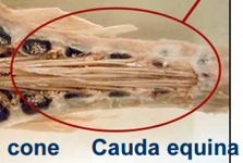
Collection of spinal roots that stream caudally from end of spinal cord and occupy vertebral canal
|
|
|
Medullary cone |
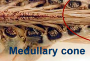
Spinal cord tapers to a point
Interarcuate space: - lots of CSF - good spot to take CSF sample or administer blocks (except in horse, give block lower) |
|
|
What does a spinal nerve do? |
Innervates the trunk elements |
|
|
What is a plexus nerve? |
Innervates the limbs Plexus = "braid" |
|
|
Cervical Intumescence |
Thickening of spinal cord = Brachial plexus - complex arrangement of the spinal nerves |
|
|
Lumbar intumescence |
Thickening of spinal cord = Lumosacral Plexus - complex arrangement of the spinal nerves |
|
|
Order of Meninges in spinal cord |
Outermost to innermost: *Epidural space (not a meninge) Dura mater (toughest) *Subdural space (vascular) Arachnoid mater (looks like spiderwebs, they trickle into the arachnoid space) *Subarachnoid/Arachnoid space (CSF found here) Pia Mater (adhered to brain/cord) |
|
|
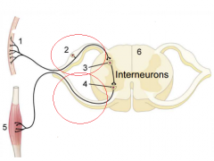
Top red circle = dorsal root (afferent/sensory) Bottom red circle = ventral root (efferent/motor) 2 = dorsal root ganglion 3 = synapse of interneuron in dorsal horn 4 = synapse at efferent neuron in ventral horn
afferent neuron has long dendrites and long axon motor neuron has short dendrites, long axon to effector
*both are myelinated in the PNS |
|
|
What is a funiculus? |
Spinal cord tract - groups of fibres that run cranial and caudal to go to different regions and carry different interneurons in the spinal cord
Division of white matter into portions |
|
|
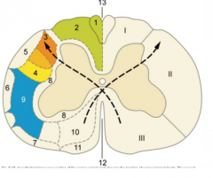
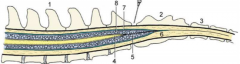
I = dorsal funiculus II = lateral funiculus III = ventral funiculus
dotted black lines = shows signals crossing into contralateral parts *must cross the central canal (bridge between the two portions) - to do this, there must be cell bodies in the central canal (i.e. interneurons!)
12 = ventral median fissure |
|
|
Where do spinal nerves exit the spinal cord? |
Intervertebral foramena (between two vertebrae) |
|
|
How are spinal nerves named? |
For the vertebrae that is cranial to them. I.e. the T1 segmental nerve is behind (caudal) to the T1 vertebrae
*EXCEPT for cervical, they are named for the vertebrae CAUDAL to them (i.e. C1 nerve is in front of C1 vertebrae, and we have a C8 nerve after the C7 vertebrae) |
|
|
Sympathetic Ganglion Chain (trunk) |
Runs between all the spinal nerves (in a way "connecting" them), parallel to the spinal cord |
|
|
Order of branching of spinal nerves |
Starting inside spinal cord:
Dorsal horn → Dorsal root → Dorsal root ganglion → unit to form spinal nerve → dorsal ramus (innervate epaxial muscles) *both sensory and motor innervation
Ventral horn → ventral root → unite to form spinal nerve → ventral ramus (innervate hypaxial muscles i.e. everything else) *both sensory and motor innervation
Also have branch off spinal nerve for ANS (both paraysmpathetic and sympathetic) |
|
|
How is a spinal nerve a mixed nerve? |
Has both efferent and afferent fibres Has both somatic and visceral fibres
2 sets afferent and efferent 2 set visceral and somatic |
|
|
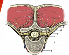
1 = spinal cord (dorsal and ventral horns) 2 = dorsal root 3 = dorsal root ganglion 4 = ventral root 5 = united spinal nerve trunk 6 = dorsal branch of spinal nerve (small) 7 = ventral branch of spinal nerve (much larger) 8 = body of vertebrae 9 = sympathetic trunk 10 = expaxial muscles |
|
|
Is there an epidural space in the brain? |
No! Only in the spinal cord |
|
|
Tentorium Cerebelli |
Invagination of tough dura mater into area between cerebrum and cerebellum
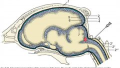
"tent over the cerebellum" = #7 |
|
|
Dura mater |
Outermost meninge in the brain Also connected to the cranial bone as periosteum |
|
|
Cerebellomedulary cistern |

Area between cerebellum and medulla oblongata (where the star is)
*Can collect CSF from here
|
|
|
Denticulate ligaments |
Extend through the arachnoid to the dura mater (i.e. they are below dura mater) *anchors spinal cord to limit side to side movement
- alternate with dorsal roots
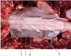
2= dorsal roots 4 = denticulate ligaments |
|
|
Filum Terminale |
#5 = filum terminale - Fibrous strand of pia mater extending past conus medullaris - gives longitudinal support to spinal cord |
|
|
Arterial supply to spinal cord |
Ventral spinal artery (largest, follows ventral fissure, mostly supplies grey matter) 2 Dorsolateral arteries (smaller, mostly supply white matter)
*outside of the vertebral canal itself
Also have some regional arteries that enter through intervertebral foramen |
|
|
Venous drainage of the spinal cord |
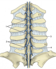
Plexus
- Join, cross over and join with bigger trunk veins such as azygous, caudal vena cava etc
1 = internal vertebral plexus 2 = intervertebral veins |
|
|
Where is CSF produced? |
In the choroid plexusi of the ventricles (one choroid plexus in each lateral ventricle and a pair in the 3rd and 4th ventricles |
|
|
CSF Circulation |
From choroid plexi in lateral ventricles → interventricular foramina → 3rd ventricle (diencephalon, surrounds thalamus) → mesencephalic aqueduct → 4th ventricle (in the hindbrain)
From 4th ventricle, CSF may go down central canal in spinal cord (they have direct communication), or circulate around subarachnoid space
CSF may enter cerebellomedullary cistern, lumbar cistern (caudel end of spinal cord) or drained into venous systems by microvilli in dorsosagital sinus (help regulate pressure of CSF)
|