Reading...
![]()
Play button
![]()
Play button
![]()
Use LEFT and RIGHT arrow keys to navigate between flashcards;
Use UP and DOWN arrow keys to flip the card;
H to show hint;
A reads text to speech;
21 Cards in this Set
- Front
- Back
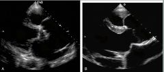
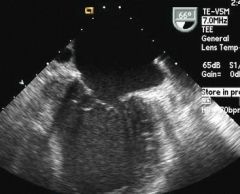
What is this
|
AI
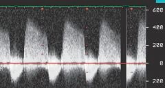
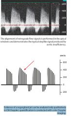
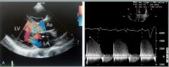
The alignment of retrograde flow signals is performed in the apical windows and demonstrates the typical steplike signal produced by aortic insufficiency. |
|
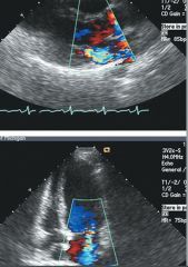
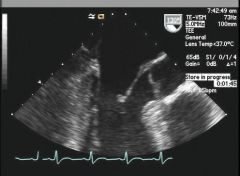
What does this DSE show?
|
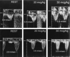
to assess severity of AS, the top panels are LVOT the bottom are AS. Gradient goes up to 3.8 with peak stress
|
|
|
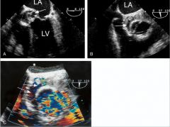
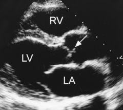
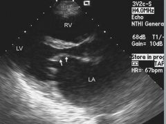
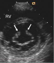
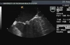
What does this show?
|
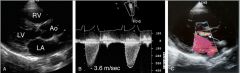
subaortic memrane. doppler reveals pk grad of 3.6 approx 50 mmhg, mild AR present
|
|
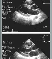
What does this show?
|
subaortic memrane. doppler reveals pk grad of 3.6 approx 50 mmhg, mild AR present
|
|
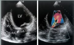
What is this?
|
Dilated AR, a severely dilated AR in the setting of prosthetic valve number A.
Number B. is the same degree of dilation in a patient with Marfan's syndrome, A lot of AR is present in both cases |
|
What is going on here?
|
Pt has a medtronic aortic prosthesis (stentless) who developed an aortic root abscess.
A. echo free space surrounding the aortic valve. B. the same on short axis view. C. Color doppler demonstrates flow within the abscess cavity and evidence of perivalvular regurgitation. |
|
What is likely happening here and what needs to be done
|
Seeing severe AR and chronic dilation and volume overload, now with severe LV dysfunction, chamber becomes spherical.
|
|
Describe this
|
Severe AR, with jet width and jet length..
|
|
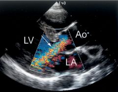
What is this trying to show?
|
An example of acute AR. Color doppler demonstrates severe AR. THERE is evidence of diastolic MR due to high diastolic left end dias pressure. B. the continious wave doppler is also consistent with severe based on the slope of the jet.
|
|
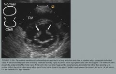
What is going on here?
|
lambl's excrescence
|
|
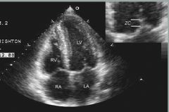
Quick choose which one is systole and which one is diastole. Second describe the zona coapta
|
Diastole on top, Systole on bottome. the zona coapta is usual 2-3 mm
|
|
Describe this and is this normal?
|
4 chamber, apical view showing the mitral valve in systole. the zona coapta is about 4mm here. note that the mitral leaflets do not typically close tip to tip but along the 4 mm length
|
|
label the leaflets
|
left to right: is P3, A2, P1
|
|
Describe this and what this is likely from and how to treat
|
likely Rheumatic mitral disease with hockey stick appearance to the anterior leaflet.
The Block score is used to treat below the valve calcification leaflet calcification calcfication kinesis of the valve Scores are 0-4 for each, max score is 16, score of 8 or less warrants valvuloplasty note the LAD dilation |
|
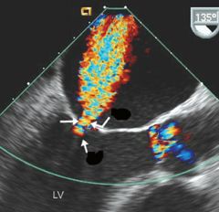
Label the black spots
|
at the neck ( the top 2 arrows) are showing the Vena contracta. the bottom arrow shows the convergence zone
|
|
what severity and why
|
moderate MR note the jet encompasis almost 25% of the LA area
|
|
what is this
|
|
|
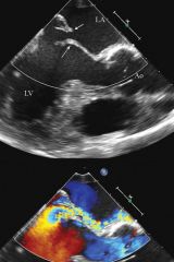
what is this
|
flail posterior leaflet with eccentric central directed jet, with coanda effect in the LA
|
|
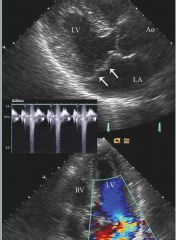
what is this
|
classic MVP, with > 2mm prolapse and thickening of the leaflets. the phonogram shows the OS at latter 40% of systole
|
|
describe what this is, hint the patient has SLE, describe the characterstics
|
Libbman Sachs endocardtis, or APLA valve disease, the lesion is on the atrial aspect on the distal mitral valve
|
|
what is this in this pathetic still image
|
mitral fibroelastoma. the small nearly spherical mass attached to the tip of the mitral leaflet
|