![]()
![]()
![]()
Use LEFT and RIGHT arrow keys to navigate between flashcards;
Use UP and DOWN arrow keys to flip the card;
H to show hint;
A reads text to speech;
52 Cards in this Set
- Front
- Back
|
ORAL CAVITY Developmental Anomalies |
Palatoschisis - Cleft palate Cheiloschisis - Cleft Lip Origin: genetic or toxic (steroid during pregnancy in primates). Involvement: Soft palate or Soft/Hard palate. Sequelae: failure of suckle, aspiration pneumonia. |
|
|
ORAL CAVITY Stomatitis and Gingivitis |
= Inflammation of the mucous membrane of oral cavity and gingiva. Lesions: macules, papules, vesicles, erosions, ulcers. Etiology: virus, chemical, trauma, toxic, autoimmune, systemic disease. Clinical signs: anorexia, ptyalism |
|
|
ORAL CAVITY Vesicular Stomatitides |
Foot-and Mouth (Picornavirus) Vesicular stomatitis (Rhabdovirus) Swine vesicular Disease (Enterovirus) Virus-induced, vesicular diseases of oral, nasal, digital epithelium Non-fatal but great economic loss. All absent and notifiable in NZ and AUS |
|
|
ORAL CAVITY Oral ulcers and erosions |
Etiology: BVD Rhinderpest (Absent and notifiable NZ and AUS) Malignant catarrhal fever Bluetongue Uremia Feline Calicivirus Feline eosinophilic granuloma complex Vit C deficiency guinea pigs (skørbug) |
|
|
ORAL CAVITY Papular stomatitides |
Etiology: Parapoxavirus Cattle: Papular stomatitis, papules in alimentary tract in immunosuppresed animals. Zoonosis: "Milker's nodules". Sheep, Goat: Contagious ecthyma/sore mouth, lesions around mouth, udder, feet and anus. Zoonosis: "Orf". |
|
|
ORAL CAVITY Necrotizing stomatitides |
Cattle, Sheep, Pigs Aka Calf Diphteria Etiology: Fusobacterium Necrophorum Signs: inappetence, pyrexia, halithosis, lesions throughout GI. |
|
|
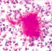
ORAL CAVITY Eosinophilic stomatitides |
Cats "Oral eosinophilic granulomas" Possibly hypersensitivity reaction but unknown Lesions: near philtrum and anywhere in the mouth. Collagen, eosinophils, mast cells and multinucleated giant cells. Rarely seen in dogs. |
|
|
ORAL CAVITY Gingival hyperplasia |
Overgrowth of gum tissue, non-neoplastic. Most common in brachycephalic breeds, is present in 30% of boxers > 5 years old. |
|
|
ORAL CAVITY Neoplasia |
Squamous cell carcinoma: dogs and cats, ventrolateral surface of tung and tonsils, malignant, locally aggressive. 60% of oral neoplasms in older cats. Melanoma: Dogs, visibly black, 90% malignant. Fibrosarcoma: Cats, neoplastic fibroblasts, 20% of oral neoplasia. Papillomatosis: Dogs, papovavirus-induced in dogs < 1 year old, cauliflower-like, regress spontaneously. |
|
|
ORAL CAVITY Teeth anatomy |
Brachydont: crown and root, enamel and cementum respectively covers the dentin. Carnivores, incisor teeth of ruminant and porcine teeth are brachydont. No enamel on roots -> gum recession -> exposed dentin -> pain. Hypsodont: Elongated, cementum/enamel/dentin from outside in, cementum and enamel invaginate into dentin forming infundibula. Horse, cheek teeth of ruminants and tusks of boars are hypsodont. |
|
|
ORAL CAVITY Feline External Resorptive Neck Lesions |
Etiology: Unknown Pathogenesis: Odontoclastic resorption of dental tissue, particularly neck or root, can disappear entirely, inflammation, pain. |
|
|
ORAL CAVITY Periodontal Disease (Dogs) |
Dental plaque (mineralized bacterial matter on tooth surface) -> inflammation of gums -> atrophy -> recession of gums and pocket formation (where more bacteria can accumulate). Teeth loosen as the periodontal ligament is destroyed by inflammation. |
|
|
ORAL CAVITY Actinobacillus lignieresii |
Woody tongue Opportunistic infection Cattle, rarely horses and small Ru. Granulomatous inflammation Fibrosis Treatment: debridement and iodine |
|
|
ESOPHAGUS Megaesophagus |
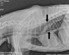
Etiology: idiopatic, innervation disorders, partial physical obstruction, stenosis, persistent right aortic arch. Can be secondary to myasthenia gravis, hypothyroidism (denervation) and more. Clinical signs: regurgitation, aspiration pneumonia. |
|
|
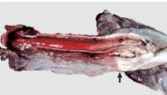
ESOPHAGUS Choke |
Esophageal obstruction Most often where esophagus can't fully expand (dorsal to larynx, thoracic inlet, base of heart, diaphragmatic hiatus). Ingestion of large food. Circumferential necrosis of the mucosa. |
|
|
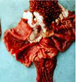
RUMEN Bloat |
Over distention by gas produced during fermentation. Mortality = 50% Primary: change of diet (clover) Secondary: obstruction/esophageal stenosis\ Postmortem: bloat line (see picture) |
|
|
RUMEN Lactic Acidosis |
Mechanism: normal ruminal microflora normally cellulytic G- bacteria. A sudden change to highly fermentable, carbohydrate-rich feed (grain) promotes growth of G+ bacteria, Strp. bovid og Lactobacillus spp/, producing lactic acid by fermentation. This decreases the luminal pH below 5 (normal 5.5-7.5) This eliminates normal flora and damages luminal mucosa. Rumenitis, detaching papillae, bacteria in bloodstream to V. portae -> abscesses in liver (A. pyogenes) |
|
|
RUMEN Vagus Indigestion |
Type 1: indflamed vagus nerve -> eructation hindered. Type 2: omasal transport failure (neoplasia, abscess) -> increased fluid in rumen. Type 3: Pyloric outflow failure -> secondary abomasal impaction. This gives wash-back of HCl into rumen, causing influx of H2O, K+, H+ and CL- into rumen and abomasum. The kidney compensates by producing bicarbonate (HCO3-) as an alternative anion -> metabolic alkalosis. Findings: hypochloremia, hypokalemia, paradoxic acuduria. Tx: IV fluids high in chloride. Type IV: Pregnancy-related. Sx:"Papple-shape", decreased rumen-contractions (normal 1-2/min), peritonitis. |
|
|
STOMACH Gastric dilation and volvulus (Dogs) |
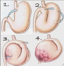
Presence of gas that cannot be passed through pylorus and can't be eructated. Vomitus can't occur. Etiology not well understood. Typical Hx: feed and postprandial exercise in large-breed dogs. From surgeon's view (ventral, pic): stomach twists clockwise around mesentery, esophagus is twisted, vascular compression, decreased drainage, hypoxia. Emergency. |
|
|
ABOMASUM Displacement (Cows) |
Normally left-sided, within 6 weeks postpartum, non-fatal, can be result of atony following grain feeding (volatile fatty acids decrease motility) and hypocalcemia. Partial outflow obstruction can lead to metabolic acidosis, rumen atony and impaired movement of ingest. Sx: Anorexia, dehydration, lack of feces, ketonuria, ping on percussion. Occationally in calves and smaller ruminants |
|
|
STOMACH Gastric Dilation and Rupture (Horses) |
Etiology: grain overload, analogous to lactic acidosis in cattle. Stomach atonous or intestinal obstruction. Sx: Sudden death Post-mortem: can occur after death, evidence of inflammation (fibrin strands) only occur in live animals. |
|
|
STOMACH/ABOMASUM Ulcers |
Ruminants, horses, foals (idiopathic), pigs, small animals (idiopathic). Predisposing factors: stress, decreased blood flow (NSAIDs). Type 1: Non-perforating, no bleeding (erosion) Type II: Non-perf., bleeding, painful Type III: Slowly perf., adhere to peritoneum, cran. abd. pain. Type IV: Rapid perf., dark vomitus (small animals), peritonitis, shock, death. Sx: pain, melena, anemia. Tx: H2 blockers (omeprazole), antihistamine (ranitidine), mucosal protectants (sucralfate), discontinue NSAIDs, hay, surgery. |
|
|
STOMACH Horse bot flies, Gasterophilus intestinalis and Gasterophilus nasalis. |
G. intestinalis: eggs on distal limbs, colonize stratified portion of stomach. G. nasalis: eggs around nostrils, swallowed, colonize glandular portion of stomach. |
|
|
ABOMASUM Parasites affecting ruminant abomasum |
Haemonchus contortus Barber's Pole Worm (twisted intestine/uterus) Small ruminants (lambs) Anemia, hypoproteinemia, edema (bottle jaw) Ostertagia ostertagia (Bo.), O. circumcincta (Ov, Cap.) Nematide, brown. Reside in abomasal glands Lesions look like leather (hyperplasia, inflammation) |
|
|
INTESTINES Developmental anomalies |
Atresia coi/ani: blind-ending segment. Megacolon: large, fecal-filled colon, caused by lack of myenteric plexuses (no peristaltic movements). Equine Overo-breed (specific spotted pattern) white foals: aganglionosis, do not pass meconium. |
|
|
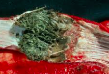
INTESTINES Equine enteroliths |
= Struvites (ammonium magnesium phosphate) - Formed around nidus, often metallic object - Often pelvic flexure or transverse colon - Vary greatly in size (up to several kgs) - Diets high in magnesium and phosphorus is predisposing. |
|
|
INTESTINES Intussuseption |
One intestinal segment "telescoped" into another intestinal segment. - Caused by inflammation and hypermotility. - Looks like accordion (harmonika). - Partial digestion of internal fold, cannot be separated. - Post-mortem occurrence can easily be separated as no inflammation (peristalsis continues post-mortem). |
|
|
INTESTINES Internal Hernias |
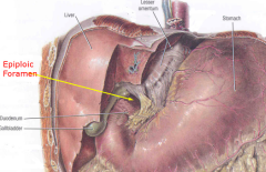
Herniation through anatomical space. Most common in Horse: Epiploic foramen. Intestines herniate, incarcerate, stangulate. |
|
|
INTESTINES External Hernias |
Umbilical Ventral (through abdominal wall muscles) Diaphragmatic Hiatal (hiatus through diaphragm) Inguinal Scrotal Perineal |
|
|
INTESTINES Volvulus and Torsion |
Volvulus Twisting around mesenteric axis. Torsion Rotation of a tubular organ along its long axis. Both result in obstruction and ischemic injury. |
|
|
INTESTINES Pundunculated lipoma |
- Older horses - Wrap around mesentery or bowel and strangulate - Benign but can be fatal upon strangulation of intestines. |
|
|
INTESTINES Renosplenic Entrapment (Horses) |
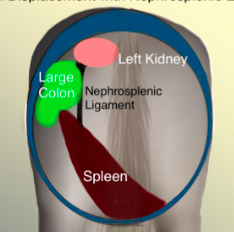
- Cause unknown (rolling?) - Left dorsal/ventral colon entrapment between spleen and left body wall. - Renosplenic ligament. - Intestinal rupture and death may occur. |
|
|
INTESTINES Strongylus Vulgaris (Horses) |
- Ingestion 3rd stage - Development to 4th stage in small intestine - Migrate via arterioles to mesenteric artery, reside 3-4 mths until 5th stage - Migrate via blood to large intestine -> adults (6 mths). - Found in cranial mesenteric artery -> arteritis, ooseus metaplasia, mural thromboses. - Colic and death from infarction secondary to verminous thrombosis. |
|
|
INTESTINES Lymphangiectasia (Dogs) |
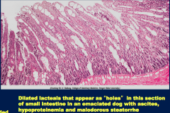
- Protein-losing enteropathy - Congenital (lymphatic vessels underdeveloped) or acquired (obstruction from granulomatous or neoplastic disease). - Diarrhea, steathorrhea (fat in stools), hypoproteinemia, ascites. - Thickened intestinal mucosa, dilated lymphatics. |
|
|
INTESTINES Rotavirus enteritis (Calf, Piglet) |
- Ubiquitous pathogen. - Calves: first 7 days of life. - Pigs: first 7 weeks of life. - Cytolytic, tropism for intestinal villi. - Sx: watery diarrhea, dehydration, spreads cranially->caudally in small intestines. - Blunting, fusion of villi following infection. |
|
|
INTESTINES Coronavirus enteritis (Calves, Cats) |
- Tropism for intestinal villi. - Calves: first 7 days of life. - Cats: 2 mths-7 years old (!). - Sx: watery diarrhea, dehydration, jejunitis, colitis, death more common than rota-. |
|
|
INTESTINES Adenovirus Enteritis (Cow, Sheep, Pig, Goat, Cervids, Horse, Exotics) |
- Species-specific. - Young/immunosuppressed. - Respiratory +/- enteritis (and liver, kidney). - Aerosols, fomites, feces. - Basophilic, intranuclear inclusion bodies in villous enterocytes. - Blunting of villi. |
|
|
INTESTINES Enterotoxic colibacillosis/E. coli diarrhea |
- Produce toxins (STa, STb, LT or verotoxins) - Calf/Piglet/Lamb: 2 days - 3 weeks old. - Watery to pasty, yellow to white, voluminous, dehydration - Infects when defenses overcome. - Attach to enterocytes via pili or fimbrae. - Secretory diarrhea (toxin causes enterocytes to secrete water and electrolytes). - Diagnosis can be made by light microscopy of freshly dead animals by noting presence of bacteria along luminal surface of enterocytes. |
|
|
INTESTINES Edema Disease (Pigs) |
- = Enterotoxemic colibacillosis. E.coli - Verotoxin. - Produced small intestine, spread via blood. - Sx: incoordination, tremors, generalised endothelial damage causing edema especially eyelids, mesenteric colon. - CMS lesions: focal malaria of medulla due to material damage. - 6-14 weeks, "best piglets affected". - Morbidity 35%, mortality 100%. |
|
|
INTESTINES Salmonellosis |
- Zoonosis. - G-, arobic/facultativt anaerobic, motile. - Invade phagocytotic cells. - Granulomatous inflammation. - Septicemic, acute enteric or chronic enteric. - Fecal-oral or trans-placental. - Generally younger animals. - Necrosis, multi-focal intestinal infarcts. |
|
|
INTESTINES Clostridial Enteritis (Calves, lambs, piglets, dogs, horses, birds) & Clostridial enterotoxemia/Pulpy Kidney Disease. |
- Enteritis: - Cl. perfringens Type A, B, C and E. - G+, anaerobic, ubiquitous, spore-forming, toxin. - Sx: sudden death, diarrhea with or without blood, reddened intestines, abdominal distention, first day of life. -Enterotoxemia/Pulpy Kidney/Overeating Disease - Type D - Increased dietary intake, good animals, sheep/cattle/goat - Sudden death, glucosuria (lambs), kidney changes not evident (lambs), hard to Dx. Vaccination recommended. |
|
|
INTESTINES Lincomycin/AB enteritis (Horses, Rabbits) |
- Cecal fermenters. - Death of normal enteric microflora and growth of Cl. perfringens Type A. Not proven. |
|
|
INTESTINES Clostridium piliformis/Tyzzer's Disease |
- Many mammalian species. - Target is liver but also heart and intestine. - Mucosal necrosis and edema. - Dx: silver stain. |
|
|
INTESTINES Clostridium difficile/Colitis X (Horse) |
- Foals: Hemorrhagic, necrotizing enterocolitis. - Horses: Necrotizing typhlocolitis (Colitis X). |
|
|
INTESTINES Campylobacteriosis (Lawsonia) |
- Most commonly pigs (Lawsonia) and poultry. - G-, curved rod, motile, obligate intracellular, can't be cultured. - Proliferativ enteropathy. - Sx: Diarrhea watery/mucoid, +/- blood, anorexia, vomiting, proliferation of crypt cells and surface erotions. - Zoonotic. - Rare in dogs, horses, sheep and more. |
|
|
INTESTINES Mycobacterial enteritis/Tuberculosis |
- M. tuberculosis and M. bovis. - Chronic wasting disease. - Ingested, uptake M-cells Ileum. - Sx: thickened colon, granulomatous lymphadenopathy +/- mineralization and necrosis. - Rarely in dogs, small ruminant, pigs. - Excreted in milk, zoonosis (AIDS). |
|
|
INTESTINES Intestinal Diseases in the Horse |
- R. equi enteritis - G+, rod, facultative anaerobic - Often seen as pyogranulomatous pneumonia in folks <6mths - Coughed up, swallowed -> enteritis Peyer's patches. - Equine granulomatous enteritis - Cause unknown, m. avium suspected. - Wasting and hypoproteinemia. Thickening of bowel. - Clostridial enteritis (Colitis X) - Cause unknown but Clostridium overgrowth suspected. - Edema, congestion, haemorrhage of cecum and colon. |
|
|
INTESTINES Intestinal Diseases in the Cow |
- BVD/Mucosal Diseases - Multifocal, sharply demarcated erosions/ulcers on tongue, palate, esophagus, rumen, abomasum + foci of necrosis intestines over GALT. - Rhinderpest - Like BVD but additional multinucleate enterocytes in intestinal lesions. - Malignant Catarrhal Fever - Necrotizing arteritis and phlebitis of subcutis, in rete miracle around pituitary gland, stomatitis, esophagitis, abomasitis etc. - Winter Dysenteria - Catarrhal ileitis and jejunitis. - Johne's Disease/Paratuberculosis - Wasting, thickening of intestinal mucosa. |
|
|
INTESTINES Intestinal diseases of the Pig |
Unweaned Piglets - Cl. perfringens Type A, C - Clostridium difficile - E. coli - Rotavirus - Transmissable Gastroenteritis/Coronavirus - Coccidiosis Weaned Piglets - Lawsonia intracellularis - Swine Dysenteria - Trichuris suis - Ascaris suum - Salmonella |
|
|
INTESTINES Parvovirus enteritis |
- Canine parvovirus type 2. - = "cat distemper, feline enteritis, mink enteritis, racoon enteritis". - Severe usually fatal, dividing cells (crypt cells, bone marrow). - Diarrhea, vomiting, panleukopenia, villous atrphy, basophilic inclusion bodies enterocytes, congenital cerebellar hypoplasia kittens, haemorrhagic intestines on necropsy, bone marrow depleted. - Another strain (Type 1) produces myocarditis and respiratory disease in young pups. No signs of lesion seen in Type 2 infection. |
|
|
INTESTINES Intestinal Diseases of the Carnivores |
- Canine parvovirus type 2. - Feline Infectious peritonitis. - Histiocytic ulcerative colitis. - Canine multifocal eosinophilic gastroenteritis. - Inflammatory bowel disease. - Diffuse eosinophilic gastroenteritis. - Feline ulcerative colitis. - Canine senile GI amyloidosis. - Parasites (lots) - Neoplasia (adenomas, polyps, adenocarcinomas, leiomyomas, lymphosarcoma) |
|
|
INTESTINES Feline Infectious Peritonitis |
- Coronavirus - Young and old cats - "Wet form": fibrinous polyserositis. - "Dry form": No effusion. - Both: multifocal lesions in most organs, pyogranulomas < 2mm, vasculitis and protein effusion (sterile), wasting disease, fatal. |