Reading...
![]()
Play button
![]()
Play button
![]()
Use LEFT and RIGHT arrow keys to navigate between flashcards;
Use UP and DOWN arrow keys to flip the card;
H to show hint;
A reads text to speech;
13 Cards in this Set
- Front
- Back
|
Nodular fasciitis: Common presentation, clinical population involved
|
rapid growth of a small lesion in 20-50y/o on the face, arm, shoulder subcutis
|
|
|
Important histologic findings in Nodular fasciitis (4 things)
|
"zonation effect"-hypocellular center with hypercellular periphery. Scattered stellate cells in loose "tissue culture" arrangement. Many histiocytes and giant cells. Prominent Mitosis.
|
|
|
Differentiation of Nodular Fasciitis from other fibrous tumors (4)
|
Other tumors are uniform, NF is variable; NF has "loose" appearance; Marked nuclear atypia, and florid inflammation are not seen in NF
|
|
|
T/F Nodular fasciitis can be seen extending to the skin in every location
|
False. NF should not extend to skin, except when on the face
|
|
|
Recurrence rate of Nodular Fasciitis
|
Extremely low, <2%
|
|
|
Immunohistochemical pattern for Nodular fasciitis:
SMA, MSA, Desmin, CD68, Keratin, S100 |
SMA +
MSA + Desmin - CD68 + in macrophages/GCs Keratin - S100 - |
|
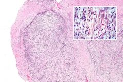
Identify
|
Nodular Fasciitis
Note the subcutaneous location, loose central area with more cellular periphery. No capsule. Plump to stellate myofibroblasts in loose arrangement with delicate vessels and intermixed histiocytes. NO inflammatory cells. Mitoses are okay. |
|
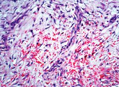
A 20 year-old woman presents with a rapidly enlarging and tender 2 cm mass on her right forearm. Given this histological appearance, what can you tell her about the prognosis and diagnosis?
|
Nodular fasciitis, extremely good prognosis with little chance (<2%) of recurrance.
|
|
|
What age group is commonly affected by proliferative myositis and fasciitis?
|
Older, >50years
|
|
|
What is the typical clinical presentation of proliferative myositis and fasciitis?
|
Rapid growth, can be tender
|
|
|
What cells are the precursors (by EM) of proliferative myositis and fasciitis?
|
Myofibroblasts and fibroblasts
|
|
|
What is the difference between proliferative myositis and proliferative fasciitis?
|
PM occurs in muscles while PF occurs in subQ
|
|
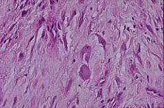
A 50 year-old man presents with a rapidly enlarging, tender, 3cm mass on his right arm. Given these histopathologic findings of the subcutaneous mass, what is your diagnosis and the patient's prognosis?
|
Proliferative fasciitis. Excellent prognosis, low recurrence rate.
|