![]()
![]()
![]()
Use LEFT and RIGHT arrow keys to navigate between flashcards;
Use UP and DOWN arrow keys to flip the card;
H to show hint;
A reads text to speech;
51 Cards in this Set
- Front
- Back
|
General functions of the brain: |
- acquiring knowledge (from perception) - storing information (from memory) >> controlling motor behaviour) 1. element movements: e.g. respiration, heart rate 2. complex voluntary/willed actions: e.g. grasping >> main acheivement: language and communication |
|
Gross brain structure: |
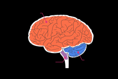
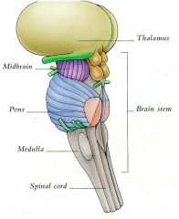
- Brain = very delicatge - protected by tough connective tissue membrane = dura matter Right and left hemispheres: bilaterally symmetrical > Each with 3 major divisions: 1. cerebrum (largest) 2. Cerebellum 3. Brainstem (smallest) |
|
|
The cerebrum: 2 main components with subdivisions |
1. telencephalon a) cerebral cortex: conscious sensation.perception, voluntary movements/higher cognitive (language) functions b) Basal Ganglia: movement planning & control 2. Diecephalon a) Thalamus: various nuclei (related to cerebral cortex) b) Hypothalamus: various nuclei for regulating appetites + endocrine functions (via pituitary glands) c) Epithalamus: Pineal gland, regulates circadian rhythms |
|
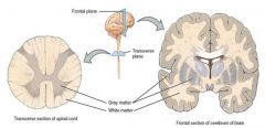
What is the brain made of? 1. Gray matter |
- contains cell bodies + dendrites & synaptic inputs from terminal axon (= site of information transfer & processing) > Neuron cell bodies may be arranged in: - layers/sheets e.g. cerebral cortex - groups/clusters: e.g. in thalamus + basal ganglia |
|
What is the brain made of?
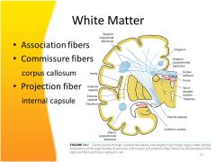
2. white matter |
- contains axons + their myelin sheaths formed by oligodendrocytes
- provides routes for direct long-range connections between neurons in different gray matter regions e.g.
> corpus callosum: carries axons between neuron cell bodies in right/left cortical hemispheres > internal capsule: carries axons between neuron cell bodies in different thalamic nuclei to different parts of the cortex
|
|
|
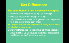
Sex differences in brain structure |
|
|
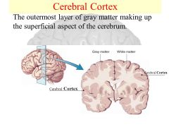
Cerebral cortex |
- the largest + most evolved human brain region > Anatomically the right + left hemispheres are: - bilaterally symmetrically + deeply folded - different regions specialised for different functions > special senses (vision/hearing) > motor control (voluntary movement) > cognitive (language memory) |
|
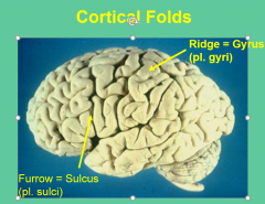
Cortical folding: General knowledge |
outer cerebral cortex is deeply folded, to pack 1250 cm2 surface area into skull Folds consist of: ridges and furrows >> deepest sulci divide the cortex into 4 lobes w/ different functions |
|
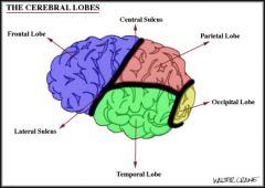
Cortical sulci, lobes and functions |
1. central sulcus divides 2 lobes - frontal lobe = motor/movement - parietal lobe = somatic sensation, touch, temp, pain 2. lateral sulcus divides these from - temporal lobe: auditory/hearing 3. Pre-occipital notch: demarcates - occipital lobe = vision |
|
|
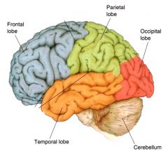
Lobes of the brain |
|
|
|
Functional localisation |
The Cortical Grey Matter contains different Neurontypes organized into distinct layers: >> Pyramidal & Granule/Stellate neurons >> Layers 1-6, from pial surface to white matter (there are subtle differences in the sizes/density of these neurons + layer thickness from one region of the cortex to the other) |
|
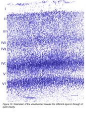
Broddman's cellular architecture: 6 major layers |
I = mainly dendrites + synapses of cortical cells IV = small granule (stellate) cell bodies II III V VI =Pyramidal cell bodies (extra blue areas have more neural bodies- high concentration + no neural cell bodies in I) |
|
|
Brodmann's areas: CYTOarchitecture |
- 52 discrete areas based on differences in layer thickness, cell size distribution
Broddman's area 44/45 = Broca's area for speech production (left hemisphere inferior frontal lobe)
Broddman's area 22 = wernicke's area for speech comprehension (left superior temporal lobe) |
|
|
Functional brain imaging: for identifying regions of increased brain activity |
1. PET however is intrusive 2. Functional magnetic resonance imaging (fMRI) - non invasive: looks at blood flow in brain + activations can be mapped onto anatomical brain image |
|

Primary cortical areas |
- bilaterally symmetrical (one in each hemisphere) - basic sensory or motor functions - are related to opposite side of the body - organised as MAPS
1. there is one primary motor area: for the initiation of all voluntary movement
2. there are several primary sensory areas: one each for somatic sensation/hearing/vision |
|
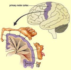
primary cortical areas: motor cortex (M1) |
- frontal lobe, pre-central gyrus - BA = 4: the largest layer - 5 pyramidal cells - MAP: MotorHomonculus (inverted + distorted) > Micro stimulation = movements on opposite side of body > unilateral damage = hemi-plegia (paralysis on osob) |
|
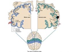
Primary cortical areas: somatosensory cortex (S1) |
- parietal lobe, post-central gyrus - BA = 1. 2. 3. - Map: Sensory Homonculus (inverted + ditorted) > unilateral damage = hemi-anaesthesia (loss of tactile, thermal, pain and joint sensation on osob) |
|
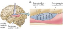
Primary cortical areas: auditory cortex (A1) |
- temporal lobe, heschl's transverse gyri on superior temporal gyrus in the lateral sulcus - BA = 41 / 42
> unilateral damage = reduced sensitivity or sounds in opposite ear & contralateral auditory hemifield
> projects directly to 2o Auditory Areas involved in speech comprehension |
|
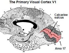
Primary cortical areas: Visual cortex (V1) |
- occipital lobe, upper + lower banks of calcarine sulcus - BA 17 thickest layer 4 + sub layers + band of myelin
> unilateral damage = hemi-anopsia (blind in opposite half of visual field)
> Retinotopic: Inverted& distorted (large regions for fovea & central vision, small for peripheryea |
|
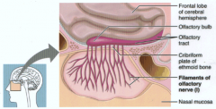
Cranial nerve 1: Olfactory nerve (cerebrum)
|
- Pure sensory - function: relates to sense of smell - foramen: enters cribiform plate of ethmoid bone - region supplied: terminates on neurones in olfactory bulb, to higher brain centers - lesions: causes loss of sense of smell (anosmia) |
|
|
Cranial nerve 2: optic nerve (cerebrum) |
- Pure sensory - Function: Vision - Foramen: optical canal at back of orbit |
|
|
cranial nerve 3: Oculomotor nerve (Brainstem) |
- Motor - Function: controls eye movement/pupil changes - innervates: 4 extraocular muscles (EOMs) 1. medial rectus 2. inferior rectus 3. superior rectus 3. inferior oblique foramen: superior orbital fissure, back of orbit |
|
|
cranial nerve 4: trochlear nerve (brainstem) |
- Motor -function controls eye movement - innervates: superior oblique - foramen: superior orbital fissure, back of orbit |
|
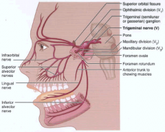
Cranial nerve 5: Trigeminal nerve (brainstem) |
- mixed: sensory and motor >> main sensory nerve for head >> motor innervates muscles of mastication + tensor tympani - foramen (3): superior orbital fissure, formen rotundum, foramen ovale |
|
|
Cranial nerve 6: abducens nerve (brainstem) |
- motor
- function: controls eye movement - innervates: lateral rectus
- foramen: superior orbital fissure, back of orbit |
|
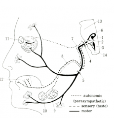
cranial nerve 7: facial nerve (Brainstem) |
- Motor, senory + autonomic Function: - M: innervates muscles of face/ear - S: innervates taste buds 2/3 of tongue to solitary nucleus - A: supplies lacrimal gland + submandibular/sublingual salivary glands - foramen: internal auditory/acoustic meatus - lesion: leads to ball's palsy |
|
|
Cranial nerve 8: vestibulo-cochlear nerve (brainstem) |
- Pure sensory 1. cochlear nerve: carries auditory info (hearing) 2. vestibular nerve: carries info regarding position/movement of head - foramen: internal auditory/acoustic meatus •Cochlearnerve ends in cochlear nuclei in medulla/pons junction •Vestibularnerve end in vestibular nuclei in medulla/pons junction |
|
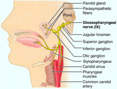
Cranial nerve 9: glossopharyngeal nerve (Brainstem) |
- mixed sensor, motor + autonomic - function M: elevates larynx for swallowing S: bitter taste posterior 3rd + somatic sensation in pharynx Parasympathetic: salivary glands Formen: jugular formen |
|
|
cranial nerve 10: vagus nerve (brainsteam) |
- sensory, motor, autonomic Function: M: controls muscles of pharynx, soft palate + larynx, controlling speech/swallowing S: somatic sensations for above + abdominal organs Parasympathetic: resting/digesting |
|
|
cranial nerve 11: accessory nerve (brainstem) |
- Pure motor - Function: innervates muscles that rotate head, lift shoulders - lesions: cause difficulty in above tasks |
|
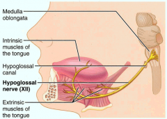
cranial nerve 12: hypoglossal nerve (brainstem) |
- pure motor - Function: innervates intrinsic and extrinsic muscles of the tongue for movement and speech prod - lesions: spasms on the same affected side of tongue |
|
|
Sensory and motor pathways in the brain are not continuous nerves but consist of... |
1. separate nuclei in the gray matter - operate as relay stations - process information before passing it on via... 2. Long range axons in white matter: - carry info to neurones at next relay |
|
|
what are sensory and motor pathways? |
sensory pathways = ascending Motor pathways = descending |
|
|
Brainstem divisions |
1. separate nuclei in the gray matter - operate as relay stations - process information before passing it on via... 2. Long range axons in white matter: - carry info to neurones at next relay |
|
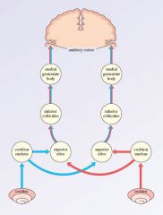
The Auditory Pathway |
>> there are multiple reylays from inner ear toA1 cortex 1. Hair cells in inner ear = receptors (on basilar membrane of cochlea) 2. contain tonotopic map of sound freq 3. innervated by cochlea ganglion cell axons (8th cranial nerve) 4. goes to cochlear nuclei in medulla (same side) 5. to inferior colliculus in midbrain (most axons cross midline) 6. to medial geniculate nucleus in thalamus 7. to A1 cortex, Heschl's gyri, superior temporal lobe |
|
|
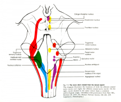
key nuclei in the medulla |
Motor: - nucleus ambiguous - hypoglossal nucleus Sensory: - nucleus solitarius - vestibular and cochlear nuclei - dorsal nucleus of vagus nerve - inferior salivatory nucleus |
|
|
The cortical Language pathway |
>> 4 seperate relay stations 1. primary cortex A1 2. Wernicke's secondary area for speech comprhension >> via huge axon pathway: arcuate fasciculus 3. Broca's secondary area for speech production 4. + cooperation with vocalization region of M1 cortex |
|
|
inter-cortical pathways |
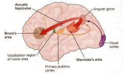
damage to Arcuate fasciculus = Conduction aphasia - understands speech but problems finding the right word -skips/repeats but otherwise fluent |
|
|
Descending pathways from upper motor neurons |
- send instructions to lower motor neurons in spindal cord/ brainstem that innervate skeletal muscles >> initiate voluntary movements: - cortico-spinal tract = fine control of body e.g. hand grasping - cortico-bulbar tract- to medulla = fine control of vocal apparatus /tongue e.g. speech production (most axons in descending pathways cross the midline) |
|
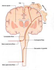
two descending motor pathways |
1. motor cranial nerve fibres (vocal apparatus) 2. motor spinal nerve fibres (limbs) |
|
|
Cerebellum: control of voluntary movement |
1. Planning: - PMA of frontal cortex - contains stored motor programs - generate complex movement sequences (e.g. in speech 2. Initiation: - M1 activates the intended motor program 3. Execution: - cerebellum monitors feed back from movement - co-ordinates it to enhance smoothness, speed, accuracy |
|
|
sensory aspects of motor control |
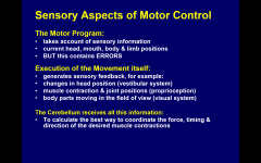
1.intention: Motor program monitors current sensory aspects
2. Action: Execution of movement - gives sensory feedback of changes
3. Cerebellum: Receives above & looks for mismatch between intention + action - coordinates, corrects errors - updates plan |
|
|
cerebellar functions |
Involved in sensori-motor coordination: •Regulates Proper force & sequences of muscle contraction (timing) •Accurate movement sizes oramplitudes (space)
cerebella lesions = ataxia -clumsy/misdirected movements
1. intention tremor + decomposition: mistiming of muscles forces/ contraction sequences 2. Dysmetria: inaccurate movement amplitudes (over/undershooting) |
|
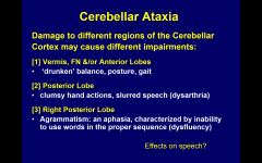
cerebellar ataxia |
|
|
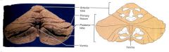
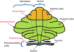
gross anatomy of the cerebellum:
1. outer cortex:
|
receives motor plans + different sensory inputs
midline: vermis + floccular-nofular lobe = receives vestibular info
lateral: 2 left/right hemispheres >> -anterior lobe: proprioceptive (knowing where body parts are in space) - posterior lobe: proprioceptive, visual auditory
|
|
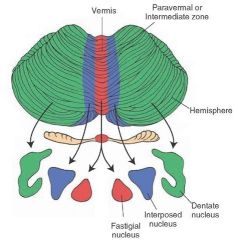
Gross anatomy of the cerebellum 2. paired deep nuclei |
receive input from one cortical region & sends outputs involved in motor coordination
- fastigial nuclei (at midline, from midline cortex) - interposed nuclei (from anterior lobe) - dentate nuclei (from posterior lobe |
|
|
cerebellar outputs |
1. spinal cord: cordinates balance, posture/gait 2. red nucleus: coordinates fine (hand, vocal) movements 3. updates motor plan via thalamus to M1 |
|
|
cerebellar connections |
1. Midline: F-N lobes + vermis - input: receives vestibular info - nucleus: Fastigal - output: vestibular nuclei - spinal cord 2. Anterior lobe: - input: receives info from brainstem/spinal cord - nucleus: interposed - output: brainstem nuclei-spinal cord 3. Posterior lobe: - input: receives info from sensory motor cortex - nucleus: dentate - output: red nucleus |
|
|
higher cognitive functions of the cerebellum |
- posterior lobes of cerebellar cortex 1. motor learning: acquisition of new motor skills 2. verbal working memory - temporary rehearsal during speech prep (inner speech) - sequencing syllable strings at normal speech rate |
|
|
speech regions involved in articulation
|
planning the movements: Broca's area - left inferior frontal cortex intonation: mirror image area, right inferior frontal cortex initiating movements: M1 smooth movement coordination: superior cerebellar cortex |
|
|
lateralization |
how some function tend to be more dominant in one hemisphere compared to the other Pre-articulated speech areas: - broca's area + cerebellum activated - M1 is not |