![]()
![]()
![]()
Use LEFT and RIGHT arrow keys to navigate between flashcards;
Use UP and DOWN arrow keys to flip the card;
H to show hint;
A reads text to speech;
49 Cards in this Set
- Front
- Back
|
Origin and epithelium cell type of the epidermis
|
ectodermal- stratified, squamous, keritinized, epithelium |
|
|
Layers of the epidermis (Come, Let's Get Sun Burned) |
1. Stratum germinativum (basale) (deep) 2. Stratum spinosum 3. Stratum granulosum 4. Stratum lucidum 5. Stratum corneum (superficial) |
|
|
Stratum Germintivum |
Single layer of cuboidal or columnar cells that rest on basil lamina. Hemidesmosomes attach them to Basil Lamina, desmosomes attach them to each other. Cell mitosis occurs here |
|
|
Stratum Spinosum |
Polygonal cells that have spiny projections that form intercellular bridges. Many desmosomes that contribute to the cohesiveness of the epidermis. Mitosis is seen here. |
|
|
Malphigian Layer |
Stratum Germitivum and Stratum Spinosum |
|
|
Stratum Granulosum |
Characterized by basophilic keratohyaline granules. Also contains membrane bound membrane coating granules. These contain glycoaminoglycans that are exuded into extracellular space and form a barrier to microorganisms foreign substances and water. |
|
|
Stratum Lucidum |
Most prominent in thick skin (often unidentifiable in thin skin) Translucent layer of acidophilic cells devoid of nuclei and organelles. |
|
|
Stratum Corneum |
Flattened keratin filled cells.No nuclei or organelles. Called horny cells or squams. |
|
|
Cutaneous Burns |
1st degree- damage to the superficial epidermis. cells of the stratum germitivum remain viable and regenerate the epidermis 2nd degree-epidermis is competely destroyed, remnants of hair follicles and sweat glands regenerate the epidermis. 3rd degree- epidermis and dermis destroyed. Skin grafts required. |
|
|
Keritinocytes |
predominant cells in the epidermis, ectoderm origin. Specialized differentiation gives rise to stratum corneum. |
|
|
Basil Cell Carcinoma |
70% of skin caners. typically occurs in ages forty and up, especially n fair skinned people. Usually on eyelids or nose. Histologically cells form nests or islands. Seldom metastasize. |
|
|
Squamous Cell Carcinoma |
20% of skin cancers effects fair skinned blondes with lots of sun exposure Late stage dermis penetration- prognosis is based on location, size, and depth of lesion. 2-5% chance of metastasis |
|
|
Langerhans cells |
dendritic cells found primarily in stratum spinosum. Not attached to adjacent cells by desmosomes. Engulf invading microorganism can migrate. May serve as reservoir for HIV |
|
|
Melanocytes |
Neural crest origin. Located in the stratum germativum. Regionally replicate and maintain melanin units. Not connected to adjacent cells by desmosomes, but can be attached to the basil lamina by hemidesmosomes. Contains tyrosinase for melanin conversion. Mealnin i primarily found in keritinocytes. |
|
|
Melanin granules |
Granules take position above the nuclei of the keritinocytes in the stratum germitivum and spinosum. Protects the dividing cells from UV radiation. |
|
|
Malignant melanoma |
2% of skin cancers. Very metastatic. Histologically, appears as nests of pigmented melanocytes that penetrate the epidermis. They will also invade the dermis where the can access the lymph and blood stream. |
|
|
Merkel cells |
Typically found in thick skin. Mechanoreceptive cells (associated with nerve endings) Neurosecratory granules. |
|
|
Papillary layer of the dermis |
Loose connective tissue that contains fibroblasts, mast cells, macrophages, and some luekocytes- forms dermal papillae/ pegs. |
|
|
Reticular layer of the dermis |
Dense irregular connective tissue, type I collagen, contains fewer cells than the papillary layer. |
|
|
Thick skin |
Also called glaborous skin, found in areas of greater wear and abrasion such as the palms of the hands an soles of the feet. Will contain all five layers of epidermis with a very prominant stratum lucidum and a thick stratum corneum. No hair. |
|
|
Thin skin |
Also called hairy skin, this is the skin that covers most of the body. It had no distinct stratum lucidum and a very thin stratum corneum. |
|
|
Hypodermis |
Not considered a part of the skin. Lies deep to the dermis and is also called superficial fascia or subcutaneous connective tissue. It is comprised of loose connective tissue and fat cells. |
|
|
Hair follicles |
Invagination of the epidermis, bulbous terminal dilutions (while growing). The bulb rests upon dermal papilla that contain organelles that nourish the follicle. |
|
|
Eccrine sweat glands. |
Simple coiled tubular glands that secrete a nonviscous fluid. Evaporation of this fluid cools the skin. Contain catabolites. Light staining simple cuboidal acini and dark staining stratifies cuboidal ducts. Associated with myopithelia cells. |
|
|
Apocrine sweat glands. |
Specialist sweat glands in axillary, areolar, and anal regions. Open into hair follicles and secrete a viscous odorless fluid (acquires distinct odor due to bacteria). In primitive mammals serves to attract opposite sex. In humans these glands become active at puberty. |
|
|
Sebaceous glands |
Develop in association with hair follicles (not found on palms and soles) However, they are found on lips, glans, penis and clitoris. Secrete sebum via holocrine secretion. More active during puberty- when clogged promote bacterial growth- acne. As cells fill with sebum the nuclei become pynotic and are lost. |
|
|
Mammary glands |
Elaborate lactiferous duct system. Enlarged during lactation. |
|
|
Nails |
Plates of keritinized epithelial cells. Clear and used for quick assessment of blood oxygenation.Nail bed is malpigian layer. |
|
|
Free nerve endings in skin |
Unmylenated axons penetrate the basil lamina and enter the stratum germitivum, spinosum Involved in temperature and pain perception. May function in crude touch (eyeballs) |
|
|
Meisseners corpuscles |
Most abundant in thick skin as well as the skin of the lips and the nipples. Specialized encapsulated receptors in dermal papillae. Involved in discriminate touch. |
|
|
Pacian corpuscles |
Found in the dermis (and hypodermis) of thick and thin skin. Especially abundant in the skin of the finger tips. Involved in perception of pressure and vibration. |
|

|
Stratum Spinosum |
|

|
Stratum Germitivum |
|

|
Statum Granulosum |
|
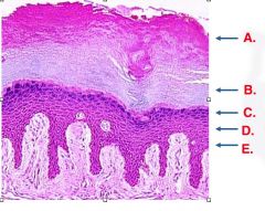
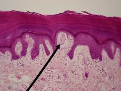
What are layers A and B |
Stratum lucidum and stratum corneum. (Since these are very prominent in this section we know that we are looking at thick skin. |
|
What are the cells shown? |
Keratininocytes |
|
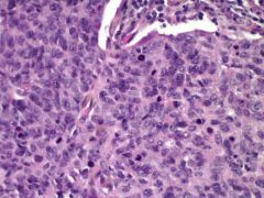
What skin cancer is shown? |
Basil cell carcinoma- basil cells form discrete islands or cell nests. |
|
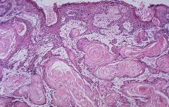
What skin cancer is shown? |
Squamous cell carcinoma, complete change in morphology. |
|
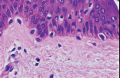
What are the clear cells in the basil layer? |
Melanocytes- recognized by their ovoid nuclei in a clear space. |
|
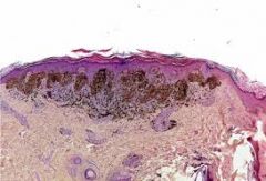
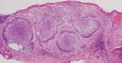
What cancer is shown? |
Malignant melanoma- will appear as abnormal nests of pigmented melanocytes that penetrate the epidermis. |
|
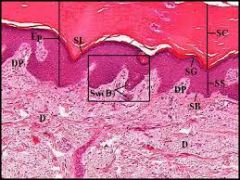
What's in the box? |
Papillary layer of dermis |
|

|
Reticular layer of dermis |
|

|
Hypodermis |
|

|
Hair follicle |
|

|
Eccrine sweat glands |
|

|
Apocrine sweat glands |
|

|
Sebaceous gland |
|
|
Meissners Courpuscle |
|

|
Pacinian Corpuscle |