Reading...
![]()
Play button
![]()
Play button
![]()
Use LEFT and RIGHT arrow keys to navigate between flashcards;
Use UP and DOWN arrow keys to flip the card;
H to show hint;
A reads text to speech;
18 Cards in this Set
- Front
- Back
|
drug eruptions are common and may mimic many other skin conditions. theys may be immune mediated or non immune. discuss immune mediated and non immune
|
immune mediated- hypersensitivity to the drug.
non immune- predictable side effects or interactions with other drugs/ unpredictable - idiosyncratic, pseudo-allergic or an intolerance. |
|
|
at what stage in life are genetic disorders of the skin usually seen ?
|
may not appear to 4-5 years of age
|
|
|
what broad things might cause nutritional skin disease
|
lack of somthing in the diet/ malabsorption/ antimetabolites/ defective use
|
|
|
what might be the obvious clinical signs of protein, FA or vitamin deficiency
|
protein- poor hair coat/ immunodeficiency leading to infection.
FAs- diffuse scaling / alopecia vitamin - scaling and or alopecia / dermatitis in some |
|
|
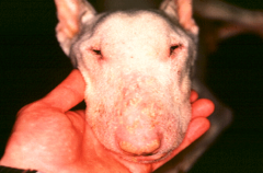
what sp is zinc responsive dermatosis seen in mostly, what is zinc needed for and what are the clincial signs seen. what breeds and age of dog are predisposed to it. hoe might the diet of pigs cause it. what disease may present with similar signs.
|
esp. pigs and dogs.
zinc is needed for enzymes which regulate RNA and DNA metabolism. ( tissue growth , maturation and repair/ Vit A metabolism/ free radical scavanging / inflammation and immune response) decreased wound healing/keratinisation defects in epidermis, hair, horn / parakeratosis ( retained Nucleus) eg periorbital crusting nordic breeds due to presumed inherited malabsorption. Puppies of any breed due to cereal diet or generic dog food. pigs food containing phytate/ high calcium/ low FAs in cereal based diets will decrease the available zinc. hepatocutaneous syndrome ( small nodular liver) |
|
|
describe the various symdromes in which zinc deficiency is though to play a role and explain the method thought to be involved
|
swine parakeratosis ( a true zinc deficiency due to diet-pigs food containing phytate/ high calcium/ low FAs in cereal based diets will decrease the available zinc.
zinc responsive dermatosis in goats and dogs ( mechanism uncertain in goats. dogs- nordic breeds due to presumed inherited malabsorption. Puppies of any breed due to cereal diet or generic dog food. SND ( superficial necrolytic dermatitis - a combination of deficiencies including zinc, AAs and FAs due to may reasons- diet insuficiency/malabsorption/ disorders of metabolism/ liver disease/ elevated glucagon. lethal acrodermatitis of bull terriers- inherited autosomal recessive disorder thought to be caused by a defect in zinc metabolism. 'acro' means extremities eg the foot. |
|
|
what is the most common cause of superficial necrolytic dermatitis ans what is a consistent feature irrespective of cause
|
liver disease. also glucagonoma.
hypo-amino-acidaemia is a consistant feature. |
|
|
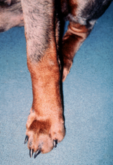
what are the symptoms of lethal acrodermatitis of bull terriers
|
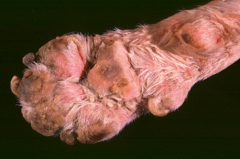
dead by age 2 - 'lethal'
affects extremities- 'acro' eg distorted swollen foot with crusted pads. stunted growth - think of zincs role small or absent thymus - again think of the role of zinc in immunity - so suceptible to skin /lung/intestinal infections |
|
|
give examples of exactly how skin lesions may indicate internal disease giving specific examples
|
influenced by hormone levels/ overall health.
endocrine, paraneoplastic or systemic diseases. A skin biopsy can be used to make a diagnosis of internal disease. eg endocrine related Alopecia ( glycocorticoids or oestrogens will cause supression/ Thyroid hormone will cause accleration. ) |
|
|
what is telogen effluvium and anogen effluvium
|
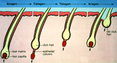
telogen effuvium may follow cytotixic therapy or other stress ( tempory hair loss due to sheding of resting hair - abruptly shifted from anogen. The loss will occur some time later ( weeks or months ) when new hairs start to grow. ( CLUB SHAPED TIP )
Anagen effluvium is similar but the onset is rapid due to sudden inhibition of cell proliferation by drugs toxins or inflammation. ( POINTED TIP) |
|
|
name the types of systemic diseases which may affect the skin
|
immune and autoimmine diseases/ circulatory diseases /drug irruptions/ genetic disorders/ metabolic, endocrine or nutritional diseases/ neoplastic and paraneoplastic syndromes.
|
|
|
define the terms anogen, catogen, telogen and effluvium
|
anogen- growth phase
catogen- transition phase telogen- resting phase effluvium -shedding |
|
|
list all types of disease that may cause alopecia, give an example of each
|
endocrine ( eg canine cushings syndrome)
genetic - colour dilution alopecia causes follicle displasia immune mediated/auto - aloppecia areata in horses and dogs mostly - monunclear cell infiltrate paraneoplastic- eg pancreatic carcinoma neoplastic - cutaneous lymphoma- atypical cell infiltrate inflammatory -COMMON-dermal inflammation and or scarring eg leishmaniasis ( protozoa)- crusting and alopecia breed associated cyclic flank alopecia eg in boxers |
|
|
give an example of paraneoplastic skin disease and describe the skin lesions and give an example of neoplastic disease which causes alopecia.
|
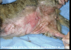
paraneoplastic syndrome- feline paraneoplastic alopecia ( rare and associated with malignant tumors on viscera , usually the pancreas. appears as ventral hair loss with shiny skin)
and many others....Cushing’s syndrome; alopecia, calcinosis cutis - eg caused by pituitary adenoma Feline exfoliative dermatitis-Thymoma Feline paraneoplastic alopecia-Pancreatic adenocarcinoma Feminisation syndrome-Sertoli cell tumour Paraneoplastic pemphigus-Lymphoma, thymoma Superficial necrolytic dermatitis-Glucagonoma neoplasia- cutaneous lymphoma - bilateral/ symmetrical and non pruritic alopecia |
|
|
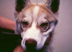
give examples of endocrine related alopecia in dogs and cats stating their cause and presentation both clinically and histologically.
|
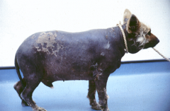
Juvenile onset pan-hypo-pituitarism ( aka pititary dwarf. seen in GSDs. autosomal recessive trait. maformed or cystic pituitary gland. notice when pup is about 2 months old due to slower growth, retained puppy coat and lack of primary hairs, gradual development of bilateral and symmetrical alopecia. histology- atrophic dermatitis with hairless telogen follicles.
cushings disease more in adults ( hyper-adrenocorticism- signs relate to gluconeogenic, lipolytic, protein catabolic, and anti-inflammatory effects of the glucocorticoid hormones .Atropic dermatitis with comedones and bilateral symetrical alopecia, thin skin and pot bellied appearance. also calcinosis cutis- cutaneous calcification- which is uncommon but caracteristic ) hypothyroidism ( mainly in dogs but over diagnosed and controversial skin signs which may be subtle - alpecia, dry skin, increased mucin makes skin thicker) sertoli cell tumor |
|
|
describe breed associated alopecia
|
Canine cyclic flank alopecia-aka seasonal flank alopecia, recurrent flank alopecia
Boxer, Bulldog, Airdale terrier, Schnauzer etc. Tardive onset, most cases regrow hair but condition is progressive in some individuals. Photoperiod – pineal gland - melatonin may influence development. Histology: Hair cycle arrest / hair follicle dysplasia Or Canine Follicular Dysplasia Syn. hair follicle dysplasia Siberian Husky, Irish Water Spaniel, Portuguese Water Dog, etc. Features vary between breeds (non-cyclical) Pathogenesis uncertain- may be linked to steroidogenesis Histology: variable hair cycle arrest; follicle atrophy / dysplasia-pigmentary incontinence / pigment clumping |
|
|
describe colour dilution alopecia in dogs. ( breeds, cause)
|
aka blue dog syndrome. A genetic alopecia.
seen in dilute coated dogs of various breeds. there is a delay in onset so breeding control is dificult. caused by inherited folicular dysplasia. the dilute hairs fail to grow and the tan colour will grow normally- opposite to black hair folicular dysplasia |
|
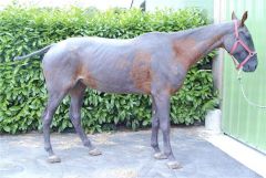
describe the cause of alopecia areata
|
Autoimmune alopecia.
Pathogenesis includes infiltration of hair bulbs by T-lymphocytes = ‘Bulbitis’ resulting in the classic ‘swarm of bees’ appearance. most often in dogs and horses. Hair loss may be localised or generalised. |