Reading...
![]()
Play button
![]()
Play button
![]()
Use LEFT and RIGHT arrow keys to navigate between flashcards;
Use UP and DOWN arrow keys to flip the card;
H to show hint;
A reads text to speech;
86 Cards in this Set
- Front
- Back
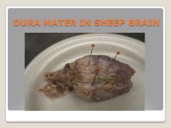
dura mater
|
hard mother”- outermost protective layer
|
|
|
arachnoid membrane
|
spongy, spider-like web of tissue
|
|
|
subarachnoid space
|
contains cerebrospinal fluid (CSF) and arteries
|
|
|
pia mater
|
delicate, innermost membrane
|
|
|
gray matter
|
nerve cell bodies
|
|
|
white matter
|
fiber bundles of neurons
|
|
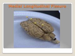
medial longitudinal fissure
|
divides 2 hemispheres
|
|
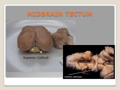
super colliculi
|
processes some simple aspects of visual stimuli
|
|
|
inferior colliculi
|
processes some simple aspects of auditory stimuli
|
|
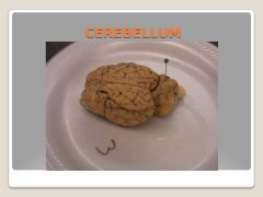
cerebellum
|
critical for motor functioning, balance, movements requiring aim and timing, some learning functions
|
|
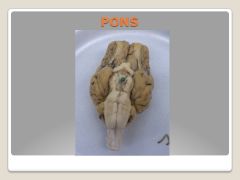
pons
|
relays sensory information from cerebellem to cortex
|
|
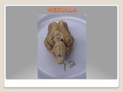
medulla
|
long, narrow structure, controls autonomic functions such as breathing, heart rate
|
|
|
brain stem
|
pons and medulla
|
|
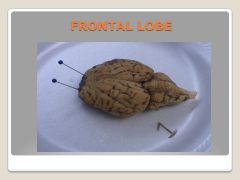
frontal lobe
|
monitors all the info about eye, head, and body positions and passes it onto the parts of the brain that control movement
|
|
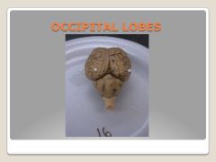
occipital lobe
|
vision
|
|
|
temporal lobe
|
auditory info
|
|
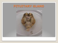
pituitary gland
|
synthesizes and releases hormones
|
|
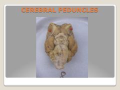
cerebral peduncles
|
motor systems from cerebral cortex that project to brainstem areas and to spinal cord
|
|
|
anterior/rostral
|
nose end
|
|
|
posterior/caudal
|
tail end
|
|
|
dorsal/superior
|
top of brain
|
|
|
posterior/caudal
|
toward the tail end
|
|
|
medial
|
toward midline of the body
|
|
|
lateral
|
away from the midline
|
|
|
How many pairs of cranial nerves are there
|
12
|
|
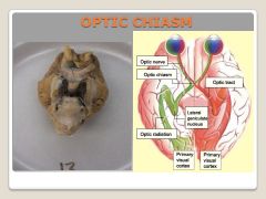
optic chiasm
|
The place where the nerve becomes the tract where 50% of the nerves cross over
|
|
|
optic nerve
|
The optic nerve is before (anterior) the chiasm and the tract is after (posterior). In general nerves are outside the brain and part of the PNS
|
|
|
Optic tract
|
tracts are insides the brain and part of the CNS
|
|

Olfactory bulb
|
I. Olfactory Bulb (Sensory) smell
|
|

Optic Nerve
|
II. Optic nerve (Sensory) vision
|
|

Oculomotor
|
III.Oculomotor(Motor)-Control of eye movements, pupil constriction (locate);
|
|

Trochlear
|
IV.Trochlear(Motor)-Control of eye movements, rolling outward and downward;
|
|

Trigeminal
|
V.Trigeminal(S & M)- Touch & pain sensations from face, Control of jaw muscles for chewing and swallowing ;
|
|

Abducens
|
VI.Abducens(Motor)-Control of eye movements, side to side (locate);
|
|
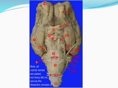
Facial
|
VII.Facial(Sensory & Motor)-Control of muscles of the face (facial expression, crying), Taste for anterior 2/3rd of tongue ;
|
|

Vestibulocochlear
|
VIII.Vestibulocochlear(Sensory)-Hearing and equilibrium;
|
|

Glossopharyngeal
|
IX.Glossopharyngeal(Sensory & Motor)-Taste/Sensations for throat & posterior 1/3rd of tongue, Control gag reflex;
|
|

Vagus
|
X.Vagus(Sensory & Motor)-Sensations from neck & throat, sensations and motor control of many internal organs including the heart, lungs, and stomach;
|
|

Spinal accessory
|
XI.Spinal Accessory(Motor)-Control of neck and shoulder muscles for turning movements of the head (locate);
|
|

Hypoglossal
|
XII.Hypoglossal(Motor)-Control of muscles of the tongue;
|
|
|
Afferent
|
going toward the CNS- afferent nerves carry sensory info from skin, skeletal muscles, joints, eyes, ears, viscera, and so on to CNS think of “a” as “approach”, “arrive”- coming into CNS – sensory;
|
|
|
Efferent
|
going away from the CNS-carry motor signals from the CNS to the skeletal muscles and viscera think of “e” as “exit”- going away from CNS- motor;
|
|

Corpus Callosum (Mid sagittal)
|
band of white matter by which each cerebral hemisphere connects with the opposite cerebral hemisphere (allows for communication between the two) (locate);
|
|

Septum Pellucidum (Mid sagittal)
|
a thin layer of tissue that separates the two lateral ventricles from each other;
|
|
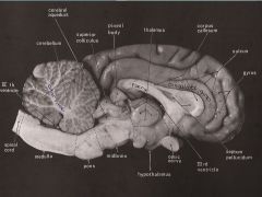
What is the Fornix (Mid sagittal)
|
band of white matter by which the hippocampus of each hemisphere connects to the hippocampus of the opposite hemisphere;
|
|

Arbor Vitae (Mid sagittal)
|
the name is Latin for “tree of life”, White nerve tissue of the cerebellum, has a tree-like outline;
|
|

Thalamus (Mid sagittal)
|
sensory relay system of the brain, sending information primarily to cortex;
|
|

Hypothalamus (Mid sagittal)
|
controls autonomic & endocrine (hormone) system; the 4 F's feeding, fighting, fleeing and fornicating; connects with pituitary for appetite;
|
|

Pineal Body (Mid sagittal)
|
aka pineal gland, responsible for secretion of melatonin – a hormone which, among other things, induces sleep;
|
|
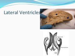
Where are the Lateral ventricles (Mid sagittal)
|
two large empty spaces directly inferior to the corpus callosum;
|
|

Where is the 3rd Ventricle (Mid sagittal)
|
hollow space directly inferior to the fornix;
|
|

Where is the 4th Ventricle (Mid sagittal)
|
hollow space directly inferior to the cerebellum;
|
|

What is the Cerebral Aqueduct and where is it (Mid sagittal)
|
short canal traveling through the midbrain, connects the 3rd and 4th ventricles;
|
|

What is the function of the Thalamus? Where is it (Coronal View)
|
below the 3rd ventricle, heart shaped; both process and relay sensory information selectively to various parts of the cerebral cortex;
|
|
|
What is the function of the amygdala (Coronal View)
|
Learning and memory (for emotionally charged events), plays role in producing fear response, stimulation (in humans) results in feelings of anxiety/fear;
|
|
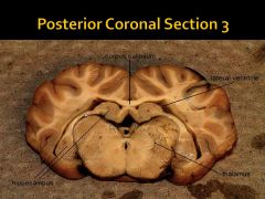
What is the function of the hippocampus? Where is it (Coronal View)
|
involved in spatial memory (ex. put a rat into a maze, learns the maze, then lesion HC- after lesion can’t remember how to get through maze), formation of new episodic memories;
|
|
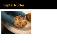
What is the septal nuclei responsible for? What happens if there is damage? Where is it? (Coronal View)
|
connected to septum pellucidum. Role in reward and reinforcement. Damage in humans can result in rage behavior and other strange behaviors (1st );
|
|

What is the caudate nucleus responsible for? Where is it (Coronal View)
|
responsible for voluntary motor responses, learning and memory (specifically in receiving feedback), may be dysfunctional in individuals with OCD; (1st and 2nd)
|
|

What is the putamen responsible for? Where is it (Coronal View)
|
involved in voluntary motor responses, reinforcement learning & implicit learning; (1st and 2nd)
|
|
|
What does the globus pallidus do? (Coronal View)
|
play an active part in pre-filtering external stimuli and may help reduce the amount of irrelevant information the brain can store;
|
|

Where is the fornix? (Coronal View)
|
Should see in the 2nd and 3rd slices;
|
|

What is the corona radiate? Where is it? (Coronal View)
|
the radiating white matter of the neocortex carry nearly all of the neural traffic from and to the cerebral cortex; all sections
|
|
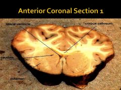
Where is the corpus callosum? (Coronal View)
|
Should see in all 3 slices;
|
|

What is the internal capsule? Where is it? (Coronal View)
|
white matter surrounding subcortical structures; runs between the caudate and putamen at some points; (1st and 2nd)
|
|
|
Where is the medial longitudinal fissure? (Coronal View)
|
should see in all 3 slices;
|
|

Where is the cingulate gyrus? (Coronal View)
|
should see in all 3 slices;
|
|

Where are the lateral ventricles? (Coronal View)
|
should see in all 3 slices;
|
|
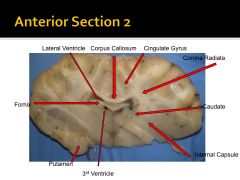
Where is the 3rd ventricle? (Coronal View)
|
should see in 2nd and 3rd slices;
|
|

Where is the pineal body (pineal gland)? (Coronal View)
|
should see in the 3rd slice;
|
|
|
Know how cerebral spinal fluid travels through the ventricles and through what openings (Coronal View)
|
CSF flows from the lateral ventricles into the 3rd ventricle via the Foramen of Monro. CSF then flows from the 3rd ventricle into the 4th ventricle via the cerebral aqueduct. CSF flows out of the 4th ventricle into the subarachnoid space of the brain or into the central canal of the spinal cord via the Foramen Magendie and the Foramina Luschka (2 of these);
|
|
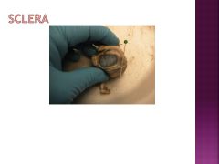
What is the sclera and where is it?
|
white protective covering of the eyeball (except cornea) – an extension of the dura mater;
|
|
What is the optic nerve and where is it (on the eye, not the brain)?
|
carries visual information to the brain;
|
|
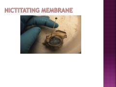
What is the nictitating membrane and where is it?
|
find it in sheep and other animals; it is a third eyelid, controlled by the abducens nerve;
|
|
|
What is the conjunctiva? Swelling causes what?
|
delicate membrane that starts at the junction of the sclera and the cornea, lines inside of eyelid and provides lubrication, conjunctivitis = swelling of conjunctiva from infection (aka pink eye);
|
|

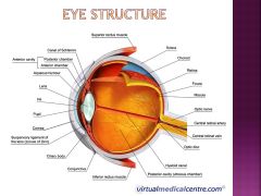
What is the cornea and where is it?
|
area of the eye anterior to the iris, transparent protective covering;
|
|
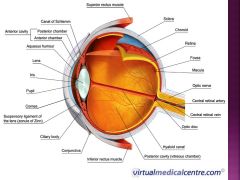
What is the iris and where is it?
|
colored part of the eye, surrounds the pupil – orange on sheep;
|
|

What is the pupil?
|
hole in the iris controlled by muscles; it widens and narrows to allow the needed amount of light into the eye, controlled by the oculomotor nerve, failure to constrict to light indicates a concussion;
|
|
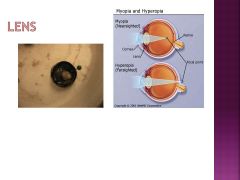
What is the lens and where is it?
|
transmits and refracts light to focus images near or far (accommodation)usually transparent;
|
|

What are the ciliary bodies?
|
controls the suspensory ligaments which adjust the shape of the lens; there are also epithelium cells here that secrete the aqueous humor;
|
|

What is the aqueous humor?
|
liquid found in the anterior chamber of the eye; secreted and removed as waste;
|
|
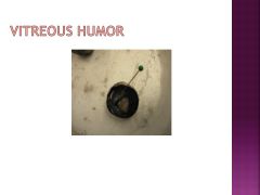
What is the vitreous body/humor and where is it?
|
gel-like substance on the inside of the eyeball; gives support and shape;
|
|
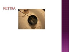
What is the retina and where is it?
|
the neural tissue containing photoreceptive cells located on the inner surface of the posterior portion of the eye;
|
|
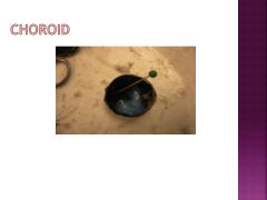
What is the choroid and where is it?
|
black substance on the back of the eyeball; behind the retina; absorbs any stray light; vascular layer ;
|
|
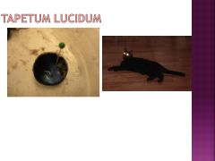
What is the tapetum lucidum and where is it?
|
blue-green iridescent membrane on back of sheep eye, reflective structure acts like a mirror, reflecting light back through the retina, allowing for better night vision;
|
|
|
What are the mammillary bodies responsible for? What disease are they associated with
|
play a role in memory, Korsakoff’s syndrome- amnesia resulting from severe alcoholism;
|
|
|
What 2 structures make up the striatum?
|
caudate nucleus & putamen;
|