![]()
![]()
![]()
Use LEFT and RIGHT arrow keys to navigate between flashcards;
Use UP and DOWN arrow keys to flip the card;
H to show hint;
A reads text to speech;
49 Cards in this Set
- Front
- Back
|
what are some of the ways animals can detect changes in the enviro?
|
smell, sound, sight, pressure, temp change, magnetic, balance, gravity and electricity |
|
|
types of sensory receptors
|
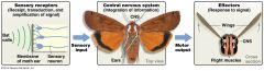
Sensory receptors are located throughout the body and are categorized by the type of stimulus -Many types of sensory receptors convey environmental changes into electrical signals -Mechanoreceptors: pressure -Photoreceptors: wavelengths of light -Chemoreceptors: specific molecules -Thermoreceptors: changes in T -Nociceptors: harmful stimuli (e.g., tissue injury) -Electroreceptors: electric fields -Magnetoreceptors: magnetic fields |
|
|
how are senses conveyed to the brain?
|
the ability to sense a change in the enviro depends on two processes: 1.Transduction 2. Transmission |
|
|
transduction |
the conversion of an external stimulus to an internal signal in the form of an action potential |
|
|
Transmission |
of signals to the CNS
|
|
|
Sensory transduction |
when cells are in the resting state, the inside of the plasma membrane is more negative than the exterior. - when ion flows cause the interior to become more positive than the resting potential, the membrane is depolarized - if changes in ion channels cause cell interior to become more negative than the resting potential, the membrane is hyperpolarized - if a sensory stimulus induces a large change in a sensory receptor's membrane potential, there is a change in firing rate of action potentials sent to brain - amt. of depolarization of the sensory receptor is proportional to intensity of the stimulus |
|
|
what are the two keys to understanding how brain interprets sensory information?
|
1. receptor cells tend to be highly specific - e.g. ear receptors respond best to certain pitches 2. each type of sensory neuron sends its signals to a specific portion of the brain - diff regions are specialized for interpreting the various stimuli e.g. sound is interpreted in the temporal lobes; sight is interpreted by occipital lobes at back of brain |
|
|
how do crustaceans maintain their sense of balance? Mechanoreception |
there are a variety of cells and organs that sense changes in pressure - Statocyst; of a crab is a fluid filled sac and lined with pressure receptor cells. *also contains a Ca-rich particle that rests on bottom - when flipped over, the particle bumps up against receptor cells, which send a signal an action potential to the brain - response is to turn crab upright |
|
|
Mechanoreception |
sensing pressure changes -pressure change affect plasma membrane, which causes ion channels to open or close... causes ion flow and depolarization *pressure -sensing systems are used for a variety of changes in the enviro such as: hearing, physical pressure on skin, movement of muscles, stretching of blood vessels, lateral line |
|
|
how do hair cells respond to changes in pressure? |
in vertebrates, ion channels are found in hair cells - pressure receptor cells - contain outgrowths (stereocilla; microvilla with actin) - found in tetrapod ears and anamniote lateral lines |
|
|
6 step process of signal transduction in hair cells
|
1. pressure waves bends stereocilla 2. potassium channels open in response to bending 3. membrane depolarizes due to influx of K+ 4. depolarization triggers inflow of calcium ions 5. synaptic vesicles fuse with plasma membrane 6. neurotransmitter is released and diffuses to afferent neuron |
|
|
what is the best studied type of pressure sensing? |
hearing; ability to sense sound, which consists of waves of pressure in air or H2O - the # of pressure waves that occur in one second is called frequency of the sound Hz |
|
|
3 structures of the ear
|
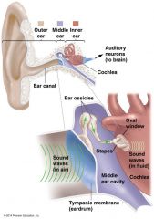
1. outer ear; collects pressure waves & funnels them into ear canal 2. middle ear 3. inner ear *all separated by a membrane |
|
|
Outer Ear:
|
collects pressure waves & funnels them into ear canal - waves strike the tympanic membrane (eardrum)* separates outer ear from inner ear; TM vibrates back and forth with the same frequency as sound waves |
|
|
Middle ear:
|
vibrations passed to 3 ear bones (i.e. Ossicles) in middle ear: 1. Malleus 2. Incus and 3. Stapes * stapes vibrates against oval window ( separates the middle ear from inner ear) *oval window oscillates in response and generates waves in Cochlear Fluid (pressure waves sensed by hair cells) -overall, ear translates airborne waves into fluid-borne waves |
|
|
amplification of the sound wave
|
Because tympanic membrane is ~15X larger than oval window, the amount of vibration induced by sound waves is increased 15X by passing through the middle ear -The 3 ossicles further amplify the vibrations from the tympanic membrane The overall effect is an amplification by a factor of 22X, which stimulates the hair cells of the cochlea |
|
|
the three ossicles
|
1. malleus 2. incus 3. stapes earbones |
|
|
cochlea |
is the auditory portion of the inner ear. It is a spiral-shaped cavity in the bony labyrinth, in humans making 2.5 turns around its axis, the modiolus. A core component of the cochlea is the Organ of Corti, the sensory organ of hearing, which is distributed along the partition separating fluid chambers in the coiled tapered tube of the cochlea
|
|
|
basilar membrane in the mammalian ear
|
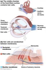
Basilar membrane varies in stiffness, and sounds of different frequencies cause the membrane to vibrate maximally in specific spots along its length -Resulting in the bending of hair-cell stereocilia -Thus hair cells in particular places on the membrane respond to certain frequencies -These differences are interpreted by the brain as different pitches |
|
|
basilar membrane |
The basilar membrane within the cochlea of the inner ear is a stiff structural element that separates two liquid-filled tubes that run along the coil of the cochlea, the scala media and the scala tympani.
|
|
|
Tectorial membrane |
smaller surface within the inner ears cochlea
|
|
|
ear diagram
|
|
|
How does hearing work?
|
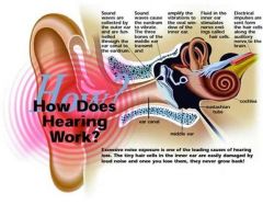
1. sound waves are collected by the outer ear and are funneled through the ear canal to the eardrum 2. sound waves cause the eardrum to vibrate. The three bones of the middle ear transmit and amplify the vibrations to the oval window of the inner ear 3. fluid in the inner ear stimulates nerve endings called hair cells 4.. electrical impulses are sent from the hair cells along the auditory nerve to the brain |
|
|
the cochlea has a set of internal membranes that divide it into 3 chambers - hair cells form rows in the middle chamber - the bottom of each hair cell connects to a structure called the basilar membrane - the stereocilia of the hair cells also touch another smaller surface called the tectorial membrane |
|
|
what do animals hear?
|
Comparatively, human hearing is not particularly acute -Humans can hear sounds between 20 Hz and 20,000 Hz -Elephants use infrasound's (sound frequencies too low for humans to hear) to communicate -Communication over long distances - Bats use ultrasonic sounds (sound frequencies too high for humans to hear) to echolocate (navigate by sound) -Also seen in marine mammals (can perceive shape better with ears than eyes) |
|
|
how does the lateral line work in aquatic animals? |
Fish & larval amphibians have a lateral line that detects changes in pressure -Contains hair cells Changes in pressure cause stereocilia (embedded in gel-like cupula) in hair cells to bend-Bending of cilia produces an action potential in sensory neurons to the brain -Aquatic animals get information about pressure changes at specific points along the head and body |
|
|
arthropod eye |
Compound eyes thought to have evolved once (in arthropod ancestors) - composed of 100's to 1000's of light sensing columns called Ommatidia *each ommatidium has a lens that focuses light onto a small # of receptor cells (2-4) than in turn send axons to the brain - Each ommatidium contributes info about one small piece of the visual field *similar to pixels on a computer * more ommatidia= better resolution= visual predators |
|
|
Mammalian eyes |
the simple (camera) eye evolved independently in two widely divergent groups: 1. cephalopods 2. vertebrates - the simple eye is a structure that focuses incoming light onto a layer of receptor cells |
|
|
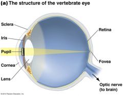
structures of the eye
|
Outermost layer of eye is a tough, white tissue called the sclera -Front of sclera forms cornea, a transparent sheet of connective tissue -Inside the cornea is a colored, round muscle called the iris *Can contract or expand to control amount of light entering eye -Hole in the center of the iris is the pupil 1.Light enters eye through cornea > pupil> strikes a curved, clear lens 2.Cornea and lens focus incoming light onto retina in the back of eye |
|
|
|
|
|
how does light pass through the eye?
|
inside the cornea is a colored, round muscle called the iris - it can contract or expand to control amt of light entering the eye Hole in the center of the eye is the pupil - light enters eye through cornea> pupil> strikes a curved clear lens - cornea and lens focus incoming light onto retina in the back of the eye |
|
|
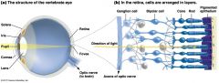
structures of the Retina
|
the retina comprises 3 distinct, synapsing cell layers: 1. light sensing photoreceptors, which form a layer at the back of retina; these cells respond to light 2. an intermediate layer of connecting neurons called bipolar cells 3. ganglion cells which form the front or innermost layer of the retina and whose axons project to the brain via optic nerve |
|
|
what do rods and cones do?
|
Photoreceptors in the vertebrate eye consist of rods and cones *Rods are sensitive to dim light but not to colors: night *Cones are much less sensitive to faint light but are stimulated by different wavelengths of light (colors) -Rods dominate most of the retina, but one small spot in the center of the retina (fovea) has only cones |
|
|
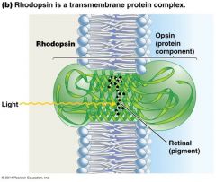
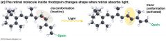
how do rods and cones detect light?
|
Rods & cones have segments packed with membrane-rich disks containing large quantities of transmembrane protein opsin -Each opsin molecule is associated with pigment molecule retinal -The two-molecule complex is called rhodopsin -Retinal changes shape (at 11th carbon) when it absorbs a photon of light, leading to a change in opsin’s conformation -Shape changes leads to action potential sent to brain |
|
|
|
|
|
why do we have a blind spot?
|
On the back of our eye, the retina is the stuff that detects the light. All the information that the retina picks up is sent to the brain through the optic nerve. The only problem is that the optic nerve needs a way to get out of the eye. The place where it leaves is where we have our blind spot.
|
|
|
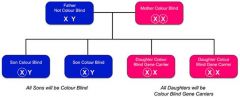
The (recessive) gene for red/green color blindness is found on the X chromosome Affects 5–10% of men, 0.5% of women Why is it more common in men? |
|
|
|
how do other animals see color?
|
How well an animal can see color depends on the number of cones and particular opsins it has -This ability may be determined by their environment *Nocturnal animals have few cones and many rods *Other organisms have ≤4 types of opsins (better color vision than humans) |
|
|
examples of color vision in animals
|
-Coelacanths live in deep water and have two opsins that perceive blue = penetrates to greater depth than other colors -Primates that eat fruit have 2–3 opsins that are sensitive to 550 nm wavelengths *Distinguish between green, yellow, and red in order to find ripe fruit -Some insects and birds see in UV which serves as a pollination adaptation because some flowers have ultraviolet patterns *Species ID? Mate selection? |
|
|
gustation
|
the sense of taste: originates in chemoreceptors
|
|
|
oflaction
|
the sense of smell; originates in chemoreceptors
|
|
|
taste buds
|
taste sensing chemoreceptors are clustered in structures known as..... - in humans, TB are scattered around the mouth ans throat, but mostly are found on the tongue - each TB contains 100 taste cells, taste receptors that synapse to sensory neurons |
|
|
what is responsible for the "salty" taste?
|
strong evidence suggests that salt and sour sensations result from ion channel activity - saltiness is primarily a result of Na+ ions dissolved in food -Na+ ions flow into taste cells through open Na+ channels & depolarize the cell membrane |
|
|
what is responsible for "acidic" taste
|
"sourness" -results from presence of protons which flow into taste cells through H+ ion channels |
|
|
pheremones
|
a chemical released by females into the enviro causing a behavioral change
|
|
|
thermoreception
|
heat energy change detected by animals - hypothalamus (CNS) regulates homeostatis with T - Pit vipers use them to sense heat energy given off by predators and prey *brains may make a thermal image |
|
|
electroreception
|
sensation of electric fiels - in sharks, ampullae of Lorenzini are specialized protions of the lateral line system |
|
|
vomeronasal organs
|
sensory organs in the nasal regio - in these organs, signals are sent to different parts of the brain e.g. a snake may follow its prey by using its vomersonasal organ |
|
|
magnetoreception
|
sensing magnetic fiels - found in bacteria, fungi, invertebrates & all vertebrates classes - Earth produces a magnetic field (from Fe core) as it rotates on its axsis how do they sense magnetic fields? - deposits of Fe inside sensory neurons in beak *disruption of Earth's magnetic field can prvent birds from navigating *sea turtles |