Reading...
![]()
Play button
![]()
Play button
![]()
Use LEFT and RIGHT arrow keys to navigate between flashcards;
Use UP and DOWN arrow keys to flip the card;
H to show hint;
A reads text to speech;
69 Cards in this Set
- Front
- Back
|
sensory pathway
|
the impulses involved in sensations follow precise pathways:
receptors sensory neurons sensory tracts sensory areas |
|
|
receptors
|
*detect changes (stimuli) and generate impulses.
Receptors are VERY SPECIFIC to the kinds of changes they respond to: EX. **retina receptors detect light rays **nasal receptors detect vapors *ALL generate electrical nerve impulses |
|
|
sensory neurons
|
transmit impulses from the receptors to the CNS.
*FOUND in both cranial & spinal nerves, but ONLY carry impulses from ONE type of receptor |
|
|
sensory tracts
|
white matter in the spinal cord or the brain that transmits the impulses to a specific part of the brain
|
|
|
sensory areas
|
most are in the cerebral cortex
**AREAS feel & interpret the sensations. *learning to interpret sensations begins in infancy and continues throughout life |
|
|
characteristics of sensations
|
how the sensory areas work with information from the receptors:
*projection *intensity *contrast *adaptation *after-image |
|
|
projection
|
the sensation SEEMS to come from the area where the receptors were stimulated BUT it is actually felt by cerbral cortex.
EX. phantom pain, the person still FEELS that the body part is there after amputation because the nerves continue to generate impulses and PROJECTION of the limb |
|
|
intensity
|
some sensation are felt more distinctly and to a greater degree than others
EX. a WEAK stimulus (dim light) will affect a small # of receptors, but a STRONGER stimulus (bright sunlight) will stimulate a larger # of receptors |
|
|
contrast
|
the effect of a previous or simultaneous sensation on a current sensation, which MAY be exaggerated or diminished.
**function of the brain, which constantly compared sensations EX. HOT day, jump in COLD pool, water feels COLDER than it actually is because the brain senses a significant difference IN temperatures. |
|
|
adaptation
|
becoming unaware of a continuing stimulus
EX. wearing a watch, at first you feel it on your wrist but cutaneous receptors for touch/pressure adapt quickly to a continuing stimulus, no change=nothing to detect, you become unaware that it is there |
|
|
after-image
|
the sensation remains in the consciousness even after the stimulus has stopped.
EX. a bright after-image seen after watching a camera flash go off |
|
|
cutaneous senses
|
the dermis and subcutaneous tissue contain sensory receptors
*free nerve endings *encapsulated nerve endings |
|
|
free nerve endings
|
*respond to ANY intense stimulus
*receptors for HEAT, COLD, ITCH & PAIN |
|
|
encapsulated nerve endings
|
*means there is a cellular structure around the nerve ending
*receptors for TOUCH & PRESSURE |
|
|
referred pain
|
pain that originates in an internal organ may be felt in a cutaneous area.
EX. *heart attack may be felt in the left arm and shoulder *gallstones may be felt in the right shoulder |
|
|
muscle sense
|
we do not need to see our muscles to be sure they are working
*conscious *unconscious |
|
|
conscious muscle sense
|
is felt by the parietal lobes
|
|
|
unconscious muscle sense
|
is used by the cerebellum (balance & equilibrium part of brain) to coordinate voluntary movements
|
|
|
sense of taste
|
*receptors for taste are found in taste buds
most are on the tongue *receptors detect CHEMICALS in SOLUTION in the mouth **chemicals =foods **solution=saliva |
|
|
taste receptors
|
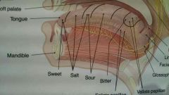
five (perhaps more) general types of taste receptors:
sweet sour salty bitter savory (a taste like grilled meat) |
|
|
impulses from taste buds
|
transmitted from the facial & glossopharyngeal (7th & 9th cranial) nerves to the taste areas in the parietals temporal cortex.
*sense of taste important b/c it makes eating enjoyable. |
|
|
interfere with sense of taste
|
medications may interfere with the sense of taste
|
|
|
sense of smell
|
gives a great deal of pleasure & practical info
|
|
|
receptors for smell (olfaction)
|
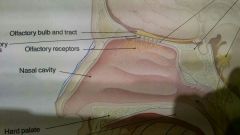
receptors are chemoreceptors that detect vaporized chemicals that have been sniffed into the upper nasal cavities
|
|
|
olfactory receptors
|
scent receptors (detect vapors) that generate impulses carried by olfactory nerves (1st cranial) thru the ethmoid bone to olfactory bulbs.
* vapors may stimulate many combinations of receptors **human brain can distinguish between 10,000 scents |
|
|
hunger & thirst
|
called visceral sensations, and both senses (hunger & thirst) are specialized in the hypothalamus
*triggered by internal changes. EX. *hunger receptors- detect changes in blood nutrient levels *thirst receptors- detect changes in the body water content (water-to-salt proportion) |
|
|
the eye
|
contains the receptors for vision and a refraction system that focuses light rays on the receptors in the retina
|
|
|
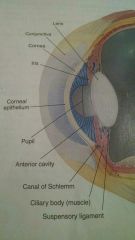
conjunctiva
|
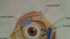
a thin membrane that lines the eyelid
|
|
|
conjunctivitus
|
inflammation of the conjunctiva membrane may be caused by allergies, bacteria, or virus
*makes eyes red, itchy, & watery |
|
|
tears
|
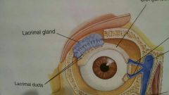
*produced by the lacrimal glands
(located at the upper, outer corner of the eyeball) *tear secretion in constant, & increased by emotional situations & irritating chemicals (onion vapors) *contains lysozyme (an enzyme) that INHIBITS the growth of most bacteria on surface of wet, warm eye *tears, mostly water w/1% sodium chloride |
|
|
orbit
|
most of eyeball is within and protected by
|
|
|
chemoreceptors in mouth
|
detect chemical changes in mouth (food) and nasal (vapors)
|
|
|
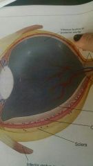
layers of the eyeball
|
**the eyeball has 3 layers
OUTER- sclera MIDDLE- choroid INNER- retina |
|
|
sclera
|
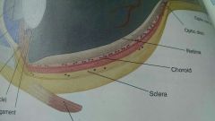
OUTER LAYER **is the thickest layer of the eyeball.
*made of fibrous connective tissue that is VISIBLE as the white of the eye |
|
|
cornea
|
*ANTERIOR (front) portion of the eyeball
**differs from the rest of the sclera, in that it is TRANSPARENT -it has no capillaries -it covers the IRIS & PUPIL inside eye -1st part of eye to refract (bend) light rays |
|
|
refraction of light rays
|
the DEFLECTION or BENDING of a ray of LIGHT as it passes through one object and into another if greater or lesser density
|
|
|
choroid layer
|
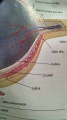
MIDDLE LAYER EYEBALL
*contains blood vessels and a dark blue pigment (from melanin) that absorbs light within the eyeball and prevents glare **IN anterior portion of choroid -ciliary body -iris |
|
|
ciliary body
|
|
|
|
lens
|
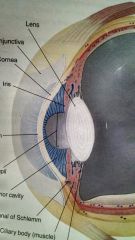
made up of clear/transparent, elastic protein
*has no capillaries *looks like an onion peel |
|
|
iris
|
the iris is INFRONT of lens
**the COLORED part of the eye **pigment is a form of melanin |
|
|
pupil
|
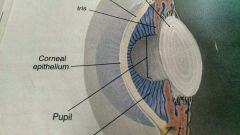
black dot on eye LIGHT RAYS enter through the pupil
**the central opening **two sets of muscle fibers in the iris change the diameter of the pupil |
|
|
retina
|
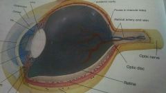
lines the POSTERIOR 2/3's of the eyeball and contains VISUAL receptors (rods and cones)
|
|
|
rods
|
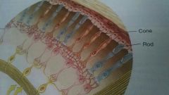
detect ONLY the presence of light
*rods are abundant toward the edge of retina *best vision in dim light or @ night depends on rods |
|
|
cones
|
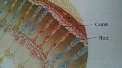
detects color which are the different wavelengths of visible light
*most abundant in the center of the retina |
|
|
two cavitities in eyeball
|
posterior cavity
anterior cavity |
|
|
posterior cavity
|
LARGER of the two cavities
*contains vitreous humor (semisolid substance) which keeps the retina in place |
|
|
anterior cavity
|
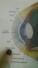
contains aqueous humor (the tissue fluid of the eyeball)
*found between the back of the cornea and front of lens |
|
|
occipital lobes
|
are the VISUAL areas of cerebral cortex
*sees images upside down and as left/right |
|
|
binocular vision
|
*EACH eye transmits a slightly different picture
*BRAIN takes images and puts them together, forming a SINGLE IMAGE and interprets them *BRAIN also turns image right side up (occipital sees images upside down and as left/right) |
|
|
ERRORS of refraction
|
not focusing on images
CORRECTION= contacts/eye glasses |
|
|
the ear physiology
|
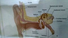
OUTER EAR
MIDDLE EAR INNER EAR |
|
|
OUTER ear consists of
|
auricle (pinna)
ear canal |
|
|
AURICAL (pinna)
|
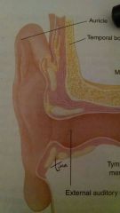
made of cartilage covered by skin
|
|
|
EAR CANAL
(aka) external auditory meatus (or) auditory canal |
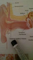
is lined with skin that contains ceruminous glands
|
|
|
MIDDLE ear consists of
|
tympanic membrane (ear drum)
eustacian tube |
|
|
TYMPANIC MEMBRANE
|
*sound waves vibrate
*CONSISTS OF 3 bones AKA *ossicles (smallest bones in body) ***malleus, incus, stapes*** |
|
|
EUSTACIAN TUBE (auditory tube)
|
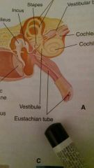
connects throat & ear
*allows AIR to enter or leave the middle ear cavity *air pressure in middle ear must be the same as atmospheric pressure in order for the eardrum to vibrate properly. |
|
|
INNER ear consists of
|
*cochlea (hearing)
organ of Corti (inside of cochlea) hearing receptors *semicircular canals *saccule *utricle **balance & equilibrium |
|
|
cochlea
|
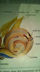
hearing receptors (shaped like a snail shell)
CONTAINS- organ of Corti |
|
|
the two senses of the ear:
|
hearing and equilibrium
|
|
|
saccule & utricle (inner ear)
|
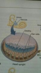
membraneous sacs between the cochlea and the semicircular canals
**provides information about the position of the body at REST |
|
|
semicircular canals
|
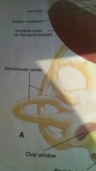
fluid filled membraneous ovals
**provide info about body in MOTION |
|
|
hearing receptors
|
specialized hair cells or stereocilia contain endings of the cochlear branch of 8th cranial nerve
|
|
|
temporal lobes of the cerebral cortex
|
auditory areas of the brain. receives impulses from both ears
|
|
|
arterial receptors
|
found in the aorta (*largest artery in the body) & the carotid arteries
*pressoreceptors *chemoreceptors |
|
|
chemoreceptors in arteries
|
detect changes in OXYGEN, BLOOD pH, & CARBON DIOXIDE
|
|
|
pressoreceptors in arteries
|
detect changes in the blood pressure in the carotid and aortic.
|
|
|
carotid artery
|
found in the neck (cervicle area)
|
|
|
hypoxemia
|
a decrease in oxygen blood level
*HYPO (decrease or low) *ox (oxygen) *emia (blood) **HYPER= increase or high **sensory to medulla to speed heart rate/speed breathing to increase oxygen intake |