![]()
![]()
![]()
Use LEFT and RIGHT arrow keys to navigate between flashcards;
Use UP and DOWN arrow keys to flip the card;
H to show hint;
A reads text to speech;
32 Cards in this Set
- Front
- Back

|
Ant. Diagastric |
|

|
Wharton duuct |
|

|
Hypoglossal |
|
|
Anatomical structure of submandibular |
Pair of elongated, flattened hooks which have 2 arms |
|
|
Superficial arm |
Lies partially inf. To post half of mandible |
|
|
Deep arm |
Hooks around post. Margin of mylohyoid to enter oral cavity, lies on lateral surface of hyoglossus, lateral to root of tongue |
|
|
Wharton duct canliculi |

|
|
|
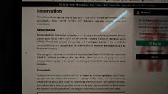
Innervation |
|
|
|
Vasculature |
Submental arteries branch of facial Submental vein |
|
|
Surgicaly excision amd nerves |
|
|
|
Relationship with nerves |

|
|

|
Mandible |
|

|
Submental artery |
|

|
C |
|
|
Secretions of sublingual are what type |
Mixed ,though predominantly mucus |
|
|
Medial border of sublingual |
Genioglossus |
|
|
What passes next to sublingual glands and between genioglossus |
Submandibular gland duct and it's nerves |
|
|
What is plica sublinualis |
It is the elevated crestbof mucous membrane called sublingual fold Each fold begins behind and laterally and move forward to meet at frenulum to join to sublingual papillae |
|
|
Sublingual ducts (8-20) |
Duct of rivinus |
|
|
Vessel suplly |
Sublingual and submental artery and same name k veins me |
|
|
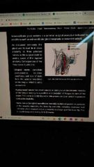
Innervation of sublingual gland |

Superior salivatory nucleus se chorda tympani carry karta fer lingual branch of mandibular ke sath fuse hota @ submandibular ganglion aur fer waha se 2 nerve jaate |
|
|
Sympathetic innervation of sublingual |
Superior cervical ganglion |
|
|
Ranula |

|
|

|
Zygomatic arch |
|

|
Facial |
|

|
I dunno how i remember this but retro mandibular |
|
|
Anatomical position of parotid |
Upar zygomatic arch Pichu sternocliedomastoid Aagae masetter jispe uske duct jaate and later pierces buccinator Nice mandible |
|
|
Two lobes of parotid seprated by facial nerve |
Deep and superficial |
|
|
Duct of parotid |
Stensen opens in front of upper second molar |
|
|
Anatomical relationship of partoid |

U cant see buccal and cervical in da image |
|
|
Innervation of parotid |

|
|
|
Clinical relevance |

|