Reading...
![]()
Play button
![]()
Play button
![]()
Use LEFT and RIGHT arrow keys to navigate between flashcards;
Use UP and DOWN arrow keys to flip the card;
H to show hint;
A reads text to speech;
128 Cards in this Set
- Front
- Back
|
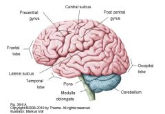
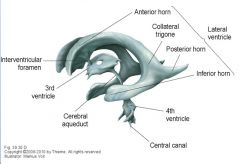
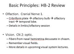
What are the four lobes of the brain?
|
Clockwise; the frontal, parietal, occipital and temporal lobes.
|
|
|
What is the name of the structure that separates the two hemispheres of the brain?
|
The cerebral longitudinal fissure.
|
|
|
What is the name of the sulcus that separates the temporal lobe from the frontal and parietal lobes?
|
The lateral sulcus
|
|
|
Which sulcus defines the border between the frontal and parietal lobes?
|
The central sulcus
|
|
|
Which two gyri are located in front and behind the central sulcus and what are they responsible for?
|
The precentral gyrus; contains the primary motor cortex, and the postcentral gyrus; contains the primary somatosensory cortex
|
|
|
Which sulcus separates the parietal and occipital lobes?
|
The parieto-occipital sulcus
|
|
|
Which sulcus lies entirely in the occipital lobe and runs backward toward the occiptal pole? Which cortex does it contain?
|
The calcarine sulcus; it contains most of the primary visual cortex.
|
|
|
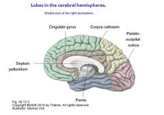
Which sulcus is located internally and separates the neocortex from the mesocortex of the singulate gyrus?
|
The cingulate sulcus
|
|
|
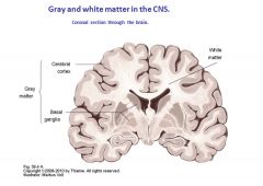
Cortical areas of the brain possesing 6 histological layers is called what?
|
Isocortex
|
|
|
Cortical areas of the brain NOT possesing 6 histological layers is called what?
|
The allocortex
|
|
|
What are the two areas the allocortex is divided into?
|
The paleocortex; includes the olfactory area AND
The archicortex; includes the fasciolar gyrus, hippocampus, dentate gyrus and parahippocampal gyrus |
|
|
From outside to inside, what are the six cellular layers of the allocortex?
|
1. Molecular layer
2. External granular layer 3. External pyramidal layer 4. Internal granular layer 5. Internal pyramidal layer 6. Multiform layer |
|
|
The molecular layer:
|
This layer is mostly devoid of cells and only contains small neurons.
|
|
|
The external granular layer:
|
This layer contains many "granule cells" (non-pyramidal cells), and a few pyramidal cells whose dendrites branch out in the granular layer and the molecular layer.
The granular cells are mostly GABAergic inhibitory neurons, and the pyramidal cells are excitatory and use glutamate as their neurotransmitter. |
|
|
The external pyramidal layer:
|
This layer contains many small pyramidal cells that are oriented with their bases toward the white matter.
The axon of each cell arises from the base and travels down into the white matter. A dendrite emerging from the apex travels upward into the external granular and molecular layers where it divides into its terminal branches (apical tuft) |
|
|
The internal granular layer:
|
This layer contains many granular cells that receive afferent input from thalamic neurons via the thalamocortical projection.
The cell fibers are tangentially oriented - forming the external "band of Baillarger" with staining. |
|
|
The internal pyramidal layer:
|
This layer contains medium and large pyramidal cells. The largest cells in this layer, the Betz cells, are found only in the region of the precentral gyrus. The thickly myelinated fibers of these cells form the corticonuclear and corticospinal tracts.
This layer also contains many tangentially oriented fibers - these form the internal "band of Baillarger" |
|
|
The multiform layer:
|
This layer of polymorph cells is subdivided into an inner, less dense layer containing smaller cells, and an outer layer containing larger cells.
|
|
|
|
|
|
|
|
|
|
|
|
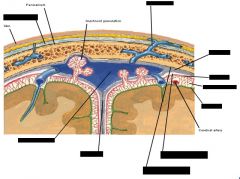
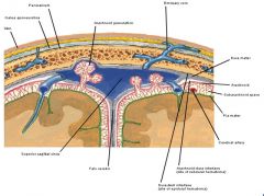
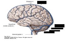
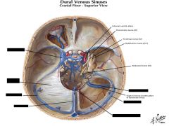
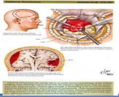
What type of hematoma develops if the bridging veins rupture?
|

The bridging veins!!!
|
|
|
|
|
|
|
|
|
|
|
|
|
|
|
|
|

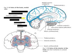
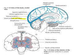
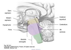
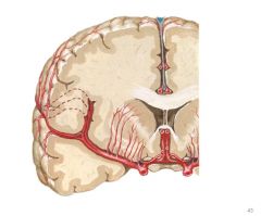
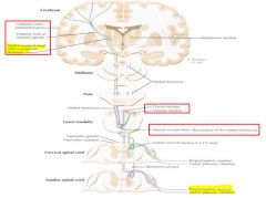
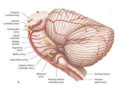
(The dark yellow region is called the diencephalon)
|
|
|

|
|
|
|
|
|
|
|
|
|
|
|
|
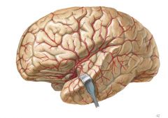
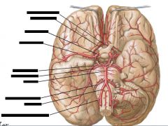
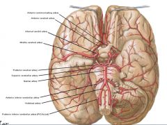
Middle cerebral artery. Runs over the insula.
|
|
MCA; note numerous small vessels (lenticulostriate) branching off and supplying interior areas.
Often a common place for cranial hemorrhage in patients with hypertension. |
|
|
|
|
|
|

|
|
|

|
|
|

|
|

|

|
|
|
|
|
|
|
|

|
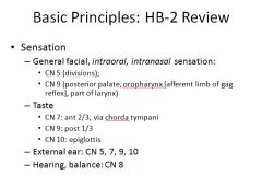
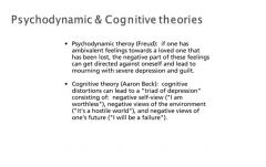
Affect descriptions:
Full = full range of emotion Euthymic = normal mood; fine Dysphoric = depressed or sad Labile = changeable mood Constricted = restricted range of emotion Blunted = even less emotion Flat = no emotion at all |
|
|

|
|
|
|
|
|

|
|

|

|
|
|
|
|
|
|
|
|
|
|
|
|
|
|
|
|
|
|
|
|
|
|
|
|
|
|
|
|

|
|
|
|
|
|
|
|
|
|
|
|
|
|
|
|
|
|
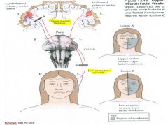
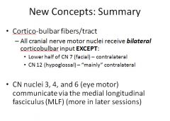
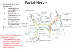
So ½ face paralysis is usually not an emergency, whereas ¼ face paralysis IS an emergency
|
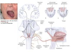
The tongue moves toward the damaged side.
The only way to tell if deviation of the tongue is due to an UML or a LML is the history and physical. If the cortex is involved, then some other motor functions should be effected. |
|
|
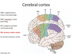
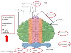
Anterior = Motor
Posterior = Sensory AMPS |
|
|
|
|
|
|
|
|
|
|
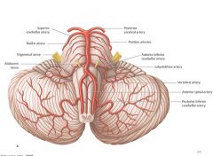
Hemorrhage of the small vessels coming off of the medial cerebellar artery can cause disfunction of the VPL nuclei
|
|
|
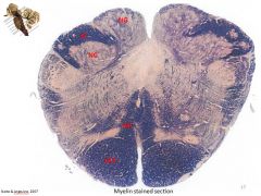
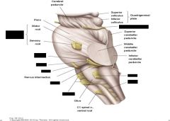
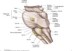
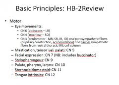
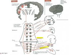
Sectioned at the medulla.
CST = corticospinal tract (myelinated motor fibers to lower levels) NC = nucleus cuneatus FC = Fasciculus cuneatus (heavily myelinated) NG = nucleus gracilis ML = Medial leminiscus |
|
|
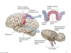
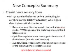
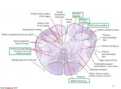
All sensory fibers from the neck down go through the VPL
|
|
|
|
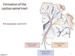
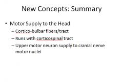
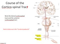
Corticonuclear fibers; also called the cortico-bulbar or just the “bulb”
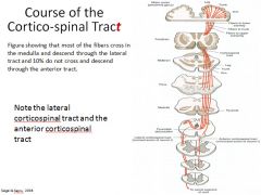
10% of fibers do not cross in the anterior cortico-spinal tract |
|
|
|
|
|
|
|
|
|
|
|
|
|
|
|
|
|
|
|
|
|
|

|

|
|
|
|
|
|
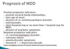
Major depressive disorder correlates most with what socioeconomic variable?
|
Marital/relationship status
|
|

|

|
|

|
|
|
|

|
|
|
|
|
|

66% contemplate and 10-15% commit suicide
|
|
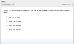
25% in 6 months
|
|
|
|
|
|

Assess for suicidal thoughts
|

|
|

|

|
|

|

|
|

|

|
|

|
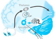
The amygdala can react to a threat before we perceive it.
|
|
|
The size/volume of what brain structure can predispose a person to PTSD?
|
The hippocampus; people with smaller ones are more predisposed
|
|
|
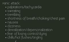
With or without agoraphobia
|
|
Panic disorder or generalized anxiety disorder
|

Specific/social phobia
|
|
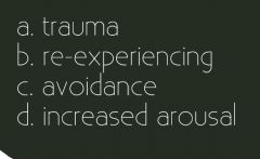
PTSD or acute stress disorder
|
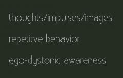
Obsessive Compulsive Disorder
|
|

Other disorders of anxiety
|
|
|
|
|
|
|
|
|
|
|
|
|
|
|
|
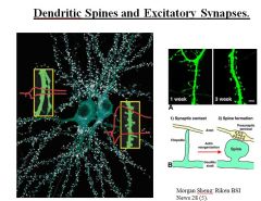
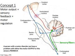
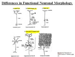
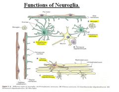
A single axon may synapse on hundreds of cells via the terminals and spines (see picture B)
|
|
|
|
|
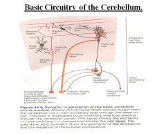
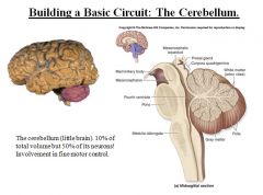
The integrator for the circuit is the Purkinje cell; it actually is responsible for brief inhibition.
This functions as a delay loop; like preventing yourself from biting your tongue when eating. |
|
|
|
|
|
|
|
|
|
|
|
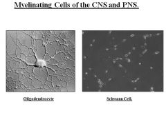
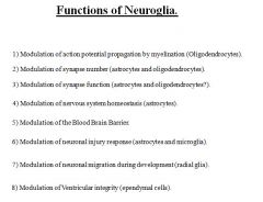
Oligodendrocytes myelinate several axons whereas Schwann cells myelinate one.
It is much worse to lose an oligodendrocyte than a Schwann cell. |
|
|
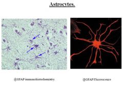
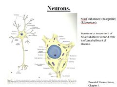
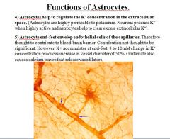
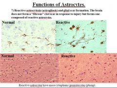
GFAP is detected in activated astrocytes, which are a hallmark of injury to the head/brain.
|
|
|
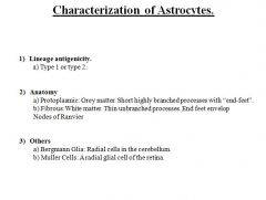
Syncytium: A series of interconnected cells
|
|
|
|
|
|
|
|
|
|
|
|
|
|
|
|
|
|
|
|
|
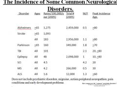
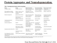
In cells with slow or no turnover, disease comes from accumulated damage
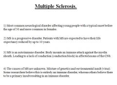
In cells with fast turnover, disease comes from neoplasia/mutation TBI = traumatic brain injury SCI = spinal cord injury * MS is the most common neurological disorder that affects young people; particularly young females. |
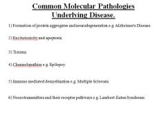
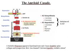
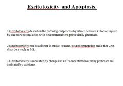
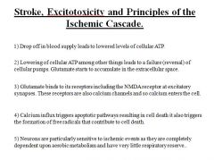
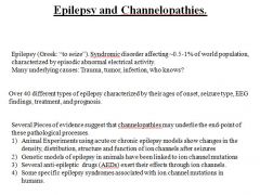
Excitotoxin: The neurotransmitter glutamate
Excitotoxicity: A neuron gets overexcited and dies; from calcium overload. Channelopathies: Lead to an aberrant firing of neurons |
|
|
|
|
|
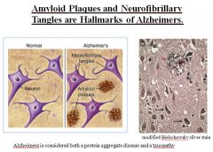
Tauopathy: The neurofibrillary tangles are made up of tau protein (microtubule proteins).
|
|
|
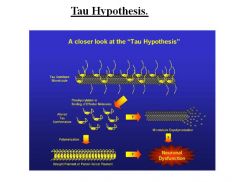
Since the tau protein is self-aggregating, it is not stabilizing the microtubule and thus, the microtubule depolymerizes.
|
|
|
|
|
|
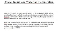
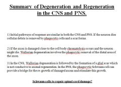
Peripheral nerves can regenerate because they don’t have the nasty inhibitory/proapoptotic factors that the CNS does.
|
|
|
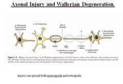
Chromatolysis: the nucleus moves off to the side and lots of Nissl bodies are formed; the cell can either recover or die at this point.
|
|
|
|
|
|
|
|
|
|
|
|
|
|
|
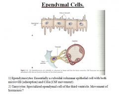
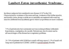
Pathology of Lambert Eaton Syndrome:
|
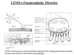
|