Reading...
![]()
Play button
![]()
Play button
![]()
Use LEFT and RIGHT arrow keys to navigate between flashcards;
Use UP and DOWN arrow keys to flip the card;
H to show hint;
A reads text to speech;
43 Cards in this Set
- Front
- Back
|
The primary function of the nervous system is survival via homeostasis. How is this achieved (3 things)?
|
1. detects internal & external stimuli from environment
2. integrates info to determine response 3. coordinates cells & tissues to respond *nervous system divided into CNS & PNS |
|
|
What does the nervous system consist of (at the cellular level)?
|
neurons (excitable cells)
their (neuron's) processes glia (supporting cells) |
|
|
What two things does a neuron consist of?
|
1. cell body (soma or perikaryon)
2. neurites |
|
|
What are the two types of neurites?
|
dendrites & axons
|
|
|
(dendrites/axons) project a short distance from the soma and contain receptors that receive chemical signals from other neurons. They taper distally & branch extensively increasing signal reception.
|
dendrites
|
|
|
What are the dendritic branches of a single neuron referred to as?
|
dendritic tree
|
|
|
(dendrites/axons) are singular projections that may extend long distances w/i the nervous system.
|
axons
|
|
|
The beginning of the axon at the soma is the _________________ followed by the _______________
|
axon hillock ("hill")
initial segment ("peak of hill") |
|
|
The initial segment is uninsulated and rich in what?
|
voltage-gated sodium channels
|
|
|
The end of the axon is the ____________, which form ___________ with other neurons or effectors
|
axon terminal (terminal button/Fr. button)
synapses *terminals contain neurotransmitter vesicles |
|
|
Info transmitted w/i the neuron is a/an (electrical/chemical) signal & info transmitted btwn neurons is a/an (electrical/chemical) signal
|
electrical (w/i)
chemical (btwn) |
|
|
The action potential (wave of depolar.) travels from the ____________ to the _________________
What happens next? |
from the initial segment to the axon terminal
neurotransmitters released from terminal to act on postsynaptic cell membrane |
|
|
How is a signal transmitted further once the receptors receive neurotransmitters?
|
Receptors generate AP that travel to the postsynaptic neuron cell body
|
|
|
T/F
neurons are more numerous than glial cells (neuroglia) |
FALSE
glial cells are much more numerous 10X! (supporting cells) |
|
|
What are glial cells responsible for?
|
-regulating chemical milieu in EC space
-myelinating neurons -phagocytosis & repair -lining fluid filled ventricles (cavities) of brain |
|
|
*Chemicals are released from what part of the neuron, allowing it to communicate with another cell?
|
Button (terminal)
|
|
|
*Which of the following CNS structures is not considered part of the brain?
cerebrum cerebellum brainstem spinal cord |
spinal cord
*CNS = brain + spinal cord |
|
|
What is the primary function of the CNS?
|
processing of sensory info & executor of responses
*more complex processing is done in brain |
|
|
T/F
Brain size correlates w/ intelligence |
FALSE
correlates directly w/ body size *2% body weight, 15-17% of cardiac output, 20% oxygen |
|
|
T/F
The orientation of the central axis of the nervous system (neuraxis) changes above the brainstem |
TRUE
|
|
|
Define the following ABOVE the neuraxis
rostral caudal ventral dorsal |

rostral – toward front of brain
caudal – toward back of brain ventral – toward bottom of brain dorsal – toward top of brain |
|
|
Define the following BELOW the neuraxis)
rostral caudal ventral dorsal |
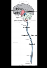
rostral – toward the cerebrum
caudal – toward the bottom of the spinal cord ventral – toward the front dorsal – toward the back |
|
|
Tracts are named based on origin and termination. What are the origin/termination of the spinocerebellar & corticospinal tract?
|
spinocerebellar:
origin- spinal cord termination- cerebellum corticospinal: origin- cerebral cortex termination- spinal cord |
|
|
Define afferent & efferent w/i the CNS:
*NOT the same as in PNS |
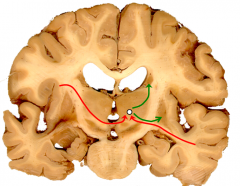
afferent= conducting TOWARD a structure
(red) efferent= conducting AWAY from a structure (green) |
|
|
Cerebrum
structure- function- |
structure- consists of diencephalon & 2 cerebral hemispheres containing *gyri (ridges) & *sulci &f fissures* (valleys), outer laminar surface is referred to as cerebral cortex
function-complex processing of sensory info & formulation of volitional motor responses * sensations must reach cerebral cortex to be CONSCIOUSLY perceived |
|
|
T/F
Like the PNS, groups of neurons w/i the CNS are called ganglia |
FALSE
called nuclei w/i the CNS |
|
|
*The internal fluid (CSF) filled spaces of the cerebrum are __________
|
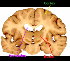
ventricles
*ventricles secrete CSF which "floats" brain w/i cranial cavity for protection & circulates over the brain & spinal cord & empties into the bloodstream via VENOUS SINUSES |
|
|
The cortex & nuceli are rich in neuronal cell bodies are referred to as ___________
|
gray matter (darker)
|
|
|
Areas under the cortext w/ collections of tracts/fasiculi are rich in myelinated axons are are referred to as ____________
|
white matter (lighter)
|
|
|
*What are the 2 main white matter structures w/i the cerebrum & what are their functions?
|
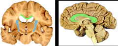
1. corpus collosum (GREEN)- major pathway for axons crossing btwn cerebral hemispheres
2. internal capsule (BLUE)- major pathway btwn cerebral hemispheres & more caudal structures (brainstem, spinal cord, thalmus, etc) |
|
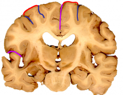
Gyri (RED) and sulci (BLUE) can be seen above. What are the deeper, more consistent sulci (PURPLE) called?
|

fissures
|
|
|
*What do the primary fissures divide the cerebrum into?
|
functional "lobes"
*areas of similar function are located near one another |
|
|
*What is a collection of neurons that forms the laminar structure on the surface of the cerebrum?
|
cortex
|
|
|
Give the structure/function of the following major nuclear group w/i the cerebrum:
*thalmus |
structure-
part of diencephalon function- somatic info sensation & motor function |
|
|
Give the structure/function of the following major nuclear group w/i the cerebrum:
*hypothalmus |
structure-
w/i diencephalon function- ANS regulation |
|
|
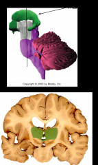
Give the structure/function of the following major nuclear group w/i the cerebrum:
*basal ganglia |
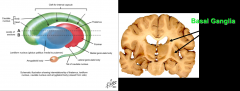
structure-
group of nuclei, associated structures include caudate nucleus, putamen, globus, pallidus, subthalmic nucleus, & substantia nigra function- motor procession |
|
|
Give the structure/function of the following major nuclear group w/i the cerebrum:
*hippocampal formation |
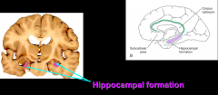
structure-
located in medial temporal lobe on each side function- consolidation of memory |
|
|
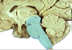
Cerebellum
*structure- *function- |
structure (ORANGE)- 2 hemispheres & vermis, attached to posterior aspect of brainstem, surface more densely convoluted than cerebral cortex (cerebrum)
function- uses complex sensory info UNCONSCIOUSLY to COORDINATE motor activity |
|
|
Brainstem
*structure- *function- |
structure (BLUE)- contains tracts btwn spinal cord & brain, neurons for both somatic motor & parasympathetic (ANS) output, modulates control of respiration, HR, & other autonomic functions
function- CNS interface with PNS (cranial nerves), provides first order processing of primary sensation & hearing, vestibular, taste *motor & sensory functions of the head & autonomic control of some body functions* |
|
|
Which component of the CNS is referred to as a "primitive brian"?
|
brainstem
|
|
|
What are the 3 divisions of the brainstem & what are their functions/locations?
|
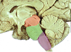
1. midbrain
(f) eye movement control, CN 3 & 4 (l) rostral most part of brainstem (RED) 2. pons (f) communication btwn cerebrum & cerebellum, CN 5, 6, 7, & 8 (l) middle portion of brainstem (GREEN) 3. medulla (f) conscious reticular formation, autonomic control (heart rate & breathing), CN 9, 10, 11, & 12 (l) caudal most part (PURPLE) |
|
|
Spinal Cord
structure- *function- |
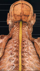
structure (YELLOW)- along spine
function- primary interface for CNS w/ body via peripheral nerves, sends & receives info from brain via axons (tracts) *major tracts that convey info btwn brain & body |
|
|
What do neurons contained in the spinal cord do (3 things)?
|
1. receive primary somatic SENSORY info
2. directly & indirectly modulate MOTOR activity of muscles 3. modulate AUTONOMIC activity of viscera |