Reading...
![]()
Play button
![]()
Play button
![]()
Use LEFT and RIGHT arrow keys to navigate between flashcards;
Use UP and DOWN arrow keys to flip the card;
H to show hint;
A reads text to speech;
79 Cards in this Set
- Front
- Back
|
where do the pontine nuclei migrate from? where do they go to?
|
from: derived from alar plate, migrate ventrally and medially
to: form the basilar pons. (will form sensory portions of associated CNs) |
|
|
what will the axons from the migrated pontine nuclei form?
|
the middle cerebellar peduncle that wraps around the brainstem in a lateral position
|
|
|
In the development of the pons, what does the basal plate form?
|
-motor (pontine cranial) nuclei of pontine tegmentum
-the midline and dorsal motor components of cranial nerve nuclei *(Pons is from rhombencephalon (metencephalon)) |
|

The pontine tegmentum is 1 of the 2 major divisions of the pons. What is the pontine tegmentum a rostral continuation of?
|
medullar reticular formation, cranial nerve nuceli, and ascending tracts.
|
|

what is contained in the pontine tegmentum?
|
pontine cranial nerve nuclei and ascending sensory tracts
(pontine cranial nerve nuclei does NOT = pontine nuclei) |
|

The basilar pons is the 2nd major division of the pons. what two tracts run through the basilar pons?
|
corticobulbar and corticospinal
(*in addition to pontine nuclei & middle cerebellar peduncle) |
|
|
In the pons, where is the pontine cranial nerve nuclei located? where is the pontine nuclei located?
|
pontine cranial nerve nuclei - pontine tegmentum
pontine nuclei- basilar pons |
|
|
at what level in the pons is the Cochlear component (SSA) of CN 8 located?
What info do the axons from the cochlea (spiral ganglia) carry to the brainstem? |
ponto medullar junction
auditory info |
|
|
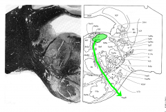
where are the cell bodies of origin located for the cochlear component?
What do axons from cochlear nuclei eventually FORM? |
dorsal cochlear nucleus and ventral cochlear nucleus
LATERAL LEMNISCUS (primary ascending auditory pathway) |
|
|
Do the cochlear component fibers cross? if so, where?
|
yes: primarily cross midline at more rostral levels in the TRAPEZOID BODY and in the dorsal and intermediate acoustic stria.
(go on to form lateral lemniscus, some axons synapse in superior olivary nucleus) |
|
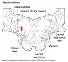
which number labels the trapezoid body?
nucleus of trapezoid body? |
8
9 |
|
|
what is the origin of the efferent cochlear bundle?
|
superior olivary nucleus
|
|
|
what reflex is the superior olivary nucleus involved in? Via which CN?
|
reflex for sound dampening via CN 7 (stapedius)
|
|
|
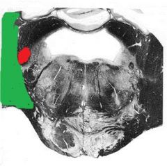
in the ponto medullary junction, what is included in the central processing nuclei?
|
nucleus of the trapezoid body, superior olivary nucleus and nucleus of the lateral lemniscus.
|
|
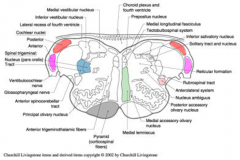
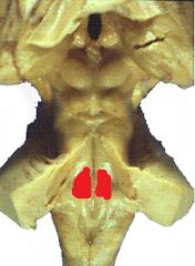
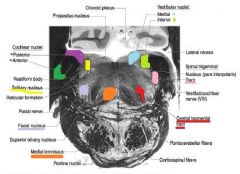
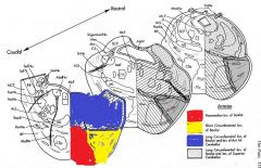
Name the colored parts.
|
red: superior olivary nucleus
orange: nucleus of the trapezoid body. |
|
|
Axons from the cochlear nuclei containing info from accessory, collect primarily where?
|
in the contralateral lateral lemniscus.
|
|
|
Q: Axons in the lateral lemniscus arise primarily from neurons located ......?
|
in the dorsal ventral cochlear nuclei
*ascend bilaterally w/ a contralateral predominance |
|
|
Where do the axons from the central processing nuclei ascend to? (3)
|
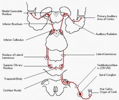
inferior colliculus
medial geniculate body (thalamus) Cortex (temporal, transvers gyri of Heschel= consious perception) |
|
|
why doesn't an ipsilateral lesion cause a hearing loss (usually)? what are the exceptions?
|
because the lateral lemniscus travels bilaterally with a contralateral predominance.
exceptions: the nerve or cochlear nuclei are involved. |
|
|
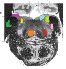
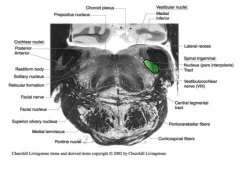
how many nuclei are innervated by the vestibular component of the vestibulocochlear nerve? what are they?
|
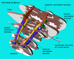
4;
medial vestibular (yellow) lateral vestibular (deiter's) (green) superior vestibular (orange) inferior vestibular (blue) |
|
|
how do the projections of the vestibular nuclei reach the cerebellum?
|
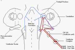
via the juxtarestiform body located in the lateral vestibular nucleus.
*juxtarestiform body = balance, coordination |
|
|
T/F
There are some direct connection of the vestibulocochlear nerve to the flocculonodular lobe. |
TRUE!
|
|
|
what is the function of the juxtarestiform body?
|
balance coordination
|
|
|
what are the two vestibular related nuclei/ structures?
|
oculomotor nuclei and thalamus
|
|
|
Via what structure do the vestibular related pathways have an impact on balance? how?
|
Both are via the medial longitunal fasciculus.
Thalamus: Projects to cortex to give conscious perception of equilibrium and orientation |
|
|
what 2 tracts relates the vestibular pathway and the spinal cord?
|
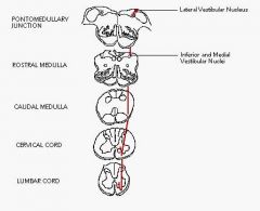
lateral vestibulospinal tract and medial vestibulospinal tract
(each from associated nuclues) |
|
|
what is another name for the medial vestibulospinal tract?
|
Medial longitudinal fasciculus
|
|
|
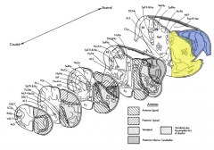
what is the function of the medial vestibulospinal tract?
function of lateral vestibulospinal tract? |
inhibitory to neck and upper trunk muscles; contributes to head-righting reflexes in relation to vision
excitatory to extensor spinal alpha motorneurons |
|
|
Besides CN 8, the caudal pons conatins the nuclei for which two CNs?
|
6 and 7
|
|
|
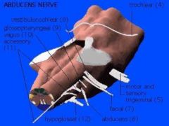
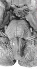
where does the facial nerve exit the brainstem?
|
just anterior to CN 8 at pontomedullar junction
(@ level of 6 & 7) |
|
|
describe the course of the facial nerve.
|
axons loop around the nucleus of CN 6 and exit ipsilaterally.
|
|
|
what indicates the presence of the abducens nucleus?
|
the bump on the floor of the 4th ventricle
|
|
|
what surrounds the abducens nucleus?
|
facial colliculus (facial fiber axons)
|
|
|
are the axons that form the loop in the pathway of the facial nerve primarily sensory or motor?
|
motor
|
|
Name the colored "bump" on the floor of the 4th ventricle.
|
Facial colliculus
(^surrounding abducens nucleus & axons of CN 7) |
|
|
how does cortical innervation from the facial nerve differ when innervating muscles of the forehead vs muscles of the lower face?
|
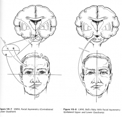
bilateral innervation to the muscles of the forehead, contralateral to muscles for the lower face.
|
|
|
If there is a lesion of the motor nucleus in the pons, what kind of paralysis should we find of the facial muscles?
|
ipsilateral forehead paralysis, contralateral paralysis to mouth and chin.
|
|
|
what kind of fibers does the intermedius branch of the facial nerve contain?
|
GVE (parasympathetic functions), SVA and GSA
|
|
|
For the parasympathetic aspect of the intermedius branch of the facial nerve, which nuclei is involved?
|
superior salivatory nucleus (GVE)
|
|
|
detail the pathway that starts at the limbic system and affects the intermedius branch of the facial nerve resulting in weeping and mouth watering to odors.
|
GVE from limbic system and olfactory areas through hypothalamus to the brainstem via the dorsal longitudinal fasciculus.
|
|
|
signals from the brainstem on the intermedius branch of the facial nerve result in?
|
reflex lachrymation and gustatory stimulation of salivation
|
|
|
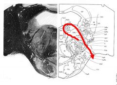
describe the pathway of the solitary nucleus (specifically gustatory nucleus). Detail it from mouth to cortex.
|
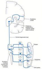
Begins with (SVA) sensory fibers from taste buds on anterior 2/3 tongue and hard and soft palates that run in chorda tympani from geniculate ganglion. Then it will project into the brainstem as intermedius nerve and enter the tractus soliatrius to synapse in the rostral portion of the nucleus. The nucleus then sends the info via the central tegmental tract to the thalamus which projects it to cortex.
|
|
|
which portion of the solitary nucleus is specifically for gustatory nucleus?
|
rostral portion
|
|
|
what tract is used by the solitary nucleus to project taste fibers to the thamalus?
|
central tegmental tract
|
|
|
which part of the thalamus are the taste fibers from the solitary nucleus projected too?
|
ventral posteromedial
|
|
|
what two parts of the ear are innervated by the intermedius nerve?
|
skin of the ear wall of acoustic meatus and external tympanic membrane.
|
|
|
what nucleus supplies the GSA for the intermedius nerve?
|
spinal trigeminal nucleus
(via geniculate ganglion) |
|
|
what is the pathway of the GSA fibers of the intermedius nerve?
|
axons innervating areas around ear enter brainstem in the nervous intermedius and descend in spinal tract of trigeminal to synapse at the spinal V nucleus.
|
|
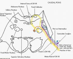
Label each nervous intermedius colored tract with the appropriate fiber (GSA, GVE, etc)
|
Red: GVE
purple: SVA blue: GSA |
|
|
via what sulcus does the abducens nerve exit?
|
inferior pontine
|
|
|
what is the function of the abducens nerve?
|
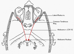
motor innervation to ipsilateral lateral rectus muscle of the eye.
|
|
|
what is one of the most frequently injured CN that also has a long intracranial course?
|
abducens nerve
|
|
|
what nuclei does the abducens nerve have a complex central connection?
|
oculomotor nuclei
|
|
What CN exhibits this path?
|
abducens
|
|
|
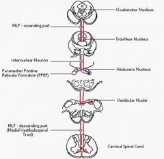
how does the abducens nerve interact with the oculomotor nucleus?
|
via medial longitudinal fasciculus
|
|
|
The pontine reticular formation contains many nuclei contributing to the recticular formation.
What do the rostral projections of the reticular formation regulate? where do they project to? |
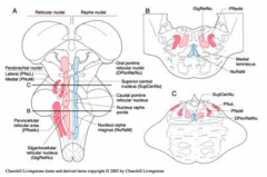
regulate consciousness and caudal projections (thalamus).
|
|
|
what do the caudal projections of the reticular formation regulate?
|
motor and sensory functions
|
|
|
what two tracts run through the substance of the basilar pons?
|
corticospinal and corticonuclear tracts
|
|
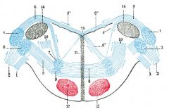
label all of the colored nuclei.
|
:)
|
|
|
at what level in the brainstem does the medial lemniscus reach its midline position?
|
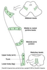
Medulla Levels (after medulla-pons junction)
|
|
|
In the pons, are the major tracts located?
|
caudal pons.
|
|
|
what tract alway runs in tandem with the ALS?
|
rubrospinal tract
|
|
|
name the three subdivision of the trigeminal nucleus. which one is most rostral? most caudal?
|
pars oralis (rostral), pars interpolaris, pars caudalis (caudal).
|
|
|
which subdivision of the trigeminal nucleus are you most likely to see at the region of the caudal pons?
|
pars oralis.
|
|
|
what are the major tracts that enter the cerebellum beginning at the pontomedullary junction?
|
inferior cerebellar peduncle (restiform body) enters the cerebellum with vestibulocerebellar projections (juxtarestiform body)
|
|
|
which tract wraps latterally around the inferior peduncle at the pontomedullar junction?
|
middle cerebellar peduncle
|
|
Name the colored regions. What part of the brainstem is this?
|
pontomedullar junction
|
|
|
what two structures make up the roof of the fourth ventricle?
|
superior cerebellar peduncles and superior medullar velum
|
|
|
what is the blood supply to the caudal pons?
|
vertebral arteries, basilar artery, posterior inferior cerebellar artery, anterior inferior cerebellar arteries.
|
|
|
What is the major blood supply to just the pontomedullary junction?
|
vertebral & medial branches of the basilar (yellow) &
posterior inferior cerebellar artery (blue) |
|
|
what is the major blood supply to just the caudal pons?
|
branches of the basilar artery: paramedian (red), and short (yellow) and long (blue) circumferential branches.
(May also be supplied by anterior inferior cerebellar artery. AICA) |
|
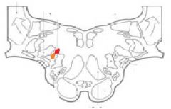
Q: What cranial nerve exits here?
|

hypoglossal nerve (CN 12)
|
|
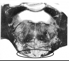
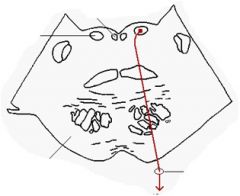
Q: A lesion in the area indicated in the above figure is most likely to affect which cranial nerve?
|
CN 6
(corticospinal tract) |
|
|
Q: The tract btwn CN 3 & 4 that carries information to coordinate eye movements is the __________________
|
medial lemniscus fasciculus
|
|
|
Obj.
List Cranial nerves of the pontomedullary junction |
CN 8 (vestibular)- cochlear component (SSA) & vestibular component
CN7 (facial)- motor & intermediate branch CN6 (abducens |
|
|
Obj.
Nuclei of cranial nerve 8 (cochlear component) & associated functions |
**dorsal & ventral cochlera nuclei- recieve axons from cochlear spiral ganglion
nucelus of trapezoid body- sound localization superior olivary nucleus- sound localization & reflex sound dampening via CN 7 stapedius & CN 5 tensor tympani nucleus of lateral lemniscus- sound localization & acoustic reflex |
|
|
Obj.
Nuclei of cranial nerve 8 (vestibular component) & associated functions |

-medial vestibular nucleus
-lateral vestibular nucleus (Deiter's) -superior vestibular nucleus -inferior vestibular nucleus (^ vestibular= balance coordination) -oculomotor nuclei- conscious perception of equilibrium & orientation |
|
|
Obj.
Nuclei of cranial nerve 7 & associated functions |
motor branch:
-facial motor nucelus: facial motor function to muscles of facial expression & (SVE) somatic motor to pharygneal arch muscles (stylohyoid, digastric post. belly, stapedius) -abducens nucleus: motor to upper & lower face intermediate branch: -superior salvatory nucleus: (GVE) Stimulate secretions (lachrymal & salivary glands, mucousa) -solitary nucleus: (SVA) taste |
|
|
Obj.
Nuclei of cranial nerve 6 & associated functions |
-oculomotor nuclei: conjugate eye movements
|