Reading...
![]()
Play button
![]()
Play button
![]()
Use LEFT and RIGHT arrow keys to navigate between flashcards;
Use UP and DOWN arrow keys to flip the card;
H to show hint;
A reads text to speech;
24 Cards in this Set
- Front
- Back
|
Descirbe the counterstrain technique for the lateral trochanter tender point.
|
Location: Found about 12 cm below the greater trochanter along the lateral surface. Treatment: Moderate abduction of the thigh. Fine-tune with hip internal or external rotation.
|
|
|
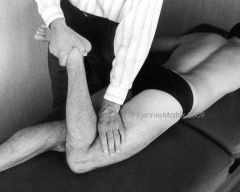
Descirbe the counterstrain technique for the biceps femoris tender point.
|
Location: Just below the proximal attachment and approximately one-third above the distal attachment. Treatment: Flexion of the knee with external rotation and slight abduction of the tibia. Plantar flexion of the foot by pressing on the calcaneus. If further reduction in tenderness is needed, the hip is extended.
|
|
|
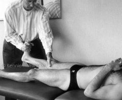
Descirbe the counterstrain technique for the semimembranosus tender point.
|
Location: Just below the proximal attachment and approximately one-third above the distal attachment. Treatment: Flexion of the knee with internal rotation and slight adduction of the tibia. Plantar flexion of the foot by pressing on the calcaneus. If further reduction in tenderness is needed, the hip is extended.
|
|
|
Descirbe the counterstrain technique for the rectus femoris point.
|
Location: At the superior end of the patella in the musculotendinous region of the rectus femoris, inferior at the patellar ligament, or in the musculotendinous region at the superior attachment of the muscle. Treatment: Mild hyperextension of the knee by dorsiflexing the foot and flexing the hip. The more proximal the tender points require more flexion of the hip.
|
|
|
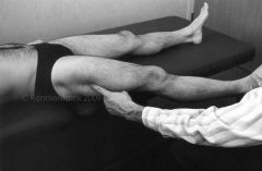
Descirbe the counterstrain technique for the popliteus tender point.
|

Location: Within the belly of the popliteus muscle. Treatment: Slight flexion of the knee with internal (medial) rotation of the tibia.
|
|
|
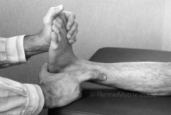
Descirbe the counterstrain technique for the tibialis anterior tender point.
|
Location: About one-third down from the top of the tibia in the tibialis anterior muscle and/or in the distal tendon where it wraps around the medial malleolus near the deltoid ligaments. Treatment: Dorsiflexion and inversion of the foot.
|
|
|
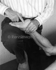
Descirbe the counterstrain technique for the fibularis brevis tender point.
|
Location: About halfway down from the superior and lateral end of the fibula and/or in the tendinous region inferior and anterior to the lateral malleolus in the sinus tarsi. Treatment: Eversion and slight plantar flexion of the foot as needed.
|
|
|
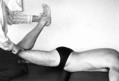
Descirbe the counterstrain technique for the gastrocnemius tender point.
|
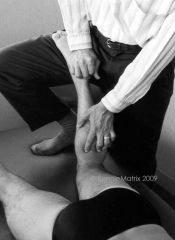
Location: Within the proximal regions of the two bellies of the gastrocnemius muscle distal to the lower popliteal margin. Treatment: Marked plantar flexion of the foot. Pushing the calcaneus toward the knee while the knee is flexed reduces tension on the gastrocnemius.
|
|
|
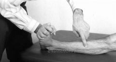
Descirbe the counterstrain technique for the quadratus plantae tender point.
|
Location: At the anterior plantar aspect of the calcaneus. Treatment: Marked flexion of the forefoot while simultaneously pushing the heel toward the forefoot to enhance release of the plantar fascia and quadratus plantae.
|
|
|
How can the medial collateral ligament be unloaded?
|
Passive flexion of the knee. The semimembranosus muscle attaches to the medial collateral ligament and medial meniscus. Passive flexion of the knee unloads the hamstring muscles and further unloads tension on the medial collateral ligament.
|
|
|
What muscle can help reduce tension on the lateral collateral ligament?
|
Popliteus. It attaches to the lateral condyle of the femur, anterior and inferior to the lateral collateral ligament. Its action reduces tension on LCL.
|
|
|
What motion will loosen MCL and LCL?
|
Passive flexion and internal rotation of the knee.
|
|
|
What position is the leg and foot typically in during an ACL tear?
|
Forefoot is plantar flexed on ground and the knee internally rotated.
|
|
|
What may result from a lateral-to-medial force to the knee?
|
"Unhappy triad": ACL, MCL and medial meniscus tear.
|
|
|
How does the body protect an injured ACL?
|
Tightening the hamstring muscles.
|
|
|
What muscle helps reduce medial meniscal entrapment and injury?
|
Semimembranosus, it retracts the medial meniscus posteriorly during knee flexion.
|
|
|
What muscle helps reduce lateral meniscal entrapment and injury?
|
Popliteus, it retracts the lateral meniscus posteriorly during knee flexion.
|
|
|
What would cause a false-negative Lachman test?
|
Tight hamstrings.
|
|
|
When is the ACL have the highest tension?
|
Last 30 degrees of knee extension and with internal rotation.
|
|
|
If hamstring muscle tightness cannot be reduced, what must you consider?
|
A significant ACL tear or other damage to the knee joint.
|
|
|
What structures protect the PCL?
|
Oblique popliteal ligament, popliteus and quadriceps muscles.
|
|
|
Where will popliteus tendonitis manifest pain?
|
Posterior knee, palpable tenderness in the region of the lateral collateral ligament and at the motor point found slightly below the middle of the popliteal crease.
|
|
|
What is done to rehabilitate ACL injuries non-operatively?
|
Immobilization of the knee in flexion and stretching of the quadriceps muscles.
|
|
|
What is done to rehabilitate PCL injuries non-operatively?
|
Immobilization of the knee in extension and stretching of the hamstring muscles.
|