• Shuffle
    Toggle On
    Toggle Off
  • Alphabetize
    Toggle On
    Toggle Off
  • Front First
    Toggle On
    Toggle Off
  • Both Sides
    Toggle On
    Toggle Off
  • Read
    Toggle On
    Toggle Off
Reading...
Front

Card Range To Study

through

image

Play button

image

Play button

image

Progress

1/37

Click to flip

Use LEFT and RIGHT arrow keys to navigate between flashcards;

Use UP and DOWN arrow keys to flip the card;

H to show hint;

A reads text to speech;

37 Cards in this Set

  • Front
  • Back

Definition of high output failure and ddx

hyperdynamic state with CO and ¯O2 extraction with pulmonary/peripheral edema because diastolic dysfunction and circulatory overload
§ hyperdynamic state results in myocardial damage over time


§ DDx:


1.anemia,


2.hyperthyroid,


3.AV fistula,


4.Paget’s disease,


5.thiamine deficiency,


6.sepsis

Def low output

o low output – most common; ¯CO (systolic dysfunction), filling pressures (diastolic dysfunction), systemic O2 extraction

HF R sided vs L sided

– useful in acute CHF as gives indication as to which chamber failing
o Rarely isolated
o Useful clinically for etiology and treatment


§ L - Anterior MI: hypotension, pulmonary edema, S3


§ R - Inferior MI: increased JVP, no pulmn edema, hypotension responsive to fluids

HF · Systolic vs Diastolic – allows specific tx


o Systolic
§ impairment of contractility (¯CO)
§ Seen well on echo – impaired stroke volume and EF
§ Often have a component of systolic dysfunction
§ usually caused by myocyte destruction: AMI,


o Diastolic (1/3)
§ Caused by impaired relaxation (low intracellular energy), increased wall thickness, increased myocardial interstitial collagen
§ more prominent at older ages
§ caused by ischemia, cardiomyopathies caused by chronic HTN, AS

PATHOPHYSIOLOGY

Primary insults in heart failure myocyte loss and volume overload which lead to pump dusfunction
· This results in a compensatory response to maintain CO:
o Activation of renin-angiotensin-aldosterone system which results in
§ Increased volume via aldosterone
· Initially natriuretic peptides released by the heart (including BNP) promote diuresis and act counter to the R-A-A hormone system
· Tx with diuretics

§ Increased vascular tone via angiotensin II resulting in increased afterload and preload (moves the myocytes along the Frank-Starling curve)
· Tx with ACEi and ARBs

o Activation of sympathetic nervous system which causes increased levels of catecholamines and results in increased afterload and preload
§ NO made by the vascular endothelium plays a role in counter-balancing this
§ Tx with b-blockers

· These compensatory mechanisms result in a number of physiological effects:
o Increased SVR/afterload
o Increased preload
o Increased HR

· This perpetuates a viscious cycle leading to further cardiac ischemia, myocyte loss, remodeling, and decreased LV function leading to further CO impairment thus starting to cycle over again.
· Eventually these “compensations” lead to decompensation

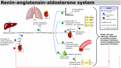
renin angiotensin system

ETIOLOGY OF CHRONIC HEART FAILURE

· Things that increase the workload of the heart:
o Hyperdynamic states
§ AV fistula
§ Hyperthyroid
§ Beri-Beri
§ Paget’s
§ Anemia
§ Sepsis

o Increase afterload (both pulmonary and systemic)
§ HTN
§ Valvular heart disease
§ Pulmonary HTN
§ COPD

· **Rememberpulmonary causes of heart failure is termed Cor Pulmonale**

· Things that decrease the muscle power of the heart
o Ischemia – CAD, AMI
o Cardiomyopathy
§ Infective – myocarditis, endocarditis, pericarditis
§ Inflammatory – amyloidosis
§ Toxicologic – EtOH
§ Drugs – adriamycin
§ Degenerative – idiopathic, HOCM
§ Post-partum
§ Anatomic – congenital heart disease
§ Tachyarrhythmia associated cardiomyopathy

ETIOLOGY OF ACUTE DECOMPENSATED HEART FAILURE

Dietary indiscretions
o Medication non-compliance
o Disease Progression
· Increased afterload
o HTN crisis
o Valvular dysfunction
o Physical, environmental, emotional stress (increase catecholamines)
o Acute PE
· Decreased output
o Ischemia
o Arrhythmia
o Acute myocarditis
o Disease progression
· Increased demand
o Thyrotoxicosis
o Pregnancy
o Anemia

NYHA classification

I- no limitations - asymptomatic during usual daily activities


II - slight limitation - mild symptoms with ordinary activities (dysp,fatigue,CP)


III-moderate limitation - symptoms noted with minimal activity


IV- severe limitation - symptoms at rest

HF CXR


· Order of appearance of X-ray findings
1. Increased heart size, cardiothoracic ratio >50%
· usually noted first b/c usually Hx of prior CHF or may have DCM, etc
2. Large hila with indistinct margins
3. Prominence of superior pulmonary veins; CEPHALIZATION OF FLOW
4. Fluid in the interlobar fissure
5. Pleural effusion
6. Kerley B lines (base on the pleural surface of the lung and extending horizontally a variable, but usually short, distance toward the center of the chest).
7. Alveolar edema

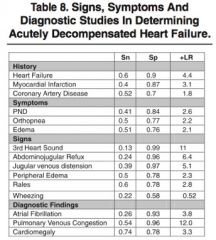
Signs and symptoms HF

NIPPV


what does it do and whats the evidence

o Increases FRC
o Increased oxygenation
o Decreased sympathetic tone
o Decreased LV preload b/c of intrathoracic pressure
o Decreased work of breathing (decreased metabolic demands of respiratory muscles)
o Relative afterload reduction


o Vital FMR, et al. Non-invasive positive pressure ventilation (CPAP or bilevel NPPV) for cardiogenic pulmonary edema. Cochrane Database of Systematic Reviews 2013, Issue 3.


reduced mortality significantly(NNT 13)
§ reduced intubation significantly(NNT 8)


Gray A, Goodacre S, et al. Noninvasive ventilation in acute cardiogenic pulmonary edema. N Engl J Med 2008;359:142-151


§ Severe APE excluded
§ Findings: no difference in mortality or intubation rates
§ There was greater mean improvements at 1 hour in terms of patient-reported dyspnea, heart rate, acidosis, and hypercapnia.


Li H et al. A comparison of bilevel and continuous posi- tive airway pressure noninvasive ventilation in acute cardiogenic pulmonary edema. Am J Emerg Med. 2013 Sep;31(9):1322-7. PMID: 23928327. \


no diff cpap/bipap

Morphine

No evidence that it venodilates centrally, there are Swan studies to suggest that it increases CVP
o Sympatholytic: decreased HR, BP, contractility, MVO2
o ADHERE registry (Emerg Med J 2008;25:205-209) shows morphine is an independent predictor of mortality, and is associated with increased ICU admission, days on a vent, and hospital length of stay.


o Amal Mattu suggests that the only reason to use it is for anxiolysis but benzos do that better anyway.
o If you are going to use it: 2 - 5 mg iv prn to effect

Nitrates

o Mainstay of treatment
o Increases nitric oxide which increased cGMP :. vasodilation
o Low dose is primary venous VD, decrease preload
o High dose is arterial VD, decreased BP, afterload (esp in HTN patients)
o Potential for coronary artery VD and reduction of ischemia
o Watch for hypotension: usually responds to fluid challenge

What level of hypotension should NTG be stopped?


How much to give?

o Depends on patient; SBP 75 and talking is OK, SBP 90 and decreased LOC not OK


o Sublingual tab/spray: rapid onset, big doses, duration 30 min
§ 400mcg/spray, consider giving sprays while starting IV NTG
o Patch has poor absorption with diaphoresis!
o IV: start 10 ug/min and increase by 10 ug/min q5min to effect (may need up to 300 ug/min; can be more aggressive with starting dose and titration is adequate BP

Nitroprusside

o Potent vasodilator: venous decreases preload, arterial decreases afterload
o More arterial dilation than nitroglycerin
o Drug of choice with hypertensive crisis and APE
o Continuous infusion, BP monitoring mandatory
o Coronary steal phenomenon = nitroglycerin better with MI b/c of possible coronary steal where less diseased vessels dilate and steal blood flow and increased risk of hypotension
o Thiocyanate toxicity possible: increased agitation, lactic acidosis (especially with renal failure); can also cause methemoglobinemia
o Convert to po therapy ASAP: ACE, NTG, hydralazine

Lasix


1. how does it work?


2. dose and how to give?

MOA: inhibits 2Na/Cl co-transporter in thick ascending loop of Henle :. increases Na and water excretion and decreases preload; also diuretic induced neurohormaonal changes of PGE2, ANP
o Effect is seen quicker than renal effects thus likely a pulmonary vasodilator (this has been shown in swan ganz studies)
o Watch for hypokalemia/magnesemia

o Dose 1mg/kg iv but may require much more with renal failure
o Can give massive doses with renal failure (max is 1000 mg); risk hearing loss
o Doses < 100mg can be pushed, > 100mg should be infused

· ACE-I in acute CHF

ACE-I good in acute setting for afterload reduction
o Captopril 6.25 mg – 25mg po
o Give dose if already on ACE-I, be very careful if not already on
o Do NOT give if critical Aortic Stenosis (afterload redution, increased gradient, reduced coronary perfusion)

Niseritide


o Recombinant BNP

o VMAC trial (Industry sponsored trial), JAMA 2002; 287:1531-40.
§ DBRCT, n=500
§ Primary E.P. = PCWP
§ Secondary E.P. = Sx at 3H
§ Results
§ ↓ PCWP (and improved other cardiac indices)
§ No improved Sx relief at 24hrs
§ No difference in mortality at 18/12 (25% for nesiritde, 21% Nitro, p=0.32)
§ Equivalent to Nitro (at best)
§ Significant hypotension, bradycardia, renal dysfx
§ Trend to higher MR
o JAMA, 2005. Pooled analysis of 860 patients
§ MR was 7.2% v 4.0% , p=0.059(niseritide –v- std Tx)

Treatment of APE in Hypotensive Patients

1.Fluids


2. Dopamine


3. Dobutamine


4. norepi


5.milrinone

Fluids fluids for APE?

o Clinically, small fluid challenge and monitor response of BP and possible worsening of oxygenation
o Small boluses 250 ml NS over 10 min
o Repeat prn if respiratory status not deteriorating
o Should increase BP if hypotension due to hypovolemia
§ REMEMBER that these patients are often hypovolemic overall they just have a redistribution of fluids to their lungs

Dopamine - for APE?

o Low doses: 2-5 ug/kg/min: dopaminergic; renal/splanhnic vasodilation
o Moderate: 5-15 ug/kg/min: beta adrenergic; increased contractility, HR
o High doses: > 15 ug/kg/min: alpha adrenergic; vasoconstrict all vessels, increases BP, HR, contractility (may ppt ishcemia, may worsen pulmn edema - venodilator can reduces worsening of Ped)
o Venoconstriction at low doses may increase wedge pressure and increase edema
o Indication = hypotension SBP 70 - 100 with s/s of hypoperfusion failing fluid challenge:10-20 ug/kg/min

Dobutatmine for APE?

Mainly B1 agonist, some B2 and some alpha
o B1 is +ve ionotrope and B2 is a venodilator
o May lead to hypotension by vasodilation if CO doesn’t increase much
o Excellent for normotensive, caution with borderline hypotension, don’t use alone in hypotensive (May norepi for vasoconstriction)
o Excellent choice for AMI: increases cardiac function without worsening ischemia by increasing heart rate
o Dose is 5 - 25 ug/kg/min: start 2 and increase to 20 ug/kg/min
o Indication = hypotension SBP 70 - 100 with NO s/s of hypoperfusion after fluid challenge

· Norepinephrine


o Mostly alpha agonsist (some beta)
o Drug of choice for volume repleated profound hypotensive (SBP < 70)
o Goal: temporary management until IABP, PTCA, surgery
o Start at 0.1 ug/kg/min and titrate up to 2-3 ug/kg/min
o Indication = hypotension SBP < 70 failing fluid challenge

MI and cardiogenic shock with acute pulmonary edema


· Needs cath lab or PTCA ASAP
· Shock trial showed no benefit of thrombolysis
· Elevate BP with dopamine then lysis is next best option if cath lab not available
· Avoid negative inotropes: no BetaBlockers!
· R/o mechanical complications of MI
o ?RV infarct
o Valve rupture (?murmur)
o Free wall rupture (?PCE w/US)
o VSD (?murmur)
o Consider AoD

ACEi systolic dysf in HF

Decrease mortality and hospitalization rates
· Every patient should be on an ACEi
· If cough, switch to ARB
· Target is ramipril 10mg/d, captopril 150mg/d

Angiotensin Receptor Blockers (ARBs) systolic dysf in HF

Used for contraindication or intolerance of ACE-I
· Increasing evidence of benefit in addition to ACE-I thus may be added for persisting CHF symptoms once other meds are maxed
· Losartan (cozaar), Valasartan (Diovan)

BetaBlockers in HF?

Cardioselective: metoprolol, carvidolol
· Good post MI to reduce mortality by reduction of neurohormnal remodelling
· Not started in acute CHF phase
· Start slow: metoprolol 12.5mg bid

Spirnolactone (Aldactone) - sys HF

· Aldosterone antagonist thus decreases Na+ resorption and increases K+ retention
· Decreases mortality in class III/IV CHF
· NEJM 1999 RALES study: EF < 35% on ACE-I; trial stopped early b/c one year death rate was 35% vs 46% (ARR 11%, NNT about 10); also showed improved symptoms with spirnolactone therapy
· Potassium sparing diuretic thus watch K+ (esp with ACE-I + spirnolactone)
· Dose: 25 - 50 mg po od
· Triamterene (Triazide), eplerenone is a similar agent
· Who should be on spirnolactone?
o NYHA class III or IV (symptomatic on less than ordinary activity or symptomatic at rest)
o EF < 35%
o Already on ACE-I

Lasix

· Symptomatic control, no reduction in mortality
· Side effects: hypokalemia, hypomagnesemia, hypotension, gout
· Metolazone (Zaroxlyn) used for synergy if resistant to lasix: ideally given 2 hrs before lasix dose

Nitroglycerin

· Symptomatic control, no reduction in mortality
· Patch or oral pills
· Tachyphylaxis a problem thus patch only worn for 12hrs/day

Digoxin

·
· Symptomatic improvement but no effect on mortality
· Has been shown to decrease hospital admission rates
· Should NOT replace ACE-I, BB, spirnolactone which do decrease mortality
· Best indication: CHF + Afib + diastolic dysfunction
· Load iv: 0.5 mg then 0.25 mg until effect or 1.5 mg total
· Maintenance: 0.25 mg per day

CCB + what other interventions?

· CCB have no role in CHF managment
· Influenza and pneumoccocus vacination
· Fluid and salt restriction counselling

ICDs


· Increasing role of ICDs in CHF and CAD patients
· MADIT II showed a decreased mortality with ICD use compared to medical therapy in patients with LV EF < 30% post MI


· SCD-HeFT: sudden Cardiac Death in Heart Failure Trial
o ICD vs amiodarone
o N=2521
o NYHA class 2-3 and LVEF < 35%
ICD Amiodarone
o 3 yr mortality 17% 22%
o 5 yr mortality 29% 36%

Chronic Resynchronisation Therapy (CRT) rationale?

· Rationale: prolongation of QRS in patients with CHF has been shown to be an independant predictor of adverse outcomes
o QRS delay occurs in up to 30% of moderate to severe CHF patients
o QRS delay, especially LBBB, causes electrical and mechanical dyssynchrony in patients with depressed LV function which lead to decrease in stroke volume, EF
· CRT = pacing of RV and LV to cause simultaneous contraction and synchrony that has been shown to improve LV function

CRT trials?

CRT trials have looked at CHF patients with EF < 35%, QRS > 120 msec and LBBB and/or evidence of dyssynchrony on echo, normal sinus node function (NOT afib), appropriate chf therapy (ace-i, bb, etc)
· Pacer leads placed in the RV and the Coronary Sinus branch vein to achieve LV stimulation (a cardiac vein!)
· Studies: MIRACLE, InSync ICD, CONTACK CD
o All 3 studies showed symptom improvement


· COMPANION trial
o NEJM 2004
o Medical therapy vs CRT vs CRT + ICD
o Combined outcome of death or hospitalization
o CRT+ICD > CRT alone > medical therapy

Diastolic Dysfunction


· BetaBlockers: rate control for better filling
· ACE-I: benefit not well defined but should be used as most have some systolic component
· Treat underlying cause: HTN, valve dz, etc
· Can’t decrease preload too much b/c they are preload dependant: careful with diuretics and nitroglycerin