Reading...
![]()
Play button
![]()
Play button
![]()
Use LEFT and RIGHT arrow keys to navigate between flashcards;
Use UP and DOWN arrow keys to flip the card;
H to show hint;
A reads text to speech;
79 Cards in this Set
- Front
- Back
|
the left and right coronary arteries arise from the aorta at the ........ behind the left and right cups of the aorta.
|
sinuses of Valsalva
McG 559 |
|
|
Name the cardiac valves:
1) (right atrioventricular valve =)...... 2) (left atrioventricular valve =)....... 3) .... 4) .... |
1) tricuspid valve
2) mitral valve 3) aortic valve 4) pulmonary valve McG 560 |
|
|
Prominent white streaks on the epicardium in cattle, often misdiagnosed as lesions, are in fact .....
|
epicardial lymph vessels
McG 560 |
|
|
In young Ru up to ... to ... weeks the ... and .... can be probe patent
|
2 to 3 weeks of age
ductus arteriosus foramen ovale McG 560 |
|
|
Postmortem blood clotting produces red clots sometimes called ......
|

currant jelly
|
|
|
Chicken fat cloths can be found in animals with ... (X4)
Eq more often have pale blood clots because of ...... |
1) severe anemia
2) systemic inflamm disease 3) leukemia 4) prolonged agonal periods Eq more often have pale blood clots because of rouleaux formation McG 561 |
|
|
Myocytes are/are not cross straited and contain a single/multiple centrally/periferally located nucleus/nuclei
|
Myocytes are cross-striated and contain multiple centrally located nuclei
(ezelsbrug: hart zelf is ook centraal gelegen) McG 561 |
|
|
the atrioventricular bundle is also called...
|
the bundle of His
McG 562 |
|
|
|
|
Can
|
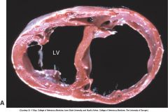
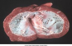
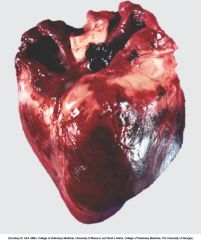
Cardiac dilation. Note the thin walls of both dilated ventricles. LV, Left ventricle
|
|
Can
|
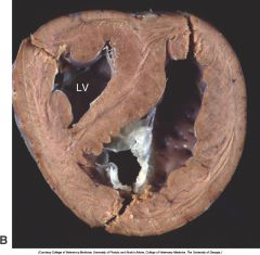
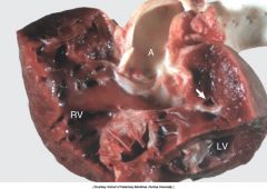
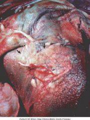
Cardiac hypertrophy (fixed tissue). Note that the right ventricular (RV) and left ventricular (LV) walls are approximately the same thickness, indicating that there is right ventricular hypertrophy.
|
|
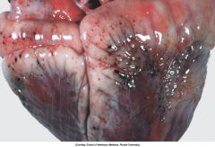
bleedings of different size.
What size = what name? |
The smaller, pinpoint hemorrhages (1 to 2 mm) are petechiae. The larger hemorrhages (3 to 5 mm) are ecchymoses.
Epicardial hemorrhage, petechiae and ecchymoses, endotoxemia, heart, cow. Note the epicardial and subepicardial hemorrhages in the fat of the coronary groove (a common site). Petechiae and ecchymoses are often attributable to severe septicemia, endotoxemia, anoxia, or electrocution. In this case, the hemorrhage resulted from injury to the endothelium from endotoxin (component of the cell wall of gram-negative bacteria). |
|
|
congestive heart failure
general pathogenesis |
cardiac disease or ↑ workload → forward failure + backward failure → ↓ renal bloodflow → ↑ renin (juxtaglom app) → ↑ aldosteron (adrenal cortex) → Na retention → ↑ plasma, edema.
Hypoxia → erythropoiesis → polycythemia → viscosity → ↑ workload → hypoxia McG 567 |
|
|
Left side heart failure.
Most common causes (X3). |
1) myocardial contractility loss, associated w myocarditis
2) dysfunction of mitral or aortic valves 3) congenital heart diseases McG 567 |
|
|
Right side heart failure.
Most common causes (X3). |
1) pulmonary hypertension
2) cardiomyopathy 3) disease of the tricuspid and pulmonary valves McG 568 |
|
|
Mitral valve and tricuspid valve.
Which one on which side? |
mitraL → left
tRicuspid → right |
|
|
↑↑ Plasma natriuretic peptides may indicate cardiac disease, because these hormones are synthesized by cardiac muscle cells.
Name 3 plasma natriuretic peptides. |
A type (atrial)
B type (brain) C type McG 568 |
|
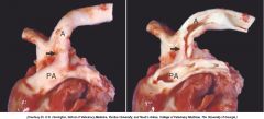
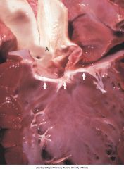
P = .....
PA = ...... arrow = ..... |
Patent ductus arteriosus (arrow) between the pulmonary artery (PA) and the aorta (A)
|
|
|
patent ductus arteriosus
- breed predisp - in which breed inherited - sex predisp |
- poodle, collie, pomeranian, chihuahua, maltese, english springer spaniel, keeshond, bichon frise,shetland sheepdog
- poodle - female |
|
|
ductus arteriosus is normally converted to the solid ...... postnatally
|
ligamentum arteriosum
McG 569 |
|
diagnosis?
Which breeds of Can greatest freq? (X3) |
Ventricular septal defect
English bulldog, English springer spaniel, Westie McG 570 |
|
Tetralogy of Fallot.
Name 3 primary + 1 secondary lesion Inherited in.... |
Primary:
- ventricular septal defect - pulmonic stenosis - dextroposition of aorta Secondary: - hypertrophy of right ventricular myocardium Inherited in keeshond and English bulldog McG 570 |
|
|
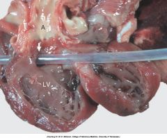
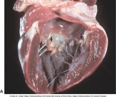
Subaortic stenosis, heart, opened left side, dog. A thick, white, broad band of fibrous connective tissue (arrows) encircles the left ventricular outflow tract below the aortic valve. The force of the blood ejected through the stenotic lesion is responsible for the “jet lesions” in the overlying aorta (A) (right half—roughened surface; left half—dilation [note the gray area]).
|
|
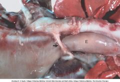
Diagnosis?
Breeds? |
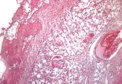
Persistent right aortic arch, megaesophagus
GS, Irish S, Great Dane Because during embryogenesis the aorta was formed from the right aortic arch instead of the left one, the aorta is now on the right. Thus in order for the ligamentum arteriosum (arrow) to connect the aorta with the pulmonary artery, it has to pass dorsally over the esophagus and trachea. The ligamentum, together with the aorta and pulmonary artery, form a vascular ring that constricts the esophagus (E), which is dilated cranial to the constriction |
|
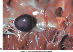
Diagnosis?
Prognosis? |
Valvular hematocyst, heart, opened left side, mitral valve, postnatal calf. A dark, blood-filled cyst protrudes from a cusp of the mitral valve. Arrows indicate chordae tendinae. Hematocysts usually occur in ruminants, do not cause any functional abnormality, and usually regress within a few months of birth.
|
|
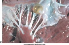
Diagnosis?
Prognosis? |
Valvular lymphocyst, heart. A lymph-filled cyst is on a cusp of the atrioventricular valve. Like hematocysts, lymphocysts usually occur in ruminants, do not cause any functional abnormality, and usually regress within a few months of birth.
|
|
MD?
Breeds? |
Endocardial fibroelastosis
Burmese and siamese cats McG 572 Subendocardial fibroelastosis, heart, left ventricle, dog. The endocardium is opaque because increased amounts of collagen and elastic fibers were deposited in the subendocardium secondary to turbulence of blood flow within the ventricles. This dog had a persistent ductus arteriosus. This lesion may have a hereditary basis in Burmese cats and is often a sequela to turbulence within ventricles in cardiac disease. |
|
fibrinous pericarditis, Eq.
Common Et? |
Streptococcal infections
(Strep. zooepidemicus) McG 577 |
|
Calcitonin
|
Source: Thyroid
Action: Decreases the blood calcium level, decreases reabsorbtion of calcium and phosphate |
|
|
calcinogenic plants (name 3) cause which syndrome?
|
1) Cestrum diurnum
2) Trisetum flavescens 3) Solanum malacoxylon 4) Solanum torvum Enzootic calcinosis (= Manchester wasting disease) McG 578 |
|
Can.
- ND - Most affected breed? - Other breeds? |
- Valvular endocardiosis
- Cavalier King Charles spaniel (100% by 10 years of age) - Cocker spaniel, beagle, dachshund, poodle, Pomeranian, schnauzer, Chihuahua, Doberman, fox terrier, Boston terrier, Pekingese, deerhound, wolfhound (general: associated with old age and small breeds) McG 579 |
|
|
Atrial thrombosis is a frequent lesion in ....
|
age Syrian hamsters
McG 579 |
|
|
Endocarditis is usually the result of bac inf, except lesions produced by migrating ....... in Eq and an occasional case of mycotic inf.
|
Strongylus vulgaris larvae
McG 579 |
|
|
Relative frequency of valvular involvement w endocarditis in animals is .....
|
mitral > aortic > tricuspid > pulmonary
McG 579 |
|
|
Pathogenesis of endocarditis is complicated but the components of Virchow´s triad are involved. Name them.
|
Endothelial injury
turbulence hypercoagulability McG 580 |
|
|
2 forms of myocardium hypertrophy
|
1) Eccentric hypertrophy → enlarged ventr chambers, walls somewhat thinner
2) Concentric hypertrophy → small ventr chambers, thick walls. McG 581 |
|
|
3 stages of myocardial hypertrophy
|
1) initiation
2) stable hyperfunction 3) deterioration of function associated w degeneration of hypertrophied myocytes. McG 582 |
|
|
Name a few diseases that cause right ventricular hypertrophy
|
Can: dirofilariosis, congenital pulmonic stenosis
Bo: "brisket disease" (high altitude disease) Eq: chronic alveolar emphysema (heaves) McG 582 |
|
|
Left ventr hypertrophy occurs in:
- dogs with..... - cats with..... - dogs and cats with ..... |
-Can: subaortic stenosis
- Fe: hyperthyroidism - Can&Fe: systemic hypertension McG 583 |
|
|
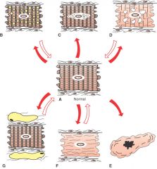
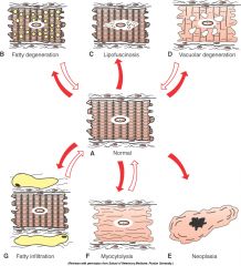
Difference between fatty infiltration and fatty degeneration of the hearth?
|
infiltr.: ↑ lipocytes interposed between myocardial fibres
degen.: accumulation of lipid droplets in sarcoplasm McG 584 |
|
|
fatty degen. of myocard. Causes?
|
Systemic disorders such as:
severe anemia toxemia copper deficiency McG 584 |
|
|
Hydropic degen. of myocard. Cause?
|
chronic admin of anthracyclines (= group of antineoplastic drugs)
McG 584 |
|
|
Lipofuscinosis of myocardium. Causes?
|
age
severe cachexia hereditary in Ayrshire cattle McG 584 |
|
|
Myofibrillar degen. of myocard.
Histo? Causes? |
Histo: pale eosinophilic sarcoplasm, lacks cross striations.
Cause: furazolidone cardiotoxicity in birds, potassium deficiency in rats McG 584 |
|
|
Name the 3 different morphologic types of primary cardiomyopathies
|
1) Hypertrophic
2) Dilated 3) Restrictive McG 589 |
|
|
Common cause of...
1) suppurative myocarditis 2) necrotizing myocarditis 3) hemorrhagic myocarditis 4) lymphocytic myocarditis |
1) pyogenic bacteria from valvular endocarditis
2) toxoplasmosis in Can and Fe 3) Blackleg in Bo. (Clostr. chauvoei) 4) parvovirus McG 591 |
|
|
Congenital rhabdomyomatosis in Su and guinea pigs is really a ....
|
hamartoma (i.e., a malformation often resembling a neoplasm that is composed of an overgrowth of mature cells and tissues that normally occur in the affected organ).
McG 593 |
|
|
Cardiac hemangiosarcoma is important in which spp?
At which location? What is a freq metastasis? |
Dogs
wall of right atrium Lung McG 593 |
|
|
What kind of tumors are most common at the base of the hearth (esp in Can)?
Breed? |
aortic body tumor or chemodectoma (paraganglioma)
Breed: most freq in brachycephalic dogs (and occasionally ectopic thyrois or parathyroid tissue gives rise to neoplasms in this area) McG 594 |
|
|
arteries are classefied into 3 types: .....
|
muscular arteries, elastic arteries and arterioles
|
|
|
Vessel walls are divided into 3 layers or tunics:.....
|
intima, media, adventitia
|
|
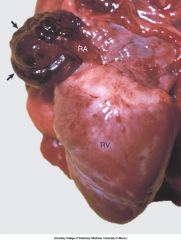
Can
Diagnosis? |
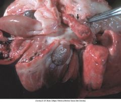
Hemangiosarcoma, heart, right atrium, dog. A dark-red hemangiosarcoma protrudes from the wall of the right atrium (RA), a predilection site in the dog for this tumor (arrows). RV, Right ventricle.
|
|
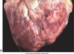
Bo
Diagnosis? |
Lymphosarcoma, heart, myocardium, cow. A, Sites of infiltrating neoplastic lymphocytes in the ventricular myocardium are evident as numerous white areas and nodules (arrows).
|
|
|
Most cases of aneurysm are idiopathic, but known causes include... (X3)
|
- copper deficiency in Su (necessary for development of elastic tissue)
- Spirocerca lupi - Strongylus vulgaris McG 598 |
|
|
What can cause medial hypertrophy of the muscular pulmonary arteries of Fe?
|
Aerulostrongylus abstrusus
Toxocara spp Dirofilaria immitis McG 599 |
|
|
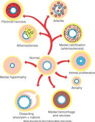
Main difference between arteriosclerosis and atherosclerosis?
|
arterio... - intimal fibrosis of (mainly) large elastic arteries
athero... - intimal and medial lipid deposits in elastic and muscular arteries (beetje kort door de bocht) McG 599 |
|
|
A high-cholesterol diet in Su , rabbit and chicken can lead to ......
while Can, Fe, Bo, Capr and rat are resistant |
Atherosclerosis
McG 599 |
|
|
The deposition of iron and calcium salts in the cerebral arteries of aged Eq is called: .........
|
Siderocalcinosis (or iron rings)
It is considered an incidental finding McG 600 |
|
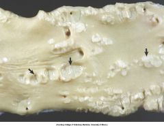
Bo
|
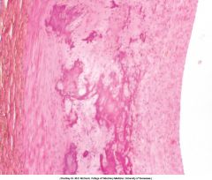
Johne’s disease, arteriosclerosis, aorta, cow. Multiple prominent, white, mineralized foci are in the tunica intima and media
|
|
Bo
|
Medial calcification, aorta, cow. Note the layer of mineralization in the middle of the tunica media. H&E stain.
|
|
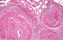
Buzzword?
Spp? |
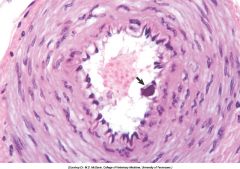
Intimal body
Eq Intimal body (arrow), intestine, muscular artery, horse. Intimal bodies are distinctive small, mineralized masses within the subendothelium of small muscular arteries and arterioles of horses. They are an incidental finding and have no pathologic significance. H&E stain. |
|
|
On histo (HE) you see:
an homogenous eosinophilic zone in the vessel walls (small muscular arteries and arterioles). Which stains do you order, and what do they tell you? |
DDx: hyaline degeneration / fibrinoid necrosis / amyloidosis
Stains: 1) Congo red and methyl violet → amyloid is positive 2) PAS → fibrinoid is positive If both 1) and 2) are negative → hyaline McG 600/601 |
|
|
Fibrinoid necrosis (or fibrinoid change) of arteries is frequent in many acute degenerative and inflamm diseases.
Esp in Su it is an important diagnostic feature of...... (X4) |
- Selenium-Vit E deficiency (heart)
- Edema disease (gastric submucosa) - Cerebrospinal angiopathy - Organic mercury toxicosis (meninges) In Can freq in cases of uremia or hypertension. McG 601 |
|
|
short pathogenesis of fibrinoid change
|
injury to intima and media (such as occurs in immune mediated vasculitis) → deposition of immunoglob / complement / plasma proteins (incl fibrin) in the wall of vessels.
Note: process is not true regressive alteration of cells → terms fibrionoid degeneration and necrosis are inappropriate (but often used) McG 46 |
|
|
In Su with Vit E/selenium def, vascular fibdrinoid necr produces 2 typical gross lesions: .....
The observed complex of vascular lesions has been termed:...... |
- Mulberry hearth disease
- hepatosis dietetica (= a massive hemorrhagic hepatic necrosis) Complex = dietary microangiopathy McG 601 |
|
Su
ND? |
“Mulberry heart disease,” suffusive hemorrhage, epicardium, right ventricle, heart, pig. Red areas of suffusive hemorrhage (“mulberry-like”) are present on the epicardial surface of the right ventricle.
|
|
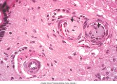
Su, heart.
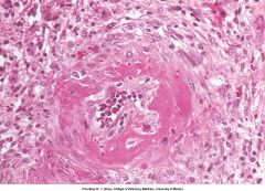
What kind of vascular change? Most likely cause? |
Fibrinoid change (=fibr necr)
Vit E / Selenium def. Selenium–vitamin E deficiency (“mulberry heart disease”), fibrinoid necrosis, myocardial arteriole, heart, pig. Note the circumferential eosinophilic deposits in the wall of the arteriole. H&E stain. |
|
Can
What does the arrow point at? |
Arterial thrombus, pulmonary artery, dog. Arterial thrombi are composed primarily of platelets and fibrin because the rapid flow of blood tends to exclude erythrocytes from the thrombus, and thus arterial thrombi are usually pale beige to gray (arrow).
|
|
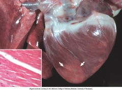
Can
What do the arrows point at? |
Myocardial infarction, heart, left and right ventricles, dog. Pale, necrotic, circumscribed areas (arrows) are present in the ventricular walls and are most prominent at the apex. Inset: The cardiac myocytes are eosinophilic (ischemic necrosis) and have lost their nuclei (karyolysis).
|
|
|
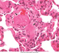
Eq.
What is the vascular lesion? What is systemic condition can this be part of? Why is it important to get fresh animals at necropsy if you want to see this? |
Fibrin thrombi
DIC (Disseminated intravascular coagulation) Fibrinolysis continues after death. Fibrin thrombi, disseminated intravascular coagulation, lung, alveolar septal capillaries, horse. Fibrin thrombi (arrows) occlude two alveolar capillaries. |
|
Su. Spinal cord.
What do the arrows point at? Which Spp + breed is commonly affected? |
Fibrocartilaginous emboli
Can, typically middle-aged large or giant breeds Retrograde movement within vasculature of fibrocartilagenous fragments of degenerated intervertebral disks is generally considered to underlie this presence. Result: posterior paresis or paralysis. McG 605 |
|
Su.
Et? ND? Pathogenesis of lesion? |
Erysipelothrix rhusiopathiae
Diamond skin disease Emboli of Erysipelothrix rhusiopathiae have lodged in cutaneous vessels and caused a localized vasculitis, which has resulted in thrombosis followed by ischemia and cutaneous infarction. In MD, describe: cutaneous infarction McG 606 |
|
|
Beagle pain syndrome.
Et? What is the lesion? |
- attributed to immune-mediated vascular injury
- Idiopathic necrotizing polyarteritis involving the coronary and meningeal arteries McG 607 |
|
|
Hemangiomas are often found in the ........
|
....skin of Can
McG 607 |
|
|
Hemangiosarcoma occurs freq in the ..... and the ..... of .....
|
spleen and right atrium of Can
McG 607/608 |
|
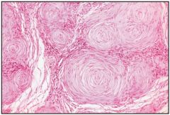
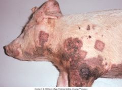
Hint: a tumor on the skin of a dog of vascular origin that is not an hemangioma.
|
Hemangiopericytoma. Histologically, it is characterized by spiral proliferations around blood vessels, with a typical “fingerprint” arrangement of the cells disposed concentrically around the capillaries that are collapsed. Capillaries are frequently hyalinized.
|
|
|
|
|
|
A venous dilation from weakened vascular walls is termed a ...... (localized) or ...... (generalized).
|
varicosity (lozalized)
phlebectasia (generalized) McG 608 |