Reading...
![]()
Play button
![]()
Play button
![]()
Use LEFT and RIGHT arrow keys to navigate between flashcards;
Use UP and DOWN arrow keys to flip the card;
H to show hint;
A reads text to speech;
13 Cards in this Set
- Front
- Back
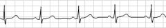
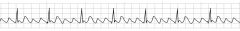
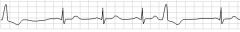
Identify the rhythm!
|
This is normal sinus rhythm. It can be identified by its rate between 60 and 100 bpm, its a regular, SA driven rhythm. Also notice the upright P waves and narrow QRS.
|
|
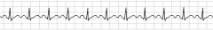
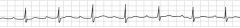
Identify this rhythm!
|
This is Sinus Tachycardia, Note that the rhythm is also driven by the SA node, is regular, and is at a rate faster than 100bpm but typically below 150bpm. It is most often caused by sympathic stimulation
|
|
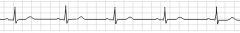
Identify the rhythm!
|
This is Sinus Bradycardia, note that it is regular, there are no premature or missing beats, the QRS is narrow with p-waves , p-q interval is normal. and most importantly the rate is <60bpm
|
|
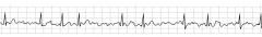
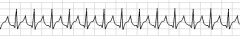
Identify the rhythm!
|
This is atrial fibrillation , notice the irregular rate, no identifiable P-waves, and the fibrillation in-between each QRS complex. Note: The atrial "kick" is lost here and >48hrs leads to increased risk of thrombosis formulation.
|
|
Identify the rhythm !
|
This rhythm is atrial flutter, A-flutter results from the development of a re-entry circuit within the atria resulting in a loop that discharges impulses at a flutter rate of 250 to 350 times per min. Note the "sawtooth" pattern .
|
|
Identify the rhythm
|
This is an agonal rhythm, it is considered an end-stage cardiac rhythm with asystole quickly ensuing. Note the QRS is wide and flattened. Patient will be pulseless.
|
|
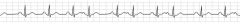
Identify the rhythm !
|
This is Supraventricular Tachycardia , generally speaking the rate is over 160bpm , notice the narrow QRS complex and the rapid, regular pattern. there may not be any visible P waves due to the overpowering QRS complexes being so close together. Just note that they are there.
|
|
Identify the rhythm.
|
This is an example of Premature Atrial Contractions with an underlying sinus rhythm. notice that because of the premature SA node firing , the rest of the cardiac conduction pathway also fires off.
|
|
Identify the rhythm !
|
This is an example of Premature Ventricular Contractions with an underlying sinus rhythm. The PVC's can be multifocal or unifocal in nature.
|
|
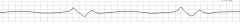
Identify the block!
|
This is a 1st Degree Atrioventricular Block. Notice the prolonged p-r interval. >.20sec. This is a result of prolonged transmission of the electrical impulse through the AV junction.
|
|

Identify the block!
|
This is a 2nd degree atrioventricular block type 1. Notice the progressively prolonged p-r interval until finally the heart simply doesn't fire past the atrium resulting in a "dropped" QRS and T wave. The heart then attempts to restart , and again progressively elongates the p-r interval.
|
|

Identify the block!
|
This is an example of a 2nd degree atrioventricular block type 2, in this case there is no elongating warning signs before the dropped beat. The atrium continues to fire at a regular pace however not all beats make it past the AV junction resulting in sporadic incomplete beats.
|
|

Identify the block!
|
This is an example of a 3rd Degree Atrioventricular block. Notice that the atrium and ventricles are firing entirely independent of each other. There is no communication in the AV junction and/or bundle of his conduction pathways , thus the atrium and the ventricles do their own thing.
|