![]()
![]()
![]()
Use LEFT and RIGHT arrow keys to navigate between flashcards;
Use UP and DOWN arrow keys to flip the card;
H to show hint;
A reads text to speech;
45 Cards in this Set
- Front
- Back
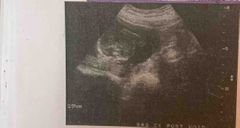
3rd trimester image is most suspicious for |
Polyhydraminos |
|
Front (Term)
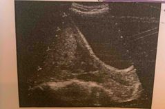
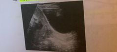
What does this 3rd trimester sonogram demonstrate |
Oligohydraminos |
|
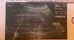
Most likely cause for this diagnosis |
Premature rupture of membrane |
|
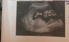
In addition to gender of fetus what does this reveal |
Anterior placenta |
|
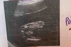
Fluid volume in this image is suspicious for |
Polyhydraminos |
|

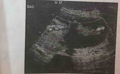
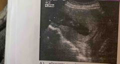
Condition most likely identified in this sagittal sonogram of cervix |
Placenta previa |
|
More commonly associated with this condition |
Painless vaginal bleeding |
|
Most accurate placenta location |
Fundal |
|
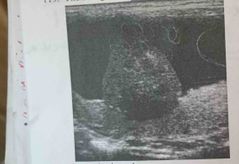
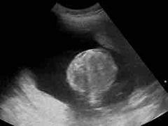
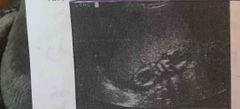
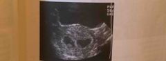
Sonogram is most likely identifies |
Chorioangioma |
|
Transverse image of uterus is most likely |
Succenturiate placenta |
|

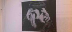
Duplex image identifies this structure |
Single umbilical artery |
|
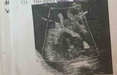
Finding is associated with |
Mulifetal gestations |
|
Lower uterine segment in this sonogram is consistent with |
Incomplete cervix |
|
Severe lower abdominal pain & vaginal spotting |
Placental abruption |
|
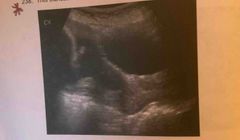
Postvoid transabdominal image of the cervix demonstrates |
A cervix free of placenta |
|

Sagittal image of the cervix in late second trimester pregnancy demonstrates |
Placenta previa |
|
Patient most likely presents with |
Painless vaginal bleeding |
|

Early 2nd trimester pregnancy demonstrates |
Placentomegaly |
|
Maternal cause for this abnormality include |
Diabetes mellitus |
|
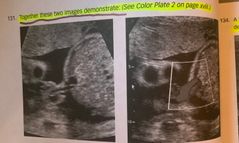
Together these two images demonstrate |
Cord insertion |
|
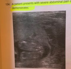
A patient with severe abdominal pain and vaginal bleeding demonstrates |
Placental abruption |
|
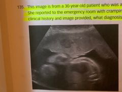
She reported cramping in her lower abdomen, due to physical domestic violence |
Placenta previa |
|
Grade of the placenta in this image |
Grade 3 |
|
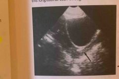
Longitudinal scan through uterus of an a symptomatic pregnant patient |
Normal retroplacental space |
|
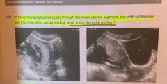
One with full bladder and the other after partial voiding, what placental position |
Anterior marginal |
|
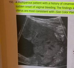
Mid pelvic pain sudden onset vaginal bleeding |
Placenta increta |
|
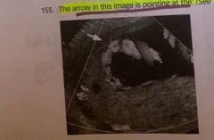
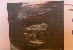
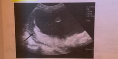
Pointing at arrow |
Placental edge |
|
Structure discovered during routine obstetric exam for gestational dating |
Chorioangioma |
|
Patient with painless vaginal bleeding |
Complete placenta previa |
|
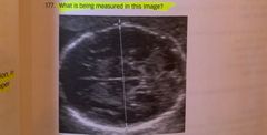
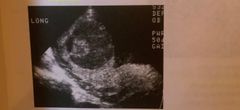
Being measured in this image |
Biparietal diameter |
|
Demonstrates |
Umbilical vein thrombosis |
|
Twin A appears,twin B appeared small and growth restricted consistent with |
Twin to twin transfusion syndrome (TTTS) |
|
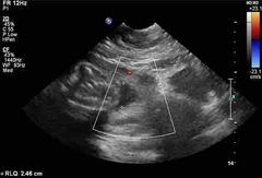
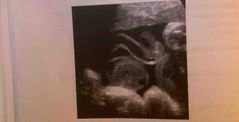
Power Doppler image demonstrates |
Cord entanglement |
|
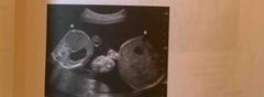
Large for gestational age 7-8 wks post LMP demonstrates |
Twins |
|
One fetus in this second trimester sonogram reveals |
Acardiac twin |
|
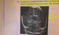
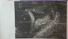
Rounded fluid filled midline structure within the fetal brain, when color Doppler is turned on the structure shows |
Vein of Galen aneurysm |
|
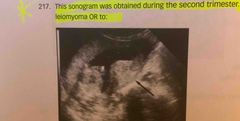
During 2nd trimester, arrow pointing to a leiomyoma or to |
A contraction |
|
Longitudinal image of the fetal thorax demonstrates |
Pleural effusions |
|
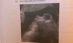
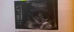
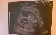
Image leads u to suspect |
Anencephaly |
|
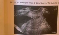
Transvaginal image of graves cervix, patient at high risk for |
Preterm birth |
|
A habitual aborted has undergone a McDonald procedure, pointing arrow this longitudinal scan through her lower uterine segment |
Cerclage |
|
Trans abdominal image image of the cervix reveals |
Funneling |
|
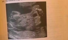
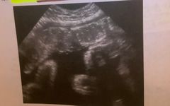
Transverse image demonstrates |
Conjoined twins |
|
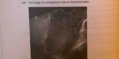
Postpartum uterus demonstrates |
Normal postpartum anatomy |
|
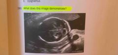
Image demonstrate |
Risk for aneuploidy |