![]()
![]()
![]()
Use LEFT and RIGHT arrow keys to navigate between flashcards;
Use UP and DOWN arrow keys to flip the card;
H to show hint;
A reads text to speech;
56 Cards in this Set
- Front
- Back
|
functions of the respiratory system |
1. exchange of gas between the atmosphere and blood 2. filtration, temp. regulation and humidification of inspired air 3. olfaction 4. production of sound |
|
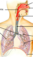
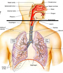
components of respiratory system top to bottom |
1. nasal caity 2. nose 3. pharynx 4. larynx 5. trachea 6. lungs 7. smaller airways 8. diaphragm |
|
|
larynx |
1. is the most narrow portion of the airway 2. most likely area for upper airway to be blocked |
|
|
upper vs lower |
|
conducting portion |
conducts air to smaller and smaller airways,delivering to places that actually does respiration |
|
|
actual respiratory portion are the |
lobules |
|
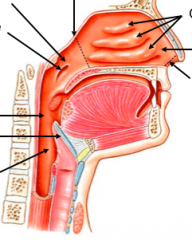
upper respiratory system right to left |
1. external nares 2. nasal vestibule 3. conchae 4. internal nares 5. nasopharynx 6. eustachian tube 7. oropharynx 8. epiglottis 9l laryngopharynx |
|
|
conchae |
three middle ridges of bone that projects into nasal cavity, disallows the amount of area that can be reached by our fingers |
|
|
turbedents |
when conchae are covered by mucus |
|
|
internal nares divides |
nasal cavity from the pharynx |
|
|
nasopharynx and the eustachian tube |
nasopharynx has an opening, the eustachian tube, which connects it to the middle ear |
|
|
epiglottis |
can move epiglottis so that it can cover when you just want air or if when you are eating food |
|
|
turbinates |
turbinates covered in mucosal membrane so turbinate swirls when you inhale, maximizees contact of air to the turbinates and humidifies the air |
|
|
blood passing the mucosa membrane... |
warms the air |
|
|
bolus |
chewed food |
|
|
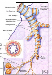
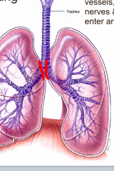
roots of the lung aka hilum where bronchi enters the lungs |
|
|
hilum |
sites where blood vessels, lymphatics, nerves and airways enter and leave lungs |
|
|
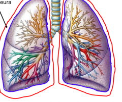
pleural cavity space between visceral and parietal pleura |
|
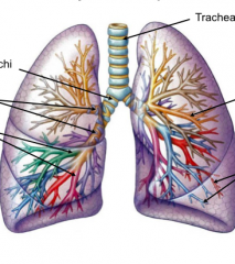
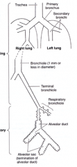
major airways top to bottom |
1. trachea 2. primary bronchi 3. secondary and tertiary bronchi 4. bronchioles 5. terminal bronchioles |
|
|
right side ten tertiary bronchi... |
thus ten segments |
|
|
left side w/ ten segments but... |
two pairs become fused together leading to 8 segments |
|
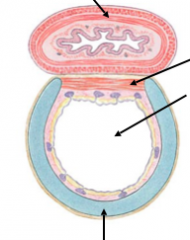
trachea superior top to bottom |
1. esophagus 2. smooth muscle 3. trachea 4. cartilage ring |
|
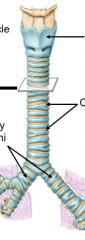
trachea |
1. larynx 2. cartilage rings 3. primary bronchi |
|
|
cartilage ring |
helps maintain shape of trachea to let air in when exhaling, it is not complete b/c when bolus is swallowed, it expands |
|
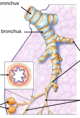
airways top to bottom |
1. primary bronchus 2. secondary bronchus 3. smaller bronchi 4. smooth muscle 5. bronchioles 6. alveoli |
|
|
alveoli |
where bulk of respiration actually occurs |
|
|
bronchioles and cartilage |
bronchioles don't have cartilage, instead there is smooth muscle to regulate diameter |
|
|
bronchi and cartilage |
they have some cartilage but aren't completely covered, more so for structural support but not for strength |
|
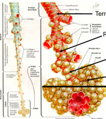
terminal airways |
1. terminal bronchiole 2. respiratory bronchioles 3. alveolar ducts 4. alveolar sac |
|
|
alveolar sacs |
where respiration occur |
|
alveoli |
1. alveoli 2. alveolar sac |
|
types of airways |
1. conducting portion 2. respiration portion |
|
|
conducting |
only conduct air |
|
|
respiratory |
both conduct and respire |
|
|
structure of Airway Wall |
1. cartilage 2. smooth muscle 3. elastic fibers |
|
|
cartilage |
1. found in larger airways 2. helps keep airways open |
|
|
smooth muscle |
1. predominates in smaller airways 2. controls diameter of airway 3. constriction reduces airflow |
|
|
elastic fibers |
1. predominate in smaller airways and respiratory portion 2. elastic recoil provides force for expiration |
|
|
Asthma Attack |
1. smooth muscle in wall of bronchioles contracts 2. contraction reduces airflow 3. bronchioles most numerous airways 4. there is great resistance to airflow |
|
|
contraction caused by |
1. parasympathetic stimulation 2. mediators of allergic reactions (histamine) |
|
|
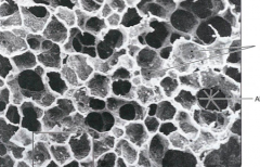
Emphysema |
1. reduced ability to expire air from lungs 2. due to breakdown of alveolar walls, producing abnormally large air spaces 3. this lowers surface area for gas exchange, there is less respiratory efficiency 4. as well as reduced elastic recoil, due to loss of elastic fibers |
|
|
emphysema caused by |
1. smoking 2. other irritants, pollution, dust |
|
|
respiratory epithelium |
1. pseudostratified epithelium 2. there is a mucus layer that floats on serous/ watery layer |
|
|
mucus |
mucus traps garbage that we breath in to the mucus layer, the cilia moves this mucus up towards the upper respiratory to either swallow it or expel to the environment |
|
|
respiratory epithelium found in |
1. nasal cavity 2. nasopharynx 3. large airways |
|
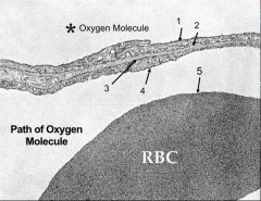
alveolar wall clockwise |
1. capillary 2. alveolar epithelial cell 3. endothelial cell of capillary 4. alveolar macrophage 5. elastic fibers 6. surfactant |
|
|
1. luminal area 2. first portion of basal lamina 3. capillary portion of basal lamina 4. endothelium 5. airspace 6. the plasma membrane of RBC |
|
|
TYPE 2 Alveolar Cells |
1. secrete surfactant 2. surfactant lowers surface surface tension of alveolar fluid 3. prevents alveolar walls from sticking together and collapsing |
|
|
Respiratory Distress Syndrome |
1. occurs children born prematurely 2. type 2 alveolar cells not yet active 3. lack surfactant 4. alveoli fail to inflate properly 5. insufficient oxygenation of blood |
|
|
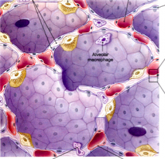
alveolar macrophages |
1. ingest debris in alveoli 2. enter airways 3. become trapped in mucus sheets 4. carried toward pharynx by ciliary action 5. expectorated or swallowed |
|
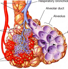
alveolar capillaries top to down |
1. sooth muscle 2. respiratory bronchiole 3. elastic fibers 4. alveolar duct 5. alveolus 6. alveolar sac 7. capillaries |
|
|
muscles of quiet breathing inspiration |
1. diaphragm - contraction flattens diaphragm - increases vertical diameter of thoracic cavity 2. external intercostals - left ribs superiorly - move sternum anteriorly |
|
|
muscles of quiet breathing expiration |
elastic recoil of lung tissue and thoracic wall |
|
|
forced breathing inspiration |
accessory muscles of respiration - sternocleidomastoid- elevates sternum - scalenes- elevate ribs 1 and 2 - pectoralis minor- elevates ribs 3-5 - pectoralis major |
|
|
forced breathing expiration |
1. internal intercostals 2. abdominal muscles - move inferior ribs inferiorly - compress viscera- move diaphragm superiorly |
|
|
tripod position |
when breathing hard, upper extremities are fixed to the ground, the pectoral muscles are used to move the test |