![]()
![]()
![]()
Use LEFT and RIGHT arrow keys to navigate between flashcards;
Use UP and DOWN arrow keys to flip the card;
H to show hint;
A reads text to speech;
19 Cards in this Set
- Front
- Back
|
Name the gas conducting portions of the respiratory tract |
Nostril, nasal cavities, paranasal sinuses, pharynx, larynx, trachea, bronchi, bronchioles |
|
|
Name the gas exchanging portions of the respiratory tract |
respiratory bronchioles, alveolar ducts, alveolar sacs, alveoli |
|
|
Where does the respiratory primordium develop |
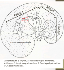
a ventral groove in the floor of the foregut |
|
|
How does the laryngo-trachial groove form? |
Beginning at the 4th pharyngeal arch, the ventral groove begins to separate from the foregut by the formation of two tracheo-pharyngeal grooves, pinching an outgrowth away from the trachea. These grooves fuse forming the tracheo-oesophogeal septum which separates the foregut from the laryngo-trachial tube. Everything cranial to this region is the pharynx |
|
|
What structures will be found in the larynx? |
arytenoid cartilage and epiglottid cartilage. Will form from neural crest derived mesenchyme, and develop into the aforementioned structures in the cranial portion. |
|
|
How are the vocal cords formed? |
Laryngeal cartilage begins to develop as folds and laryngeal ventricles are developed. Folds constrict to regulate laryngeal lumen. Ventricles begin as left and right diverticula in the wall of the larynx. |
|
|
What embryonic tissue forms the trachea |
Endodermally derived with contributions from mesenchyme (neural crest derived) which forms cartilaginous rings. The endodem will become ciliated, pseudo stratified and respiratory rich in goblet cells |
|
|
What are the periods of respiratory development? |
Embryonic: primordium of lungs and bronchi formed Fetal: ramified bronchi are formed, preliminary gas exchange structures Postnatal: alveoli develop and lungs take on adult form |
|
|
What are the subdivisions of the fetal respiratory period? |
Pseudo glandular Cannalicular Sacular Alveolar |
|
|
What are the events of the Embryonic respiratory period? |
Lung buds divert from the larynotracheal tube forming Principal Bronchi Principal Bronchi branch into lobar bronchi Lobar bropnchi push into the developing pleural cavity sorrounded by mesenchyme that will become pleura Lobar bronchi branch into segmental bronchi **Right Tracheal bronchi forms in cattle, sheep , pigs
|
|
|
How does the pleural cavity separate from the pericardial cavity? |
Pleuropericardial folds (mesoderm) grow medially into the pleuro pericardial cavity, fusing centrally to separate the developing lungs from the heart. Pericardial cavity then constricts in size around the heart, lungs develop caudally to encricle the heart. |
|
|
Describe pseudoglandular phase of the fetal respiratory period |
"gland-like" branching of the segmental bronchi into terminal bronchioles (may branch up to 20 times) Terminal bronchioles are lined with endodermal epithelium. |
|
|
Describe canalicular phase of the fetal respiratory period |
Terminal bronchioles divide even further into respiratory bronchioles More divisions of respiratory bronchioles become secondary tubules called canalicules lined with cuboidal epithelium |
|
|
Describe sacular phase of fetal respiratory development |
Canaliculi form terminal saccules Terminal saccules divide into blind ended saccules |
|
|
Describe alveolar phase of fetal respiratory development |
Terminal sacules differentiate into alveoli Type I alveolar cells Cuboidal Type II alveolar cells produce surfactant alveolar differentiation occurs most distally first *Mature alveoli form after birth |
|
|
Describe postnatal respiratory events |
Mature alveoli form from primitive alveoli (alveolar phase) Extensive growth of respiratory bronchioles, saccules, and alveolar development Amniotic fluid and glandular fluid expelled Any remaining fluid absorbed by epithelial cells |
|
|
What is the difference in the lobar patterning of Pigs and cows? |
Pigs have a tracheal bronchi supplying right cranial lobe (also cattle, sheep). Also middle, accessory and caudal lobes on R side. Left cranial lobe is furthered divided into cranial/caudal. Also Left caudal lobe. Cows: Also have a tracheal bronchi supplying (cranial and caudal) R cranial lobe. Left lobe is (cranial/caudal) cranial and caudal lobe |
|
|
What are the molecular signals that cause pulmonary branching? |
Fgf 10 secreted by mesenchyme surrounding lung causing epithelial growth toward the centre of secretion BMP-4 secreted from apical epithelial cells initiates branching Shh inhibits Fgf10 and Tgf b1, which produces collagen and fibronectin, split the bronchi into buds. Newly sprouted buds will respond to FgF10 secreted by mesenchyme and split. |
|
|
Clinical Relevance |
Respiratory anamolies are rare: Ectopic lung tissue in abdominal cavity Cysts in the lung as a result of dilated terminal bronchi that fill with glandular fluid |