Reading...
![]()
Play button
![]()
Play button
![]()
Use LEFT and RIGHT arrow keys to navigate between flashcards;
Use UP and DOWN arrow keys to flip the card;
H to show hint;
A reads text to speech;
16 Cards in this Set
- Front
- Back
|
Where do primitive germ cells originate from?
|
-the primordial germ cells migrate by ameboid movement from the yolk sac into the hindgut & finally reside in the undifferentiated gonad
|
|
|
Describe the development of the primitive genital system at the undifferntiated stage
|
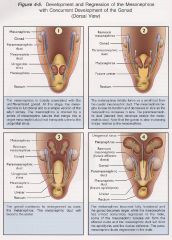
-the genital ridge forms medial to the embryonic kidneys
-when the primitive germ cells arrive in the genital ridge, they stimulate local connective tissue to proliferate -this results in the formation of compact strands of tissue called primitive sex cords -these proliferating sex cords cause the genital ridges to enlarge and push toward the developing kidney (mesonephros) -during its development the embryo utilizes three morphologically distinct renal systems -the first, called the pronephros (pronephric kidney) is a non-functional remnant of a primitive form of kidney found in lower animals -early in embryogenesis, the pronephros regresses and is replaced by a functional, bilateral pair of intermediate kidneys known as mesonephros (mesonephric kidney) -the mesonephros produces urine that is drained by a bilateral pair of ducts called the mesonephric ducts -these ducts were formerly called Wolffian ducts -the mesonephric ducts extend caudally & empty into the urogenital sinus -this final renal form is known as the metanephros (metanephric kidney) -it will develop functional nephrons and will serve as the functional form of kidney in adult mammals -at the same time the mesonephros is developing, a new pair of ducts beside the mesonephric duct begins to develop -these ducts are called the paramesonephric ducts or Mullerian ducts -form on either side of the mesonephric duct, thus paramesonephric |
|
|
What is a teratoma?
|
-some primitive germ cells don’t make it to the gonadal ridges
-most die, but some survive in ectopic location & form tumors called teratomas -teratomas contain irregularly arranged masses of many tissues, including hair, teeth, cartilage & bone, cardiac & skeletal muscle, thyroid, pancreas, gut, and kidney; these represent derivatives of all embryonic germ layers -usually they are benign cystic tumors but they may become malignant |
|
|
Describe the development of the male reproductive system
|
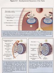
-the substance that controls the pathway toward either male or female development is called testis determining factor (TDF) and is controlled by the Y chromosome
-when testis determining factor is synthesized by the sex cords within the primitive gonad, the development of the male reproductive system is stimulated -the absence of TDF results in the development of a female reproductive system -between 5-15 mesonephric tubules penetrate into the primitive gonad and make connections with the primitive sex cords via the rete testis -the rete testis is a network of tiny ducts that connect the seminiferous tubules to the efferent ducts -the efferent ducts are derived from the mesonephric tubules -the mesonephric duct will give rise to the epididymis & ductus deferens -the first testis unique cells to differentiate are the Sertoli cells, which are derived from coelomic epithelium. -in the embryo, Sertoli cells secrete AMH (anti-Mullarian hormone) which initiates degeneration of progenitors of female reproductive ducts or paramesonephric duct -some of the mesenchymal cells that surround the gonadal cords in the testis differentiate as intersitital Leydig cells |
|
|
Where do the accessory reproductive glands develop from?
|
-the prostate gland, bulbourethral glands arise as multiple small diverticula from the roof of the urogenital sinus immediately caudal to the mesonephric duct
|
|
|
Describe the descent of the testes
|
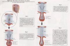
-the testes lie in a retroperitoneal position and are attached caudally to the ligamentous gubernaculums
-the gubernaculums extends caudally and resides in the area of the future scrotum -as the fetal body grows, the testes are pushed against the peritoneum -this “pushing-out” causes the peritoneum to wrap around the gubernaculums and the testes The descent of the testicles has 3 phases: ●growth and elongation of the fetal body away from the testes ●rapid growth of the extra abdominal gubernaculums -the distal potion of the gubernaculums is that portion which has passed through the inguinal ring and forms an outgrowth into the future scrotum -the rapid growth of the gubernaculums in the scrotal region is the “force” responsible for mechanically moving the testes into the inguinal canal ●shrinkage of the gubernaculums within the scrotum -once the testicle is through the inguinal canal, the enlarged gubernaculums begins to shrink in size -while there appears to be a specific substrate that governs gubernacular growth (“descendin”) factors that cause gubernacular regression have not been identified |
|
|
When do the testes descend in the different mammalian species?
|
-descent of the testes from the body cavity into the scrotum occurs by mid-gestation in the bull & the ram and during the last quarter of gestation in the boar & the human
-in the stallion, the testes enter the scrotum either just before or after birth |
|
|
What abnormalities can occur if the testis doesn't descend?
|
-failure of the testes to descent into the scrotum is called cryptorchidism
-bilateral cryptorchidism results in sterility -cryptorchidism testes are able to produce testosterone -thus, the cryptorchid male possesses secondary sex characteristics that are normal and has normal reproductive behavior |
|
|
Describe the development of the female reproductive tract
|
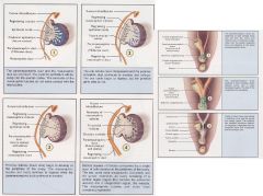
-in the absence of testis determining factor (TDF), certain cells of the sex chords differentiate into primitive follicular cells and the bulk of the genital ridge becomes ovary
-in absence of TDF, the epithelial cords (sex cords), fragment into cellular clusters, each enclosing a primitive germ cell -these clusters of germ cells penetrate less deeply into the interior of the future ovary than in the male -thus, primordial follicles are formed along the outer surface of the ovary and will eventually become the cortex of the ovary -rete tubule formation in the ovary is not pronounced and direct connection between the rete tubules and the mesonephric tubules does not occur -therefore, there is no tubular outlet for the gametes -thus, the female embryo will be born with a pool of oocytes from which folliculogenesis will occur for her reproductive lifetime -the ducts of the female reproductive tract are provided by the paramesonephric ducts -the cranial part of the paramesonephric duct remains open to the peritoneal cavity, but the caudal end butts against the dorsal wall of the urogenital sinus and will fuse with it -the uterus & vagina result from a fusion of the paramesonephric ducts -the cranial vagina, cervix & uterus originate from the paramesonephric ducts (and thus mesoderm) -the caudal vagina and vestibule originate from the ectoderm that is a portion of the urogenital sinus -both male & female gonad & duct system originate behind the peritoneum (retroperitoneal) |
|
|
What is freemartinism? Why does it occur?
|
-freemartinism is a heifer born twin to a bull
-the heifer calf is sterile, but the bull calf is normal and is fertile -results from a unique condition during the formation of the placenta in the cow -in the bovine, the extraembryonic membranes of each conceptus fuse to form a common chorion -these membranes (chorion) share the same cotyledons -thus there is a common blood supply between the male fetus & female fetus -because of this shared blood supply, each conceptus will be exposed to the same hormonal milieu (ie. the female will be exposed to testosterone and anti-mullerian hormone from the male fetus) -this common blood supply is established by about day 39 of gestation -in the bovine, the development of the testes occurs before the development of the ovaries -the testes produces anti-mullerian hormone and this hormone inhibits the growth of the paramesonephric ducts (Mullerian ducts) -since the female twin is exposed to anti-mullarian hormone as the female reproductive tract is developing, the paramesonephric ducts do not develop completely -this incomplete development results in reproductive tracts that are “blind” and canalization of the tract (formation of a canal or lumen) is not complete -in addition, the potential ovaries cease to grow and do not develop the appropriate complement of germ cells -therefore, the ovaries are incapable of producing estrogen and often produce substantial amounts of testosterone as well as androstenedione -the response of the freemartin to this elevated level of testosterone & androstenedione may range from asymptomatic to significant “bullish” behavior -the external genitals often show some masculinization, such as enlarged clitoris or increased ano-genital distance -the internal reproductive tracts are ambiguous -lower part is female, but ducts closer to the gonads resemble deferent ducts |
|
|
Describe the development of the external genitals
|
-extending dorsally from the base of the genital tubercle on either side of the cloacal plate are the cloacal folds (genital folds)
-after separation of the rectum from the urogenital sinus, the cloacal folds become anal & urethral folds ❤FEMALES -in females, the urethral folds & genital swellings enlarge to form the labia -ventrally they partially circumscribe the genital tubercle, which will form the clitoris ❤MALES -in males the urethral folds meet in the midline & fuse, initiating the closure of the penile urethra -the genital swelling remains as the scrotum -masculinization of the external genitals is testosterone dependent -within these tissues, the enzyme 5-α-reductase type 1 converts testosterone to dihydrotestosterone, which is the essential activator for growth & differentiation of precursors of the penis & scrotum |
|
|
Describe the phenotype of an XXY individual. What species does this occur in?
|
-Klinefelter syndrome
-due to meiotic X-chromosome non-disjunction during oogenesis -phenotypically male -male internal reproductive tracts (ie. mesonephric ducts), lack oviducts & uterus & have male external genitals -small testes and lack mature sperm -develop secondary signs such as increased fat accumulation on hips & long extremities due to reduced level of testosterone -reported in tortoiseshell colored cats |
|
|
Describe the phenotype of an YO individual.
|
NOT VIABLE
|
|
|
Describe the phenotype of an XO individual. What species does this occur in?
|
-Turner syndrome
-phenotypically female -sterile with short stature, lateral webbing of neck & cardiac malformations (coarctation of the aorta) -reported in horses → phenotypically female but small for age & breed, ovaries small & inactive, they are sterile |
|
|
What is pseudohermaphroditism? What is a male pseudohermaphrodite? female pseudohermaphrodite? What species does this occur in?
|
❤Pseudohermaphroditism
-a condition where the secondary sex characteristics of an individual do not match the gonads (ovary or testis) -the external reproductive organs may look like typical penis or clitoris or may look “intermediate” *Male pseudohermaphrodite -phenotypically female with testes (occurs in goats that are homozygous for “polledness” -genetically female (XX) but possess an autosomal gene with Y effect on their chromosomes which control polledness *Female pseudohermaphrodite -phenotypically male with ovaries (occurs in certain dog breeds ie. cocker spaniels, pugs, Weimaraner, German Shorthaired pointer etc.) -these animals have an XX genotype with an SRY translocation |
|
|
What is a true hermaphrodite?
|
→the term true hermaphrodite is used when both ovarian and testicular tissue are present
-can be in the form of one ovary on one and testis on the other side or as an “ovotestis”, which can be unilateral or bilateral -an ovotestis displays characteristics of a testis and an ovary in the same gonad |