![]()
![]()
![]()
Use LEFT and RIGHT arrow keys to navigate between flashcards;
Use UP and DOWN arrow keys to flip the card;
H to show hint;
A reads text to speech;
21 Cards in this Set
- Front
- Back
|
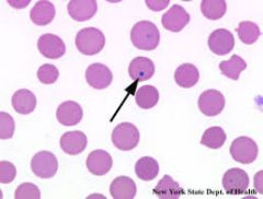
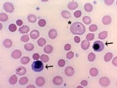
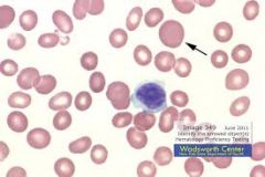
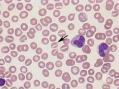
Howell-Jolly Bodies
-RBC's nucleus is supposed to be eaten by macrophage, but sometimes pieces are missed (will eventually be found by another macrophage) |
|
|
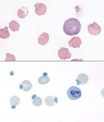
Heinz bodies -oxidized hemoglobin -easily seen w/ Vital Stain (new methylene blue) -very sensitive to environment
|
|
|
-young, larger, bluish (due to ribosomes- will lose) -normal: very low numbers -sick/anemic: high numbers
-seen w/ Romanowski's stain
*not seen in horse (RBC width dist. instead) |
|
|
*only seen with Vital Stain
-probably same as chromasia -aggregated ribosomes/ER
*no reticulocytes in horses! |
|
|
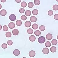
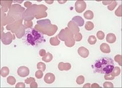
Nucleated RBCs
-normal in small numbers |
|
|
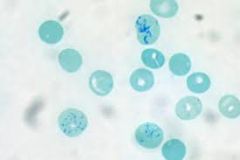
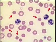
Basophilic stippling -due to ribosomes; deficiency of ribosome-metabolizing enzyme (pyrimidine 5' nucleotidase) -lead poisoning in dog |
|
|
Anisocytosis |
|
|
Poikilocytosis |
|
|
macrocytosis |
|
|
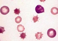
Creantion
-processing issue; slide not allowed to dry |
|

|
Schistocytes
-coagulation issue |
|
|
Spherocytes
-immune-mediated hemolytic anemia; macrophage takes a chomp trying to get antibodies |
|
|
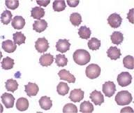
Acanthocytes
-pseudopodia-like thing -sign of RENAL disease; changes in outer lipid layer |
|

|
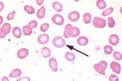
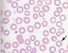
Target cells
-hemoglobin trapped in center due to lipid membrane change |
|
|
Stomatocytes
-change in inner lipid layer of membrane |
|
|
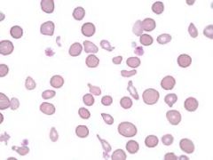
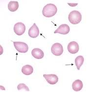
Dacryocytes
-lost flexibility, can't maintain shape -uncommon in animals (seen w/ bone marrow dz in humans) |
|
|
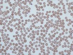
Hypochromasia
-iron deficiency -smaller cells because are continually dividing |
|
|
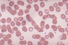
Rouleaux
-w/ saline, will disperse! If dispersed, not agglutination |
|
|
Agglutination
-antibodies react to self antigens |
|
|
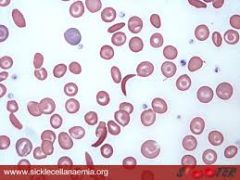
Sickle cells
-common in deer; change in oxygen retention of cells |
|
|
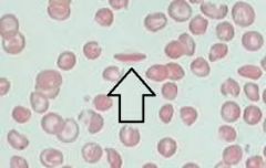
Hemoglobin polymerization -seen on normal smears, no known significance |