Reading...
![]()
Play button
![]()
Play button
![]()
Use LEFT and RIGHT arrow keys to navigate between flashcards;
Use UP and DOWN arrow keys to flip the card;
H to show hint;
A reads text to speech;
30 Cards in this Set
- Front
- Back
- 3rd side (hint)
|
UrinConsary System Anatomy
|
Consists of the kidneys, ureters, bladder, and urethra.
|
|
|
|
Kidney Anatomy and function
|
The functional unit of the kidneys is the nephron. Each kidney contains more than a million nephrons which filter waste products from the blood, reabsorb water and nutrients from the tubular fluid, and secrete excess substances in the form of urine. The nephron filters about 190 L of water out of glomerular blood each day.
|
|
|
|
Urine Formation Steps 1-2
Glomerulus and Bowman's Capsule |
The formation of urine begins in the glomerulus, a tuft of capillaries with very thin walls and a large surface area. The blood pressure within the glomerulus is higher than in the Bowman's capsule which surrounds it. This causes the filtration of fluid into the bowman's capsule.
|
|
|
|
Urine Formation Steps 3-4
Proximal Convoluted tubule and Loop of Henle |
From the Bowman's Capsule, the initial urine proceeds into the proximal convoluted tubule where a large amount of water and all nutrients are reabsorbed into the blood capillaries surrounding the tubule. It then flows into the Loop of Henle which has a descending limb, a loop, and an ascending limb. Salt and water are reabsorbed in the loop of henle.
|
|
|
|
Urine Formation -to the bladder
|
After the loop of henle, the distal convoluted tubules permit the excretion of concentrated urine by actively secreteing substances such as potassium and hydrogen which helps the body maintain electrolyte balance and acid-base balance of blood and body fluids. Last, the urine passes from the collecting tubules whose openings are in the papillae, into the calyces, and on to the funnel shaped renal pelvis and tubular ureters. Peristaltic waves move the urine down teh ureters and into the bladder.
|
|
|
|
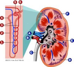
A. Renal vein
B. Renal Artery C. Ureter D. Medulla E. Pelvis (Major and Minor Calyces) F. Cortex |
1. Ascending Limb of Loop of Henle
2. Descending Limb of Loop of Henle 3. Peritubular capillaries 4. Proximal Tubule 5. Glomerulus (Bowman's Capsule and Glomerular Capillaries) 6. Distal Tubule |
|
|
Ureters
|
The ureters enter the bladder through an oblique funnel that functions as a valve to prevent backflow of urine into the ureters during bladder contraction.
|
|
|
|
Bladder
|
The bladder acts as a reservoir for the urine before it leaves the body. The openings of the two ureters lie at the posterior corners of the triangle-shaped floor (the trigone) and the urethral opening is situated at the anterior lower corner. The average bladder holds about 250mL of urine.
|
|
|
|
Other Kidney Functions
|
The kidney is also important in the production of RBC's and in the control of blood pressure. Erythropoietin stimulates the rate of production of RBC's and is released by the kidneys. The juxtaglomerular apparatus in the kidney also releases renin which acts to release angiotensin which controls blood pressure.
|
|
|
|
Congenital Anomilies Related to the Kidneys
|
- Number
- Size - Rotation - Position - Fusion - Renal Pelvis - Ureters |
|
|
|
Anomilies: Size - Hypoplasia or Hyperplasia
|
Hypoplasia - Under developed (small) kidney. The is a defective development of renal tissue. You will see atypical small vessels, reduced number of calyces, and it is usually unilateral.
Hyperplasia - Larger kidney. NOT hypertrophy which is larger cells, but is more cells produced. Both are seen well with sonography, and may be seen on normal abdomen xrays or with contrast in IVP. |
|
|
|
Anomalies - Number - Agenesis
|
A congenital absence of an organ. No kidney shadow will be observed on imaging studies.
Treatment is not needed for size or number anomalies. |
|
|
|
Anomalies - Rotation - Malrotation
|
Poles of the kidneys are rotated abnormal to regular rotation. May look like a pathologic condition on imaging studies due to strange appearance of renal paranchyma, pelvis, or calyces when actually the kidney is fine, just rotated. No treatment is necessary.
|
|
|
|
Anomalies - Position - Renal Ectopia
|
An abnormally positioned kidney with in place or position. Can be found in the pelvis or in the thoracic area. Can also be crossed ectopia where both kidneys are on one side. Usually seen on IVP or CT. Full abdominal images are needed in order to search for an ectopic kidney. No treatment is needed unless there are problems.
|
|
|
|
Anomalies - Fusion - Horseshoe Kidneys
|
This is when the lower poles of the kidneys are fused. Usually seen on KUB or CT. No treatment is necessary unless there is a problem such as kinking of the ureters.
|
|
|
|
Ureteroceles
|
A ureterocele (sac of urine) is a cystic dilatation of the distal ureter near its insertion into the bladder. The ureterocele may be inside or outside of the bladder and can be caused by an obstruction or may cause an obstruction itself.
|
|
|
|
Treatment of Ureteroceles
|
Most common treatment is stent placement. But, may have to be surgically removed. Seen on IVP.
|
|
|
|
Renal Trauma
|
Issues with collection can cause extravasation. You can see this with contrast studies where you have extravasation of urine and possibly bleeding into tissues. Will probably do a CT, ultrasound and Nuc med study to test further. If it is a large trauma they may do a nephrectomy (remove kidney). If it is small they may just watch it.
|
|
|
|
Inflammatory Disorders of the Kidney
|
- Pyelonephritis
- Tuberculosis - Cystitis |
|
|
|
Pyelonephritis
|
Inflammation of the kidney or renal tissue which is seen in acute and chronic forms. Is related to a bacterial infection in that it is a pus-generating disease.
|
|
|
|
Acute Pyelonephritis
|
Acute inflammation of the renal parenchyma and renal pelvis. Is diagnosed with IVP sometimes but 75% of the time these are normal. Diagnosed more with urinalysis with hematuria and increased WBC's.
|
|
|
|
Signs/Symptoms and Treatment of Acute Pyelonephritis
|
Pt would have flank pain, pain with urination, frequent urination, and urgent urination. Affects the interstitial tissues between the tubules so will appear as "Squeezed" looking collecting structures or look like a chronic nephrogram.
Treatment is antibiotics. |
|
|
|
Chronic Pyelonephritis
|
A chronic inflammation of the kidney. Inflammation since birth in many pts. Will have same symptoms as acute but will be chronic symptoms. Often caused by obsturction of some sort.
|
|
|
|
Signs and Treatment of Chronic Pyelonephritis
|
Will look like blunting of the calyces. Seen on IVP and CT. Treatment is to eliminated the obstruction if possible and to control the hypertension that is caused by the backup into the vascular system.
|
|
|
|
Renal Abscess
|
May be seen with pyelonephritis. S/S are fever, WBC increase, chills, aches, pain, blood in urine, etc...Will drain it to see if it is urine, pus, or hemorrhage.
|
|
|
|
Renal Tuberculosis
|
The spread of infection (TB) to the kidneys causes an ulcerative destructive process which occurs in the tips of the papillae and causes enlargement of the calyces. This leads to cortical scarring and parenchymal atrophy. It may cause calcifications and can also involve the ureters and bladder.
|
|
|
|
Diagnosis and Treatment of TB of the Kidneys
|
Seen on KUB, IVP, and CT as calcifications around the papillae.Seen as small rounded calliceal ends or as calcified granulomas that take over the whole kidney. At this extreme, it can be seen without contrast on a plain film.
Treatment - antibiotics, antiviral meds, and kidney removal. |
|
|
|
Cystitis
|
Inflammation of the urinary bladder which is seen most commonly in women and children (hygiene is the #1 preventor). It is caused by inadvertent spread of bacteria present in fecal matter. Can also be caused by catheterization. Diagnosed via IVP or cystogram.
|
|
|
|
Radiographic appearance of cystitis
|
Bacteria is eating away at the mucose of the bladder so it will appear "lumpy" and irritated with thicker looking walls. If it is let to go on without fixing, you may also see air in the walls of the bladder as it gets trapped there and it can lead to kidney complications if it is not taken care of.
|
|
|
|
Signs, symptoms, and Treatment of cystitis
|
Urinary frequency, burning, and urgency are common signs. Antibiotics, prevention (hygeine and educaiton), increased fluid, cranberry juice - acid makes an unhealthy environment for bacteria.
|
|