Reading...
![]()
Play button
![]()
Play button
![]()
Use LEFT and RIGHT arrow keys to navigate between flashcards;
Use UP and DOWN arrow keys to flip the card;
H to show hint;
A reads text to speech;
28 Cards in this Set
- Front
- Back
|
How does a right lateral radiograph affect the kidneys?
|
Increases the distance b/w renal shadows
|
|
|
True or false. Both kidneys should be present on a lateral radiograph.
|
False, May not see the right kidney due to silhouette w/ adjacent liver, may not see left kidney due to feces and ingesta-Don't freak out!
|
|
|
What is the normal size of a kidney in a dog and cat?
|
Dog: 2.5-3.5 x length of L2
Cat: 2.4-3.0 x length of L2 MEASUREMENTS ARE ALWAYS ON VD!!!!! |
|
|
What is involved in performing an excretory urograph?
|
-Enema
-Patient must be well hydrated! (so can filter through contrast) -Always do survey films -Inject 400mg/lb IV as a blus -Acquire films immediately 5,10,20 and 40 minutes (both lateral and VD) |
|
|
What contrast media is used for excretory urograms?
|
Iodinated water-soluble contrast medium
|
|
|
What is gained by performing an excretory urogram?
|
-Improves morphologic assessment of kidney
-Poor test of function but will tell you a little bit about the filtration rate |
|
|
What are 4 contraindications for excretory urogram?
|
1) Azotemia & dehydration (if kidneys aren't working will retain media=trouble)
2) Pheochromocytoma 3) Multiple myeloma 4) Prior allergic reactions to contrast media |
|
|
**What are the 4 phases of an excretory urogram?
|
1) Arteriogram-immediately
2) Nephrogram-within 20 seconds of injection 3) Pyelogram-begins 3-5 minutes post injection 4) Cystogram-as contrast is cleared into the lower urinary tract |
|
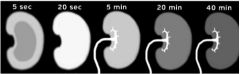
What are the phases of the excretory urogram from left to right?
|
1. Arteriogram
2. Nephrogram 3. Pyelogram 4. Cystogram 5. Cystogram |
|
|
What does the arteriogram phase of the excretory urogram show? How long is it?
|
-Documents initial blood flow to the kidney
-Very short, seldom imaged |
|
|
What does a normal nephrogram show?
|
Contrast is filtered and accumulates w/in the renal tubules
-Should see good initial opacification that gradually fades over the study period -Opacification of kidneys should be uniform and symmetrical |
|
|
What does the contrast outline in a pyelogram? What can be evaluated?
|
The collecting system is oulined, will be able to evaluate the renal pelvic and diverticuli
|
|
|
How big should a normal renal pelvis, recess, and proximal ureter be?
|
Pelvis: <2mm wide
Recesses: < 1mm wide Proximal ureters: <2.5 mm in diameter |
|
|
What does a cystogram show?
|
-Contrast material entering the lower urinary tract
-See segmental images of the ureters and outline of bladder |
|
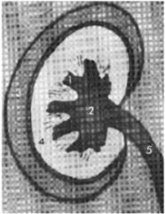
Label the numbers
|
1=diverticuli (recesses)
2=renal pelvis 3=cortex 4=medulla 5=ureter |
|
|
What does it mean if you are performing an excretory urogram and you see contrast being eliminated in the feces?
|
Means kidneys aren't working so contrast is being eliminated by alternative routes
|
|
|
How do you know if contrast induced renal failure occurs when performing an excretory urogram?
|
See opacification of kidneys that persists or becomes more intense over time
|
|
|
What are 4 pathogeneses that possibly account for contrast induced renal failure?
|
1) Hypotension
2) Acute tubular necrosis 3) Tubular obstruction 4) Acute renal failure |
|
|
What are 2 alternative routes of excretion of contrast media used for excretory urograms?
|
Liver & GI
|
|
|
What does a poor nephrogram look like?
|
Poor initial opacification followed by progressively decreasing opacity
|
|
|
What are 2 differentials for a poor nephrogram?
|
1) Primary polyuric renal failure
2) Inadequate contrast medium dose |
|
|
How much contrast is normally eliminated in the feces?
|
Normally no more than 2% (not much increase in density)
|
|
|
What 6 kidney characteristics should always be evaluated on a radiograph?
|
1) Number
2) Size 3) Shape 4) Margination 5) Location 6) Opacity |
|
|
What are 4 differentials for a kidney condition where the kidneys are still a normal size & shape?
|
1) Amyloidosis (have to biopsy for diagnosis)
2) Glomerulonephrosis 3) Acute pyelonephritis 4) Familial renal disease -May be normal -Small and smooth -Small and irregular |
|
|
What are 5 differentials for bilateral renal enlargement?
|
1) Lymphoma
2) FIP 3) Hydronephrosis 4) Polycystic kidney disease 5) Rare primary renal neoplasia |
|
|
How can you tell that there's right renal enlargement in a radiograph?
|
-Increased focal opacity in right craniodorsal region
-Medial & ventral displacement of the descending duodenum and ascending colon -Left ventral displacement of the small intestine |
|
|
What is displaced with left renal enlargement?
|
Ventral and medial displacement of the descending colon and adjacent small intestine
|
|
|
What are 2 differentials for increased opacity of the kidneys?
|
Calculi
Mineralization (nephrocalcinosis) |