![]()
![]()
![]()
Use LEFT and RIGHT arrow keys to navigate between flashcards;
Use UP and DOWN arrow keys to flip the card;
H to show hint;
A reads text to speech;
143 Cards in this Set
- Front
- Back
|
Edh |
Swirl sign |
|
|
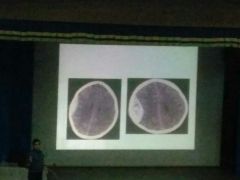
Acute SDH |
Right sided |
|
|
Left sided chronic hypodense SDH Right sided subacute isodense |
|
|
|
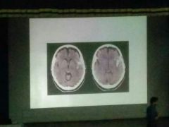
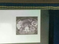
Intraventricular blood - SAH |
Visualize cisterns fissures |
|
|
Sylvian fissure hge SAH focal |
|
|
|
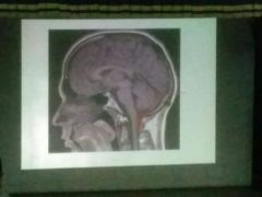
Herniation of brainstem /cerebellum Arnold chiari malformation - both type 1 n 2 have tonsilar herniation ~posterior pituitary is seen in sagittal t1 weighted MRI ~loss of this bright spot - central diabetes insipidus |
|
|
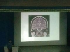
T2 coronal MRI |
|
|
|
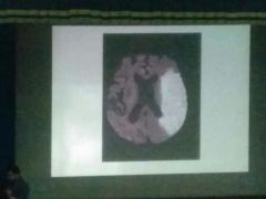
Dwi adc |

|
|
|
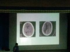
Infarct |
|
|
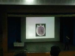
Epidermoid cyst |
|

Canavan disease |
Ring enhancing lesion Spectroscopy NAA higher than choline and creatine_normal Choline higher than creatine |
|
|
Scolex |
|

|
Extra peak - TB diagnostic Lipid Lactate peak with ring enhancing lesion |
|

|
Normal |
|

2nd image - Tuberous sclerosis |
Ts |
|

|
X |
|

|
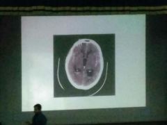
1 dense MCA sign |
|

|
1 delta sign - ncct - venous sinus thrombosis 3 empty delta sign - cect - venous thrombosis 2. Pseudo empty delta sign - SAH - NCCT |
|

|
2 - oligodendroglioma 1 gBm - involves both hemispheres of cerebrum - butterfly tumor 3 craniopharyngioma 4 |
|
|
Cerebellar vermis medulloblastoma |
|

|
Tonsilar herniation Posterior fossa tumor Ependymoma - along lining of 4th ventricle it extends |
|

|
NF 2 B/l acoustic neuroma |
|

|
Classical Basal meningitis |
|

|
Dawson finger MS |
|
|
Xx |
Popcorn pattern cavernous sinus angioma |
|

|
1 DWI - caudate and lentiform nucleus Infarct 3 perfusion MRI shows umbra and penumbra regions |
|

|
Dwi - watershed Infarcts |
|

|
Normal Diffuse Cerebral edema - white Cerebellar sign/dense Cerebellar sign/reversal sign- sign of global ischemia - poor prognosis - loss of white grey differentiation |
|

|
Aaa |
|

|
Anencephaly |
|

|
Dandy Walker |
|
|
Normal- ice cream cone appearance Incudomalleal joint Middle ear HRCT |
|

|
Apple in plate appearance of cochlea Normal HRCT |
|

|
Developmental venous angioma Mc developmental venous anomaly Medusa head appearance |
|

|
Pancake brain Alobar holoprosencephaly |
|

|
Salt and pepper appearance (any paraganglioma) Glomus jugulare |
|

|
Tram track appearance |
|

|
1 molar tooth appearance 2 bat wing appearance on CT jobert syndrome |
|
|
Tigroid pattern Metachromatic leukodystrophy |
|

|
Hot Cross bun |
|

|
Face of panda Wilson |
|
|
Dandy Walker variant Keyhole appearance Cisterna magna communicates with 4th ventricle If cyst present - Dandy Walker malformation |
|

|
light bulb appearance Acute Infarct on DWI |
|

|
Bag of worms No signal in MRI - signal void AV malformation |
|

|
Face coronal section CT scan PNS IOC for PNS lesion Spur at beginning of nasal septum - crista galli |
|
|
Opacification of right max, frontal and r nasal cavity - antrochoanal polyp - expansion also seen |
|

|
CT PNS coronal plane Soft tissue window Hyperdense secretions - fungal sinusitis of R maxillary sinus (Hyperdense compared to left) Bone erosion |
|

|
Water's view - maxillary sinus Waters with open mouth - sphenoid sinus best view for sphenoid sinus - lateral view |
|

|
Air fluid level |
|

|
USG eye - retinal detachment Classical - Y or V pattern |
|

|
Normal mammogram |
|

|
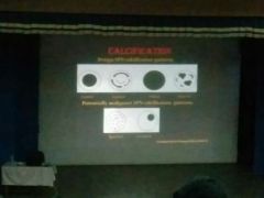
Macro calcification Popcorn Birad 2 Routine screening |
|

|
Pleomorphic microcalcification |
|

|
1 anechoic - cyst - posterior acoustic shadow 2 hypoechoic - lateral edge shadow - marker of fibroadenoma |
|

|
USG breast irregular taller |
|

|
Hypoechoic irregular - malignant |
|

|
Pleural effusion |
|
|
Normal CT lung window T5 level |
|

|
CT lung mediastinal window Below aorta - SVC |
|

|
Left pneumothorax with mild left sided collapse |
|

|
Deep sulcus sign |
|

|
CECT Pericardial effusion Pleural effusion Pericardial thickening |
|

|
Inflamed pleura Split pleura sign Empyema |
|

|
Pulm embolism Mint polo sign |
|

|
Pulm oedema |
|

|
Bronchogenic cancer causing right upper lobe collapse Golden S sign |
|

|
Miliary nodules Hematogenous dissemination of disease |
|
|
Honey combing ILD esp Idiopathic pulm Fibrosis |
|

|
Air crescent sign Aspergilloma Hydatid cyst Tumor ball in cavity Blood in cyst |
|

|
Tree in bud pattern - multiple nodular -TB (mcc) - bronchiolar impaction -other causes - RSV (esp) , CMV pneumonia |
|

|
Conglomerated necrotic lymph nodes of mediastinum TB - necrotic Ln peripherally enhancing |
|

|
Cavitatory lesion in lung TB/malignancy |
|

|
Pulm alvelolar proteinosis |
|

|
Pneumopericardium -Outlining of air by pericardium -continuous diaphragm sign - specific for pneumomediastinium |
|
|
Staph pneumonia Pneumatocele |
|
|
Corona radiata sign - malignant Cavity - thick walled, malignant |
|
|
Benign and malignant calcification patterns |
|

|
Left lung collapse Mass in trachea |
|

|
CT angiography |
|
|
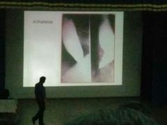
Achalasia |
|

|
Esophageal varices - serpigonous filling defect |
|

|
Hiatus hernia |
|

|
Dysphagia lusoria Aberrant right subclavian artery origin |
|

|
Supine and erect Normal erect x ray abdomen |
|
|
Multiple air fluid levels |
|

|
Air under right dome of diaphragm ~sensitivity only around 70% ~Then send for CT ~IF NO CT, LEFT LATERAL decubitus - most sensitive x ray position (90%) |
|

|
Wriggler sign - pneumoperitoneum supine xray |
|

|
Severe small bowel obstruction String of bead appearance Air bubbles get trapped at mucosal outline |
|

|
Small bowel feces sign On CT Distal obstruction of small bowel |
|

|
CT abdomen Axial 3 pancreas 7 IVC 9 portal vein |
|

|
Comb sign - ileocaecal involvement Crohn's disease - dilation of vasa recta which supply mesentry TB - mimics Crohn's |
|

|
CT virtual colonoscopy - can see proximal bowel also |
|

|
Double bubble sign -duodenal atresia -annular pancreas |
|

|
T tube cholangiogram |
|

|
1 Multiple stones in gall bladder 2 MRCP image showing filling defect in GB ie stone |
|

|
Cartwheel sign in liver Hydatid cysts - USG |
|

|
1. MR 2 MRCP - fluid containing structures seen in white |
|

|
-USG liver -cystic lesion - posterior acoustic enhancement (hypoechoic) |
|

|
-Focal isoechoic hepatic lesion -Mc asymptomatic hepatic lesion - hemangioma - hyperechoic -mixed echoic lesion - can be hemangioma - confirm: Triphasic CT or Triphasic MRI - peripheral nodular enhancement with centripetal filling |
|

|
1 ncct 2 onwards cect Any lesion in liver which shows enhancement in arterial phase and washout in portal venous phase - HCC no biopsy needed |
|
|
Sever Acute necrotizing pancreatitis |
|

|
-Acute cholecystitis - thickened gall bladder membrane -Posterior acoustic shadow |
|
|
Outer wall diameter >7mm (USG and on CT - >6mm) Dilated tubular structure Streakiness of periappendicular fat 1. Arrow head sign 2. Caecal bar sign |
|

|
On FAST - hepatorenal pouch of Morrison showing blood |
|

|
Apple core appearance - ca colon Double contrast barium enema |
|

|
Exudative form of peritoneal TB Can also be seen in malignancy |
|
|
Intussusception |
|

|
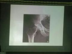
Holt oram syndrome Absent radius ASD |
|

|
Dinner fork deformity Colles |
|

|
Arthritis mutilans Pencil in cup |
|

|
Trap door/fallen leaf appearance Simple bone cyst |
|

|
Expansile lytic lesion in tibia with no sclerosis GCT |
|
|
GCT |
|

|
1 tram track sign Bamboo sign 2 dagger sign Earliest investigation to identify ankylosing spondylitis - STIR MRI |
|

|
Spondylosis - degen changes in spine Spondylolysis - break in pars articularis Spondylolisthesis - slipping of one vertebra over another |
|

|
MR vertebra T2 weighted L5 - S1 Prolapsed Inter vertebral disc |
|

|
Look at posterior vertebral margin - 1 malignant collapse : convex 2 osteoporotic/benign : concave |
|

|
MR shoulder Normal |
|
|
Bald man sign Supraspinatus rupture |
|

|
MR knee with marked ACL |
|

|
ACL tear with buckling of PCL |
|

|
Double PCL sign Bucket handle sign of medial meniscus |
|
|
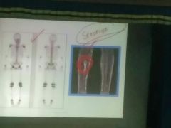
STIR MRI - IOC for stress # tibia |
|

|
Interosseous memb ossification Fluorosis |
|

|
IVP CT urograph CT urograph with 3D reconstruction |
|
|
Crossed fused ectopia |
|

|
2 - duplicate system |
|

|
Grade 5 b/l VUR |
|

|
Normal and posterior urethral valve PUV - keyhole sign |
|

|
2 Cemented/putty kidney - renal TB 1 cork screw ureter |
|
|
Right kidney abnormal Striate nephrogram - Pyelonephritis acute |
|

|
A/w xanthogranulomatous pyelonephritis CT - bear paw Infection - proteus Emphysematous pyelonephritis - more dangerous |
|

|
RCC - stellate pattern |
|

|
Normal obstetric USG Fill Form F before doing obstetric scan according to PCPNDT act |
|

|
Anencephaly |
|

|
Doppler of uterine A at 28 wks Diastolic notch - PIH |
|

|
Normal umbilical A Doppler |
|

|
High S/D ratio - uteroplacental insufficiency |
|

|
Reveral of Diastolic flow - death will occur within 48 hrs |
|

|
Torsion of right testis |
|
|
PCOS morphology |
|
|
Fishnet appearance Hemorrhagic cyst D/D - endometriosis if it fails to resolve by 6 months |
|
|
Snow storm appearance in hydatidiform mole |