Reading...
![]()
Play button
![]()
Play button
![]()
Use LEFT and RIGHT arrow keys to navigate between flashcards;
Use UP and DOWN arrow keys to flip the card;
H to show hint;
A reads text to speech;
21 Cards in this Set
- Front
- Back
|
NEGA Protocol Toes:
|
AP, both obliques, lateral
|
|
|
NEGA protocol Foot:
|
AP, lateral, and both obliques
note: AP is usually 15 degree aangle on foot to open joints |
|
|
NEGA Protocol calcaneus:
|
* tangential view angle cephalic 40-45 degrees - joint space visualized
* true lateral * obliqued posn/d similar to foot but coned to calcaneus |
|
|
NEGA protocol ankle:
|
* AP, lateral, internal & external oblique views
* the ankle mortice s/b well visualized & symmetric on internal oblique film note: turn leg not just foot |
|
|
how many bones are in the foot?
|
26 total
14 phalanges (toes); 5 metatarsals (instep); 7 tarsals (ankle) |
|
|
the phalanx, and metatarsals are composed of body and 2 articular ends - which is the head and which is the base?
|
the base is proximal; the head is distal
|
|
|
name the 7 tarsals:
|
calcaneus, talus, navicular bone, cuboid, medial coneiform, intermediate cuneiform, lateral cuneiform
|
|
|
what 4 bones articulate w/ the talus?
|
tibia, fibula, calcaneus, and navicular bone
|
|
|
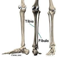
does the tibia or fibula bear body weight?
|
the tibia bears body weight
|
|
|
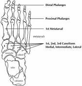
know anatomy of foot
|
|
|
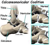
sample of calcaneus/ navicular region
|
|
|
Labelled image of the foot
|
|
|
|
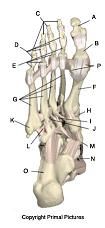
Label - DORSAL VIEW
A. distal phalanx of the hallux B. proximal phalanx of the hallux C. distal phalanges D. intermediate phalanges E. proximal phalanges F. 1st metatarsal G. lesser metatarsals H. medial cuneiform I. intermediate cuneiform J. lateral cuneiform K. styloid process L. cuboid M. navicular N. talus O. calcaneus |
|
|
|
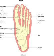
label plantar view foot:
A. distal phalanx of the hallux B. proximal phalanx of the hallux C. distal phalanges D. intermediate phalanges E. proximal phalanges F. 1st metatarsal G. lesser metatarsals H. medial cuneiform I. intermediate cuneiform J. lateral cuneiform K. styloid process L. cuboid M. navicular N. talus O. calcaneus P. sesamoids |
|
|
|
label plantar view foot:
A. distal phalanx of the hallux B. proximal phalanx of the hallux C. distal phalanges D. intermediate phalanges E. proximal phalanges F. 1st metatarsal G. lesser metatarsals H. medial cuneiform I. intermediate cuneiform J. lateral cuneiform K. styloid process L. cuboid M. navicular N. talus O. calcaneus P. sesamoids |

|
|
|
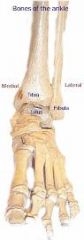
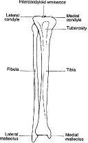
label bones of ankle AP
|
|
|
|
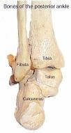
Label Posterior Ankle
|
|
|
|
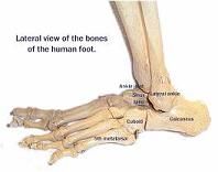
lateral view of ankle
|
|
|
|
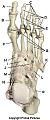
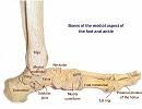
bones medial aspect of foot
|
|
|
|
image of leg bones
|
|
|
|
image of leg bones
|
|