![]()
![]()
![]()
Use LEFT and RIGHT arrow keys to navigate between flashcards;
Use UP and DOWN arrow keys to flip the card;
H to show hint;
A reads text to speech;
67 Cards in this Set
- Front
- Back
- 3rd side (hint)
|
Neuroscience definition |
A mix between biology, psychology, physiology and biochimistry that studies the nervous system (illnesses and health): - structure - connection - function Of the brain |
Interdisciplinary field / 3 attributes |
|
|
Histology |
Studying microscopic structures and tissues. - need to be Fixed - ice, formalin chemical product - slicing machine is a Microtome - different stains of colors highlight different structures |
Not visible to the naked eye |
|
|
CT scan |
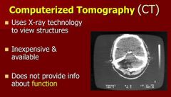
Computerized Tomography - provides only the structure |
Computerized tomography |
|
|
PET scan |
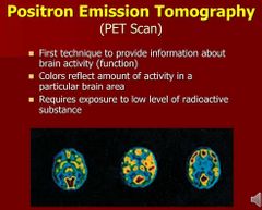
Positron Emission Tomography - shows function of the brain but not exactly in real time. It also requires a radioactive substance to work. |
Positive/Expansion/related to this field |
|
|
MRI and fMRI |
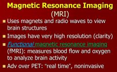
Magnetic Resonance Imagining - structure. In very great details! |
|
|
|
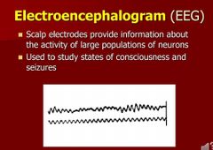
EGG |
|
|
|
|
Evoked potential |
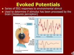
Measures perception directly through the brain (no indication given from the patient) |
|
|
|
Stimulation |
To discover the function of an area. Can be done during neurosurgery, throu implanted electrodes (done at Doctor's hopsital to treat Alzheimer). |
|
|
|
Pharmacological methods |
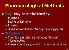
- Drugs - Microdialysis: extraction of material to assess chemicals present in a small area |
|
|
|
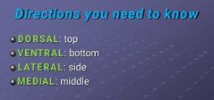
Directions |
|
4 |
|
|
Planes sections |
Coronal: divides the front from the back of the brain. Sagital: divides through the medline. Gives a side view. (Divides the left fron the right) -Midsagital: cut through 2 hemispheres Horizontal (axial): divides the top grom the bottom of the brain |
4 |
|
|
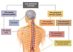
Organization of the Nervous system |
|
CNS / PNS 2 / 2 + 2 |
|
|
Peripheral Nervous System (PNS) |
Somatic = Volontary Autonomic (ANS) = involuntary |
2 |
|
|
Somatic Nervous System |

Afferent Efferent = Exit (to help remembering) |
Not automatic + movement of the signals |
|
|
Autonomic Nervous System (ANS) |
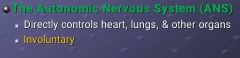
Involuntary Include the sympathetic and parasympathetic NS |
Not voluntary |
|
|
Sympathetic Nervous system |

|
ANS |
|
|
Parasympathetic Nervous System |

|
Not fight or flight |
|
|
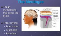
Meninges |
|
3 layers, harder outside |
|
|
Cerebrospinal Fluid (CSF) |
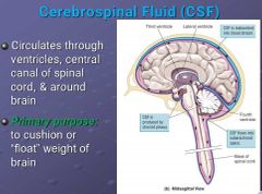
Produced by Chloroid plexus (inside ventricles). Purpose: float the brain/protect |
|
|
|
The Hindbrain (brainstem) |
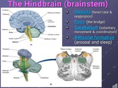
- Medulla oblongata - Pons - Cerebellum - Reticular formation
BASIC LIFE SUPPORT: Breathing, heart beath |
4 component |
|
|
Medulla |
Part of the brainstem.
Controls heart rate and respiration. |
Brainstem |
|
|
Pons |
Part of the brainstem.
Is a bridge between the right brain and the left |
No function |
|
|
Cerebellum |
Part of the brainstem. Volontary movement and coordination. |
|
|
|
Reticular formation |
Part of the brainstem. Arousal and sleep |
My favorite thing |
|
|
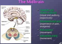
The Midbrain |
|
4 components |
|
|
Superior and inferior colliculi |
Part of the midbrain Visual and auditory respectively |
2 senses |
|
|
Periaqueducal gray |
Part of the midbrain. Pain and analgesia (pain relief) |
Tylenol |
|
|
Red nucleus |
Part of the midbrain Movement |
Like Cerebellum |
|
|
Substantia nigra |
Part of the midbrain Movement |
Like Cerebellum |
|
|
Thalamus (forebrain) |
Routes for incoming sensory information to cortex |
Senses |
|
|
Hypothalamus (Forebrain) |
Hunger, thirst, sex and body temperature (temperature control) |
Area of needs/desire and C° |
|
|
Basal Ganglia (Forebrain) |

Volontary movement |
Like somatic NS, function. 4 components: CN, P, GP,SN |
|
|
Limbic System (Forebrain) |
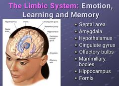
Emotion and Motivation, Learning, Personality
- Septal area: rage, attack
- Amygdala: fear and anxiety, sometimes rage too
- hypothalamus: body temperature, thirst, hunger, sex
- Cingulate gyrus: loneliness area, fomo, social pain, empathy
- Olfactory bulbs: smell
- Mamillary bodies: part of hypothalamus, part of memory, and spatial environment
- Hippocampus: processing memories
- Fornix: pathway |
Emotion, learning and memory 8 components: SA, A, H, CG, OB, MB, H, F |
|
|
Cerebral Cortex (Forebrain) |
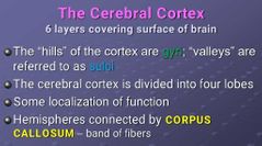
Higher cognitive function |
Hills vs valleys 4 lobes Localization of function Einstein's most developed part of the brain. |
|
|
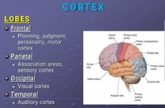
Lobes (Part of Cortex) |
|
4 lobes: F, P, O, T |
|
|
Frontal lobe (Cortex) |
Planning, judgment, personality, motor cortex |
|
|
|
Parietal lobe (Cortex) |
Association areas, sensory cortex |
Functions |
|
|
Occipital lobe (Cortex) |
Visual cortex |
Function |
|
|
Temporal lobe (Cortex) |
Auditory cortex |
Function |
|

|
|
7 components |
|
|
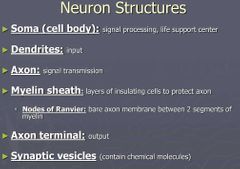
Soma/cell body (Neuron) |
Signal processing, life support center |
|
|
|
Dendrites (Neuron) |
Input |
Branch like receptors |
|
|
Axon (Neuron) |
Signal transmission |
Longest part of the neuron |
|
|
Myelin sheath (Neuron) |
Layers of insulating cells that protects the axon |
Hot dog bun |
|
|
Nodes of Ranvier (Neuron) |
The segment on axon between 2 myelin sheath |
The space in between |
|
|
Axon terminal (Neuron) |
Output |
Branch like ends |
|
|
Synaptic vesicles |
Bags containing the neurotransmitters |
At the end of the process |
|
|
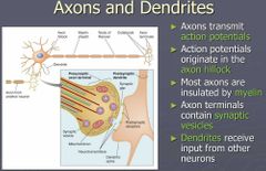
- Axons transmit... - Action potentials originate in the... - Most axons are insulated by... - Axon terminals contain ... - ... receive input from other neurons |
|
|
|
|
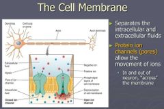
The Cell Membrane
- Separates the ... and ... f... - Proteins ion channels ...
|
|
|
|
|
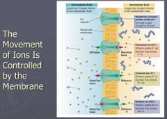
-The movement of ions is controlled by the cell membrane. - extracellular fluid charge? - intracellular fluid charge? - large protein molecule charge? - Potassium ion charge? - Sodium ion charge? - Chloride ion charge? |
|
|
|
|
Equilibrium potentials for each ions? - Na+ (Sodium ion) - K+ (Potassium ion) - Cl- (Chloride ion) |
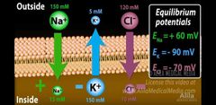
Equilibrium potentials dictate te movement of the ions through the membrane. When it goes with his gradient it calls diffusion. Sodium ions (Na+) diffuse inside the membrane |
|
|
|
Resting (membrane) potential |
- 70 mV
This is the difference between inside the membrane and outside the membrane |
Voltage + definition |
|
|
Sodium potassium pump |

For every 2 potassium ions (K+) pumped into the cell, it pumps out 3 sodium ions (Na+). This make it that there are more sodium ions outside than there is potassium ions inside. So even if both are positive, the ousite is More positive. Plus the large negative protein inside the membrane keep the inside of the cell negatively charged. |
Charges inside vs outside of the cell |
|
|
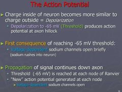
The movement of ions accross the membrane creates... |

The action potential starts at the hillock. It reaches a threshold (-65 or 55 mV), due to a change in voltage (depolarization), it then opens voltage dependent channels and propagates all the way down the axon until the sodium ions channels stops and calcium channels open instead, creating the synaptic vesicles for Exocytosis. |
|
|
|
Ions channels |
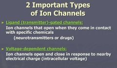
Allows the movement of ions through the cell membrane. Creating Action Potential.
- Ligand-gated (transmitter) channel: needs a neurotransmitter/chemical to open.
- voltage-dependent channels: open and close depending on the nearby electrical charge (intracellular voltage) |
2 types |
|
|
Depolarization |

|
Like a battery |
|
|
Steps to Action Potential |
|
Treshold, opening, expansion |
|
|
Action potential graph and relative refractory period |
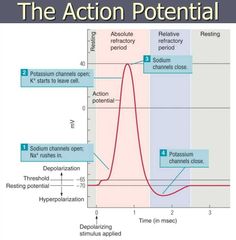
Relative Refractory Period: period of time when the neuron cannot emits a signal again because of hyperpolarization.
- Sodium channel open to let sodium ions in at the -65 (or -55) mV threshold.
- Potassium channel, opens to leave potassium ions out of the cell around 30mV.
- Sodium channel, closes at 40mV. Signals calms down.
- potassium channel, closes below -70mV and creates hyperpolarization |
|
|
|
The synapse |
|
Definition |
|
|
Exocytosis
|
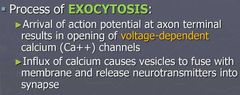
Translation: Existing the cytoplasm When the action potential arrives at the axon terminal there is no more voltage dependent sodium channels. When -65mV is reached, voltage dependent Calcium channels opens, and the influx of calcium causes vesicles to fuse with membrane and release neurotransmitters into synapse. |
Existing the cytoplasm |
|
|
Neurotransmitters released in Exocytosis |
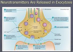
Calcium channels open to Calcium Ions (Ca++) which causes synaptic vesicle to release from microtubules. The more calcium the more neurotransmitters get released.
Synaptic vesicles (bags) recycle and get reused. |
What causes the Exocytosis |
|
|
Receptor type |
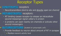
Autoreceptors: monitors neuron's activity level and provide feedback that can essentially slow down the action potential. For example it analyzes that there are a lot of seritonin in the synapse and order to slow down. |
3 types |
|
|
Ionotropic receptor |
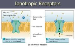
Also known as Ligand-gated channels = binding of neurotransmitters. |
|
|
|
Metabotronic receptors |
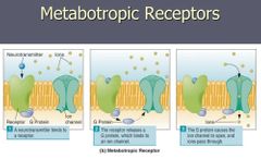
|
|
|
|
EPSPs |
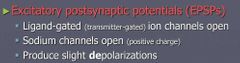
|
Effects of neurotransmitters binding |
|
|
IPSPs |
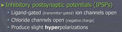
|
Effects of neurotransmitters binding |
|
|
Deactivation of Neurotransmitters |
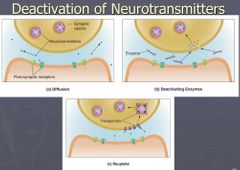
- Diffusion - Enzyme (breakdown) - Reuptake |
3 methodes |