![]()
![]()
![]()
Use LEFT and RIGHT arrow keys to navigate between flashcards;
Use UP and DOWN arrow keys to flip the card;
H to show hint;
A reads text to speech;
56 Cards in this Set
- Front
- Back
- 3rd side (hint)
|
Trail Making Test (TMT) |
two parts: 1. ss connects numbers from 1-25 scattered on paper 2. involves task switching. there are 13 numbers, 13 letters. connect number and letter in alternation. ie. 1-a-2-b-3-c-... used as a general measure of attention. can only tell if there is a deficit present but not what deficit it. |
|
|
|
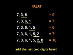
Paced Auditory Serial Addition Test (PASAT) |
ss hear series of numbers and must add the two most recently presented ones together ex. 4 7 = 11 4 7 3 = 10 4 7 3 9 = 12 used as a general measure of attention. can only tell if there is a deficit present but not what deficit it. |
|
|
|
Test of Attentional Performance (TAP) |
battery with a wide range of subtests intended to capture different and independent attentional processes. able to test for specific deficit. subtests: 1.alertness 2.divided attn 3.express saccade (disengagement) 4.overt/ covert shifting of attn. 5.visual search 6.go-no-go (response suppression) 7.incompatibility (resistance to interference) 8.crossmodal integration 9.neglect 10. attentional flexibility |

|
|
|
Tests of Everyday Attention (TEA) |
Listen to lecture 3:06 |
|
|
|
Traumatic Brain Injury (TBI) |
Any kind of trauma to the head, can range from mild to severe. Common cause of attn deficits. Suggested that mental slowing caused by these due to a reduction in availability of attn resources |

|
|
|
concussion |
Caused by brain rocking back + forth in skull 85% of head injuries are concussions Problems include slowness, difficulty in concentrating, difficulty in doing two things at once, and increased distractibility |

|
|
|
Examples of brain diseases causing attention deficits |
Alzheimer's, Parkinson's, Huntington's, Pick's and Schizophrenia |
|
|
|
How are frontal lobe and parietal lobe deficits different? |
Frontal: tend to be more related to executive function. ex. mental flexibility, inhibition and concentration Parietal: tend to be more related to spatial attention. |
|
|
|
Common causes of concussions |

|
|
|
|
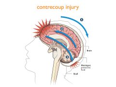
Contrecoup Injury |
both sides of brain, front + back, hit skull |
|
|
|
Why don't woodpeckers get concussions? |
1. cranium is smooth, no friction. 2. straight on head collision better than angle impact (angle puts strain on corpus collosum) |
|
|
|
Why don't helmets prevent concussions? |
Can prevent fractures of skull but can't prevent brain from moving in head. |
|
|
|
How do we diagnose concussions? |
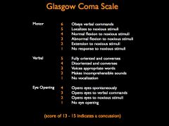
Glasgow Coma scale is one way used. although its not a very good indicator. |
|
|
|
Early signs of a concussion |
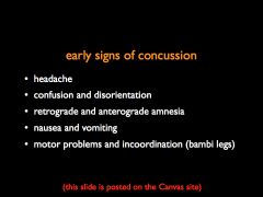
|
|
|
|
Why don't people realize they have a concussion? |
at the time of impact people feel fine because brain is traumatized (in shock) |
|
|
|
Why is it important to rest after a suspected concussion? |
A second impact right after the first can be fatal (second impact syndrome). Brain is very vulnerable and needs time to heal. |
|
|
|
What happens in the brain when a concussions occurs? |
When hit, leads to ion channelopathy. Ion channels stop working and acetylcholine (toxic) cannot get taken back inside the cell, leading to damage to neighbouring cells. |
Ion channels |
|
|
Why does it seem like women are more suscpetible to concussions? |
1. men lie?! under report concussions 2. different neck and trap. muscles (larger in men)? 3. genetic difference in brain shaking tolerability? |
|
|
|
Why are brain scans a bad indicator of concussions? |
scans won't show anything unless bleeding. bad b/c Dr. might disregard because nothing looks abnormal |
|
|
|
Why is DTI a good technique to diagnose concussions? |
injury due to axons, way to small to show on MRI or CT. DTI can show white matter (axon) differences |
|
|
|
Effects of TBI on attention |
1. poor concentration and lack of mental energy 2. increased interference by distractors 3. slower detection of targets during search 4. divided attn capacity impaired 5. increased stroop interference 6. higher frequency of action slips 7. reduced activation of executive attn areas 8. mental slowing (extreme fatigue) |
|
|
|
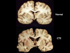
Chronic traumatic encephalopathy (CTE) |
"punch drunk" Progressive degenerative disease found in people who have had a severe blow or repeated blows to the head. Characterized by larger ventricles, degradation, similar to Alzheimer's |
|
|
|
How is Alzheimer's like a type 3 diabetes? |
according to study, if you cut off insulin to brain you see same effect as Alzheimer's |
|
|
|
Brain diseases associated with frontal cortical impairment |
Parkinson's: reduces activation in basal ganglia Huntington's: effects frontal cortical areas Schizophrenia: decreases frontal cortical activation Depression |
|
|
|
Attention Deficit Disorder (ADD) |
Neurological disorder characterized by increased distractibility and inability to focus attn in consistent manner. |
|
|
|
How to treat ADD? |
Treated with stimulants (ex. adderall and ritalin) Increases level of dopamine and NE in prefrontal cortex |
|
|
|
What causes ADD? |
suspected that it's caused by: 1. dysfunction of the thalamus because of its role in filtering 2.diminished function of the right hem. 3. deficit in the availability of attn resources 4. under-production of under-utilization of NT's |
|
|
|
Brain activation during concussion recovery |

|
|
|
|
How are spatial deficits brought about? (most commonly) |
by stokes and effects the parietal cortex |
|
|
|
Hemianopia |
vision compromised, only 1 side of visual field |

|
|
|
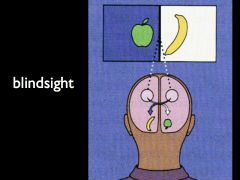
Blindsight |
Sometimes can't see things in part of visual field accurate at guessing when given options Caused by problem with primary visual pathway (cones), but rods still work |
|
|
|
Primary visual pathway |
from cone cells more direct root to brain (visual cortex) |
|
|
|
Secondary visual pathway |
from rod cells a more primitive pathway leading to the brainstem involved in reflexive behaviour unaware of this secondary pathway |
|
|
|
Graham Young |
Blindsight patient Able to detect motion and orientation but can not physically see characteristics if he turns his head to see, he is aware is aware of his disorder |
|
|
|
Spatial Neglect |
attentional deficit usually caused by stroke. Characterized by lack of awareness of objects in left visual hemifield. May also be unaware of left half of objects, and can still perceive sensations. |
|
|
|
How is blindsight different from neglect? |
Blindsight patients are aware of their deficit and therefore can move head to compensate. Neglect patients are not aware of their problem. |
|
|
|
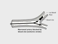
Ischemic stroke |
caused by blockage - plaque built up in arteries of brain, problems with clots. |
|
|
|
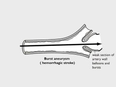
Hemmorrhagic stroke |
burst aneurysm - weak section of arterial wall burts |
|
|
|
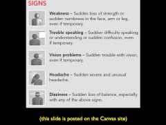
Signs of stroke |
|
|
|
|
Somatoparaphrenia |
unaware of own body |
|
|
|
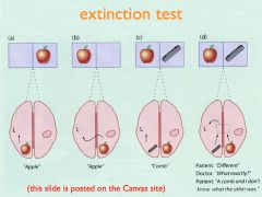
Visual Extinction |
occurs with parieto-occiptical lesions if two objects presented simultaneously, they do not "see" the object presented contralateral to lesion. can identify object, if nothing is in other field. |
|
|
|
Diagnosing Neglect (Tests) |
Line bisection test Line cancellation test Line drawing test |
|
|
|
Line bisection test |
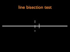
where is the middle? |
|
|
|
Line cancellation test |
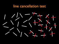
bisect white lines |
|
|
|
Line drawing test |
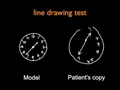
copy line drawing |
|
|
|
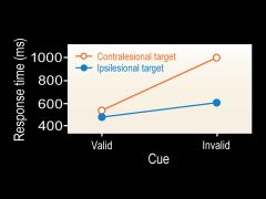
Posner Task - Extinction Findings |
1.when cue + target in right (good) side, can respond. 2.when cue + target in left (bad) side, slower response. 3.when cue is on right, target left, takes longest to respond |
|
|
|
Can people recover from neglect? |
yes! ex. patient who drew self portrait started drawing more accurately as he recovered |
|
|
|
How do we know that patients with neglect are processing stimuli unconsciously? |
tests show stimuli in bad field can prime understanding of stimuli in good field |
|
|
|
True or False: neglect extends to mental images as well. |
True. ex. Peggy Palmer would only draw half a daisy from memory |
|
|
|
Why does neglect occur? |
1. right hem. is attention center. ie. L parietal lesion does not lead to neglect 2. dynamic hem. imbalance - R hem damage results in dominant L hem and right side bias
|
|
|
|
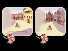
Plaza study |
method: ss asked to describe an image of a famous plaza in italy from a specific position (ie. facing south) findings: would only report half of the plaza, but when "turned around" would report the other half |
|
|
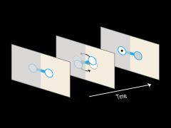
What does this study suggest? and why? |
spatial neglect can be object-based. When the object gets flipped, subjects reported correctly where dot was when presented in the initially good visual field, but slower to respond when initially presented in the bad field. |
|
|
|
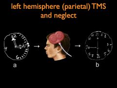
What happens if we lessen L hem dominance in patients with neglect? |
caloric stimulation (ice cold water in left ear) = recovery of neglect temporarily by shocking the left hemisphere TMS can also reduce neglect (lasts longer) |
|
|
|
Simultagnosia |
symptom of Balint's syndrome characterized by the inability to perceive different pieces of information in the visual field simultaneously because the person cannot direct attn to more than 1 small location at a time. Disappears if objects are connected |

|
|
|
Balint's Syndrome |
attnetion deficit characterized by a collection of related symptoms that include optic ataxia, ocular apraxia, and simultagnosia. Caused by bilateral dorsal parietal lesions. |
|
|
|
Attentional Dyslexia |
cause by left parietal damage. could read well but impaired ability to report single letters. errors due to mislocations (like illusory conjunctions) |
|