![]()
![]()
![]()
Use LEFT and RIGHT arrow keys to navigate between flashcards;
Use UP and DOWN arrow keys to flip the card;
H to show hint;
A reads text to speech;
24 Cards in this Set
- Front
- Back
|
PRIONS
|
- Consist of protein only - Small self replicating proteins without any nucleic acid - Derived from 'normal' proteins associated with membranes of the central nervous system (CNS) - Have different (abnormal) conformation from normal but the same amino acid composition |
|
|
DISEASES ASSOCIATED WITH PRIONS
|
Associated with Creutzfeld Jakob disease (CJD) and 'Kuru' in humans, scrapie in sheep and mad cow disease (bovine spongiform encephalitis - BSE)
|
|
|
PRIONS WHEN INGESTED
|
- Accumulate in CNS membranes - By an autocatalytic process they convert pre-existing normal forms of protein to their abnormal form (meaning the reproduce without making new copies of themselves) - The abnormal forms cannot be destroyed by the body and build up leads to destruction of CNS tissue in brain |
|
|
VIRUSES
|
- Not cells - Static structures (have no metabolic activity of their own) - Rely on host biosynthetic machinery for their protein synthesis - All classes of organisms can be infected by viruses (even bacteria - bacteriophages) - Enormous diversity (especially among bacteriophages) - Not susceptible to antibiotics |
|
|
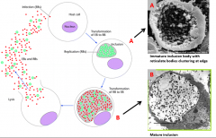
CHLAMYDIAE: LIFE CYCLE
|
|
|
|
CHLAMYDIAE: KEY SPECIES IMPORTANT IN DISEASE
|
- Chlamydia trachomatis (ocular (eye) infections, genito-urinary tract infections) - Chlamydia psittaci (respiratory tract infections in humans, gut and systematic infections in birds) - Chlamydia pneumonia (respiratory tract infections) - Chlamydia abortus (leads to abortion in sheep, rare cases can cause abortion in women when exposed to sheep) - Chlamydia caviae (infection in guinea pigs, model to study human disease) |
|
|
RICKETTSIAE
|
- Gram-negative bacteria - Small cells (0.7-2 micrometres) - Pleomorphic (cell morphology vary from cocci to filaments) - Slow growing obligate intracellular parasites - Unlike chlamydiae rickettsia can survive outside cell but cannot replicate (lack the ability to generate many important metabolites and have a requirement for coenzyme A, NAD and ATP) |
|
|
RICKETTSIAE: KEY SPECIES INVOLVED IN DISEASE
|
- Rickettsia rickettsii (causes spotted fever) - Reckettsia typhi, R. prowazekii, R. Tsutsugamushi (typhus) - Coxiella burnetii (Q-fever) - Ehrlichia chafeensis (ehrlichiosis) - Rickettsia felis (emerging human pathogen, typhus like disease) - All expect coxiella burnetti (inhalation) are transmitted to humans via arthropod bite (fleas, ticks, lice and mites, no direct person to person spread) |
|
|
MYCOPLASMAS
|
- Smallest prokaryotic cells capable of growing on cell-free media (free living) - Fastidious in their nutritional requirements (need complex growth media, limited biosynthetic capability) - Small genome size (0.1 - 1 micrometre a quarter of the size of E.coli genome) - Generally parasitic organisms that inhabit plant and animal hosts (but unlike chlamydiae and rickettsiae can replicate independently of host cells) |
|
|
MYCOPLASMA STRUCTURE
|
- Lack peptidoglycan in their cell walls (wall has no rigidity leading to pleomorphic cells, do not stain as gram-positive but are closely related to gram-positive bacteria) - Some species contain cholesterol in their cell walls (usually found in mammalian cell membranes and is absent in other bacteria) - Outer layer is triple-layered structure comprising of proteins and lipids |
|
|
THREE GENERA OF MYCOPLASMAS
|
- Mycoplasma - Ureaplasma - Acholeplasma |
|
|
MYCOPLASMAS IN DISEASE
|
- Mycoplasma pneumoniae (important cause of atypical pneumonia, can also cause genital infections - non-specific urethritis and other joint and inflammatory conditions) - Other mycoplasmas are important pathogens of animals and birds - Can be treated with some antibiotics (tetracycline or erythromycin) but not antibiotics acting on cell walls (beta-lactams) |
|
|
NON-ENVELOPED ANIMAL VIRUSES
|
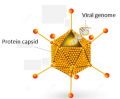
- Adenovirus - Rhinovirus - common cold |
|
|
ENVELOPED ANIMAL VIRUSES
|
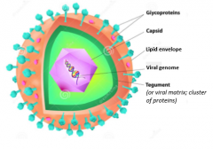
- Herpes Simplex virus - Varicella-Zoster virus - Influenza A virus - Hepatitis B |
|
|
BACTERIOPHAGE
|
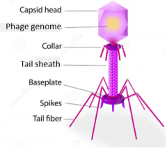
- T4 (dsDNA virus that infects Escherichia Coli - E.coli) - Lambda |
|
|
VIRAL REPLICATION CYCLE
|
- Virus' deliver their nucleic acid genome to host cells and 'hijack' the biochemical machinery (causing it to replicate the virus) - Ultimately the host cell becomes filled with virus particles - virions - Virions burst out the cell and disperse to infect new cells |
|
|
VIRAL REPLICATION CYCLE: ADENOVIRUS
|
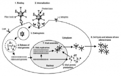
- Integrins are transmembrane receptors with alpha and beta subunits - Adenovirus is a human infecting virus |
|
|
BACTERIAL CELL WALL
|
- Structure of cell wall distinguishes two different categories of bacteria: gram-positive and gram-negative (distinguished by a staining method based on ability of cell wall to retain or lose the dye stain crystal violet - gram staining) |
|
|
GRAM STAINING METHOD
|
- Hans Christian Gram (1884) - Application of primary stain (crystal violet) to a heat-fixed smear of bacterial culture - The addition of iodine which binds to crystal violet forming a larger complex helping it 'trap' into the cell - Rapid decolourisation (alcohol or acetone) and counterstaining with safranin |
|
|
GRAM POSITIVE BACTERIA
|
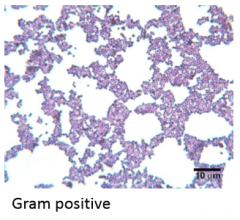
- Thick mesh-like cell wall, mostly peptidoglycan - PNG - Stained purple by crystal violet |
|
|
GRAM NEGATIVE BACTERIA
|
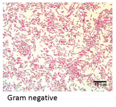
- Have a thinner PNG (peptidoglycan) layer - Does not retain crystal violet - Can be counter-stained pink by safranin |
|
|
CHLAMYDIAE
|
- Small cells which weakly stain as gram-negative - Contain lipopolysaccharide (LPS) in their cell walls - Range between 0.3-1 micrometre depending on stage in life cycle - Obligate intracellular parasite (a parasitic microorganism that cannot reproduce without entering a host cell) with poor metabolic capabilities - Genome 1/4 size of E.coli - Cell wall thought not to contain peptidoglycan although genes for peptidoglycan synthesis present in genome |
|
|
CHLAMYDIAE: ELEMENTARY BODY (EBs)
|
- Infectious form - Metabolically inactive - Small (0.3 micrometre diameter) - Resistant - capable of withstanding environmental conditions outside of host cell |
|
|
CHLAMIDYDIAE: RETICULATE BODY (RBs)
|
- Replicative form - Larger than elementary body (0.8-1.2 micrometre) - Metabolically active - Capable of replication (by binary fission) - Fragile - cannot survive in external environment |