![]()
![]()
![]()
Use LEFT and RIGHT arrow keys to navigate between flashcards;
Use UP and DOWN arrow keys to flip the card;
H to show hint;
A reads text to speech;
45 Cards in this Set
- Front
- Back
|
Anatomical Position
|
The body is described as being in this position. The body is assumed to be standing, the feet together, the arms to the side, and the head and eyes and palms of the hands facing forwards. |
|
|
Superior
|
above; toward the head or upper part of structure; this term does not used on limbs |
|
|
Inferior |
below; away from the head or toward the lower part of structure; this term is not used on limbs |
|
|
Proximal |
closer to the point of attachment (of limb to trunk of body) |
|
|
Distal |
farther from the point of attachment (of limb to trunk of body) |
|
|
Superficial |
toward the body surface; more external |
|
|
Deep |
away from the body surface; more internal |
|
|
Medial |
toward the midline |
|
|
Lateral |
away from the midline |
|
|
Anterior (ventral) |
toward or at the front of the body |
|
|
Posterior (dorsal) |
toward or at the back of the body |
|
|
Coronal plane |
vertical plane that divides into anterior and posterior parts |
|
|
Transverse plane |
horizontal plane the divides into superior and inferior parts |
|
|
Sagittal plane |
vertical plane that divides into right and left parts |
|
|
Arm is between which 2 joints? |
? |
|
|
Leg (between which two joints?) |
? |
|
|
Clavicle |
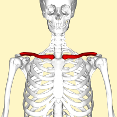
AKA the collarbone. S-shaped bone that extends between the manubrium of the sternum and the acromion of the scapula. |
|
|
Scapula |
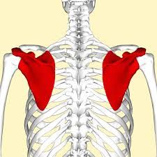
The shoulder blade. The scapula articulates with the round head of the humerous at the glenoid cavity of the scapula, the shoulder joint. |
|
|
Humerus |
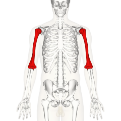
Bone in upper arm. The humerus articulates with the glenoid cavity of the scapula. |
|
|
Ulna |
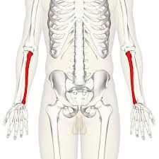
The thinner and longer of the two bones in the forearm, on the side opposite to the thumb |
|
|
Radius |
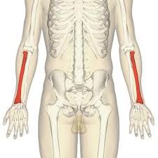
One of the two large bones of the forearm. It extends from the lateral side of the elbow to the thumb side of the wrist and runs parallel to the ulna, which exceeds it in length and size
|
|
|
Carpal bones |
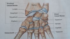
Carpal = wrist Wrist formed by 8 bones. 2 rows with 4 proximal carpal bones, and 4 distal carpal bones. Proximal: the scaphoid, the lunate, the triquetrum and the pisiform. Distal: the trapezium, the trapezoid, the capitate, and the hamate |
|
|
Metacarpal bones |
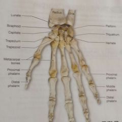
5 bones that articulate with the distal carpal bones and support the palm of the hand. Expressed by roman numerals: I - V |
|
|
Phalanges (or Phalanx) |
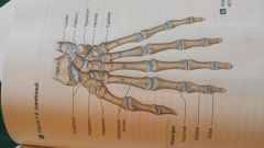
The metacarpal bones articulate with the phalanges, or finger bones. There are 14 in each hand. The thumb (or pollex) has 2 phalanges, and each finger has 3. |
|
|
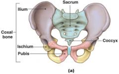
Coxal Bone |
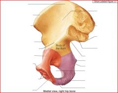
AKA the Hipbone (or pelvic bone) Two hipbones make up the pelvic girdle. Hipbone is made up of 3 bones: ilium, ischium, and pubis. (these 3 meet inside the acetabular fossa) |
|
|
Illium |

The largest of the 3 bones. Provides an extensive area for the attachment of muscles, tendons, and ligaments. |
|
|
Ischium |

illium fuses with the ischium. The ischium is the strongest of the hip bones. |
|
|
Pubis |
|
|
|
Femur |
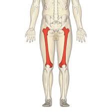
the longest & heaviest bone is the body. The femur articulates with the tibia of the leg at the leg joint. The rounded head of the femur articulates with the pelvis at the acetabulum. |
|
|
Patella |
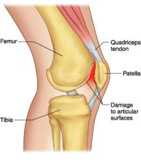
AKA knee cap. It is a large sesamoid bone that forms within the tendon of the quadriceps femoris (a group of anterior thigh muscle that extends the knee). The bone strengthens the quad's tendon, protects the anterior surface of the knee joint, and increases the contraction force of the quad. |
|
|
Tibia |
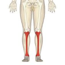
The large medial bone of the leg. The medial and lateral condyles of the femur articulate with the medial and lateral condyles of the end of the tibia. The intercondylar eminence separates the 2. ^ |
|
|
Pelvis |
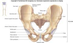
Subdivided into the greater (false) pelvis and the lesser (true) pelvis. |
|
|
Fibula |

Parallels the lateral border of the tibia. |
|
|
Tarsal Bones |
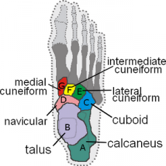
tarsus (aka ankle) contains 7 tarsal bones: the talus, the calcaneus, the cuboid, the navicular, and three cuneiform bones. The talus: the second largest bone in the foot. It transmits the weight of the body from the tibia anteriorly, towards the toes. The calcaneus: aka the heel bone, is the largest tarsal bone and may be easily examined. When standing: weight is transmitted from the tibia to the talus to the calcaneus, then to the ground. The cuboid: articulates with the anterolateral surface of the calcaneus. The navicular: (located on the medial side of the ankle) articulates with the anterior surface of the talus. The distal surface of the navicular articulates with 3 cuneiform bones. The three cuneiform bones are arranged in a row with articulations between them, located anterior to the navicular. They're named: medial cuneiform, intermediate cuneiform, & later cuneform bones. The distal surfaces of the cuboid and the cuneiform bones articulate with the metatarsal bones of the foot. |
|
|
Metatarsal bones |
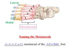
5 long bones that form the distal portion of the foot. Expressed in roman numerals I - V. The first 3 metatarsal bones articulate with the 3 cuneiform bones, and the last 2 articulate with the cuboid. They help support the weight of the body during standing, walking, and running. |
|
|
The Vertebral Column |
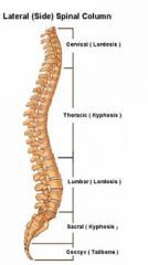
Consists of 26 bones: 24 vertebrae, the sacrum, and the coccyx. It is divided into regions: Beginning at the skull, there us the cervical, thoracic, lumbar, sacral, & coccygeal. Each region has it's own function. There are four spinal curves: cervical, thoracic, lumbar, and sacral curve. The vertebral provides a column of support, bearing the weight of the head, neck, & trunk. It transfers that weight to the appendicular skeleton of the lower limbs. |
|
|
Cervical Vertebra |
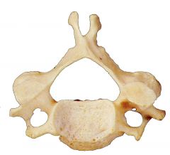
Smallest & most superior vertebrae Extends from the occipital bone of the skull to the thorax. Supports skull, stabilizes relative positions of brain & spinal cord; allows controlled head movement |
|
|
Thoracic Vertebrae |
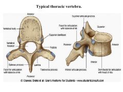
Medium sized; heart shaped body; Supports weight of head, neck, upper limbs, organs of thoracic cavity; articulates with ribs to allow changes in volume of thoracic cage There are 12; |
|
|
Lumbar Vertebrae |
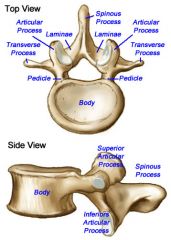
Largest of the vertebrae; oval surfaces Support the weight of the head, neck, upper limbs, organs of thoracic and abdominal cavities |
|
|
The Sacrum |
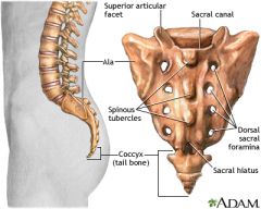
Consists of the fused components og five sacral vertebrae. Continues fusing until 25-30 yrs old; prominent transverse lines mark the former boundaries of individual vertebrae. This structure protects reproductive, digestive, & excretory organs and attaches the axial skeleton to the pelvic girdle. The broad surface allows muscles to attach. Curved; with a convex dorsal surface |
|
|
The Coccyx |
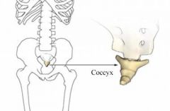
Consists of 3 to 5 coccygeal vertebrae that finish fusing around age 26. It provides an attachment site for a number of ligaments and for a muscle that constricts the anal opening. |
|
|
The Atlas (C1) |
Articulates with the occipital condyles of the skull, the atlas holds up the head. There is a joint in between that allows nodding. The atlas is different because 1. lack of body, 2. the possession of a semicircular anterior & posterior vertebral arches (each containing anterior & posterior tubercles), 3. the presence of oval superior articular facets & round inferior articular facets, and 4. the largest vertebral foramen of an vertebra. |
|
|
The Axis (C2) |
During development, the body of the atlas fuses to the body of the second cervical vertebra called the atlas. This fusion causes a prominent dens of the axis. There is no intervertebral disc between the atlas & the axis. A transverse ligament binds the dens to the inner surface of the atlas, forming a pivot: this allows rotation of the head side to side ("no") |
|
|
Vertebrosternal Rib |
jjj |
|
|
Vertebrochondral Rib |
nj |