![]()
![]()
![]()
Use LEFT and RIGHT arrow keys to navigate between flashcards;
Use UP and DOWN arrow keys to flip the card;
H to show hint;
A reads text to speech;
46 Cards in this Set
- Front
- Back
|
Pleuritis What it is? Sx? |
Sharp chest pain that worsens with breathing [Inflammation of lining of lungs] Sx: Cough, Chest Tenderness, SOB
|
|
|
Pleural Effusion Often show sx when? Sx: |
Moderate/ Large effusion, or if inflammation present Chest Pain, SOB, Coughing, Fever |
|
|
Signs of Pleural Effusion |
Increased TF, Decreased Breath Sounds, Audible Friction rub; Non-symmetric rise of thoracic cage --> Hoover's Sign; Dull Percussion |
|
|
Signs concerning effusion |
Massive Effusion comes with inc. intrapleural pressure Chest Pain: Exudative Effusion Transudative Effusion does not cause pleural irritation |
|
|
Common Causes of Pleural Effusion |
CHF, Cirrhosis, Pneumonia, Cancer, PE, Autoimmune conditions, ESRD |
|
|
Diagnostic Studies in Ple. Effusions |
CXR, US, CT Scan |
|
|
Two types of pleural effusions |
Uncomplicated and Complicated
|
|
|
Uncomplicated Pl. Effusion Contains Fluid.... |
Free of inflammation/infection Causes sx only if large enough Rarely cause permanent lung problems |
|
|
Complicated Pl. Effusion Contains Fluid... What happens if untreated? How to prevent this process? |
with Significant inflammation or infection If untreated --> May harden to form a constricting ring around lung --> "Organization" impairs breathing To Prevent: Drainage from chest tube |
|
|
What procedure is performed for new and unexplained pleural effusions? |
Thoracentesis |
|
|
Normal Pleural Fluid : pH |
7.60-7.64 |
|
|
Normal Pleural Fluid: Coloring/what it is? |
Clear Ultra filtrate of plasma from parietal pleura |
|
|
Normal Pleural Fluid : Protein content Glucose content similar to? What ion concentrations are similar to interstitial fluid? Number of WBCs? Lactate Dehydrogenase percentage of plasma? |
Protein: Less than 2% Glucose similar to plasma K, Ca, Na similar interstitial fluid <1000 per mm^3 LDH: <50% of Plasma |
|
|
Transudative Pleural Fluid: Describe Ex: |
Similar to fluid normally in pleura Rarely require drain CHF causes transudative PE |
|
|
Exudative Pleural Fluid: Describe Ex: |
Excess Protein, blood, evidence of inflammation/infection May require drainage d/o size/severity Pneumonia, Lung Cancer can cause EPE |
|
|
Exudative Effusion Lab Features are known as? |
Light's Criteria
|
|
|
What are Light's Criteria? |
Pleural Fluid To Serum +Protein > 0.5 +LDH >0.6 +LDH > 2/T3 upper limit of serum LDH
|
|
|
1/4 of Transudative Eff. are mistaken as exudative by LC; How to definitely tell? |
Difference btwn Albumin levels [in Blood, Pleural Fluid] is > 1.2 g/dL Pt has transudative effusion |
|
|
More criteria that show exudative effusion Pleural Fluid Protein? Pleural Fluid cholesterol? pH of what suggests drainage? |
>29 g/L >45 mg/dL <7.30 |
|
|
In pleural fluid WBC > 10,000 suggests? RBC> 10,000 suggests? Lymphocytosis suggests?
|
WBC: Pneumonia, Pancreatitis, Malignacy, TB RBC: Malignancy, Infarcation
TB, Lymphoma, RA, malignancy
|
|
|
Transudative Effusins finding are: |
About normal, suggest absence of pleural disease Glucose equal to serum glucose pH between 7.40, 7.55 <1000WBC |
|
|
Effusion appearances: Malignancy Pneumonia Empyema TB Lung Infarct Pancreatitis |
+Serous/Turbid/Bloody + Clear to Turbid +Turbid to Purulent +Serosanguineous +Serous to bloody +Turbid-serosanguineous |
|
|
Other notes on Ple. fluid/effusion: Milky, opalescent fluid suggests? Resulting from? Bloody fluid can come from? Indicates need for?
|
Chylothorax -->Lymph obstruction by malignancy/thoracic duct injury [by trauma/surgery] Trauma, Malignancys, postpericardiotomy, asbestos related Need for spun hematocrit |
|
|
Hematocrit level of >50% defines a |
HEMOTHORAX |
|

|
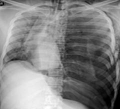
Large, R, Pleural effusion |
|
|
Collection of Free Air in chest outside lung? |
Pneumothorax |
|
|
Spontaneous Pneumothroax occurs in Primary and Secondary ways. Describe each |
1) Absence of traumatic injury to chest/ known lung disease 2) Result of an underlying condition
|
|
|
Risk Factors for PneumoThorax
|
Gender, Smoking, Age, Genetics, Hx, Lung Disease, Ventilation |
|
|
Simple Pneumothorax Type of collapse Require Emergency Txt? Side Effects? |
Partial Collapse of a lung Pressre not enough to cause CV Dysfunction May be severe enough to dec. Blood O2 --> SOB Can be small/stable/ doesn't require ETXT |
|
|
Signs & Sx of PT |
Chest Pain: Sudden Sharp on same side of lung; Stays constant through I & E
SOB: Mild/severe |
|
|
Test for PT? [3] |
Stethoscope, CXR, CT |
|
|
TXT PT? |
Observe, CXRs Bed Rest, O2 supplement
If larger collapse --> Needle/Chest Tube Insertion [attached to suction device] Possible Surgery: Laparoscopic |
|
|
Spontaneous [1?] PT occurs in |
Tall and thin, Smokers, Rec. Drug Users No apparent Injury/Trauma |
|
|
Secondary Spontaneous occurs in? |
Older pts usually From underlying lung disease: CF, COPD, CA, TB, Asthma, Pneum., etc... |
|
|
PneumoMediastinum |
Free Air in Mediastinal structures Occurs with elevation if intrathoractic pressure Substernal Chest Pain Radiates to neck, back, shoulders Exacerbated by Deep I, coughing, supine position |
|
|
Pn. Med. finding on P/E |
Subcutaneous empysema Hamman Sign [Crunching, Rasping sound] |
|

|
PneumoMediastinum |
|
|
Traumatic PT Happens from? Subset called? Results from? |
Direct Trauma to chest wall from blunt/penetrating trauma +Iatrogenic PT -Dx or Thera. med procedures [Needle Aspiration, Lung bx, IV catheter into neck vein] |
|
|
What happens in Tension PT? Pressure does what? |
Air builds under pressure and collapses totally 1 or both lungs Pressure slows/stops blood return to heart from veins --> BP drops Death can result from no txt |
|
|
Signs/ Sx of Tension PT? |
Respiratory Distress, Hypoxia, Hypotension Cardiopulm. compromise Pressure build pushes mediastium to opposite side and obstructs return to heart --> Circul. instability |
|
|
Classic Signs of Tension PT |
Deviation of Trachea Hyper Expanded chest [moves little with I] Increased Percussion Central Venous Pressure usually Raised
|
|

|
Tension PT Also: |
|
|
Empyema is? |
Collection of pus in pleural space
|
|
|
Risk Factors for Empyema? |
Bacterial Pneum, Chest sx, Lung Abcess, Trauma |
|
|
Sx of Empyema |
Dry cough, chest pain, excessive sweating, fever, malaise, SOB, Weight loss
|
|
|
Signs of Empyema Test with: |
Dec. Breath Sounds ; Friction Rub audible CT, CXR, Fluid Analysis, Thoracentesis |