![]()
![]()
![]()
Use LEFT and RIGHT arrow keys to navigate between flashcards;
Use UP and DOWN arrow keys to flip the card;
H to show hint;
A reads text to speech;
30 Cards in this Set
- Front
- Back
|
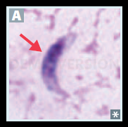
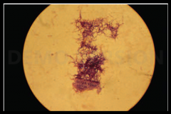
Naegleria fowleri in spinal fluid |
|
|
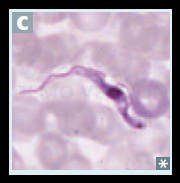
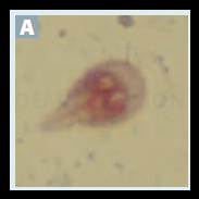
Trypanosoma brucei. Blood smear. |
|
|
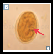
Toxoplasma gondii. Tachyzoite in biopsy material. |
|
|
Cryptosporidium. Oocysts on acid-fast stain. |
|
|
Entamoeba histolytica. Trophozoites (with RBCs in the cytoplasm). |
|
|
Entamoeba histolytica. Cysts (with up to 4 nuclei) in stool. |
|
|
Giardia lamblia. Trophozoite in stool. |
|
|
Giardia lamblia. Cyst in stool. |
|
|
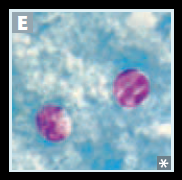
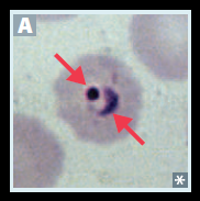
Plasmodium.Trophozoite ring form within RBC |
|
|
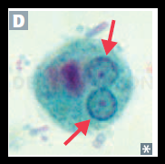
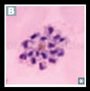
Babesia. Blood smear, ring form C1 , “Maltese |
|
|
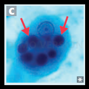
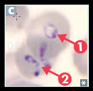
Plasmodium. Schizont containing merozoites. |
|
|
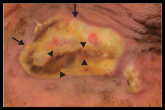
Corynebacterium diphtheriae. Pseudomembranous pharyngitis (grayish-white membrane). |
|
|
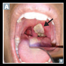
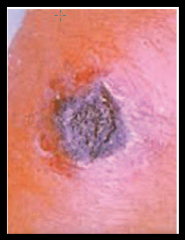
Anthrax. Ulcer with black eschar/crust. |
|
|
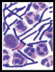
Bacillus anthracis. Gram-positive rods. |
|
|
Mycobacteria. Acid-fact staining. |
|
|
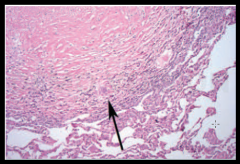
Caseating granuloma. Central necrosis (pinkish region in upper left) with multinucleated Langhans giant cell (arrow). |
|
|
Actinomyces. A. israelii on Gram stain. |
|
|
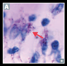
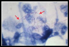
Nocardia. Arrows show Nocardia on acid-fast stain. |
|
|
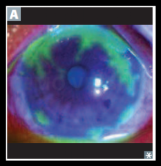
HSV-1. Keratoconjunctivitis |
|
|
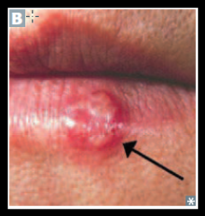
Herpes labialis |
|

|
Atypical lymphocytes seen on peripheral blood smear in EBV infection |
|
|
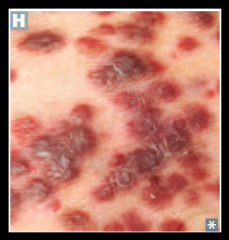
Dark/violaceous flat and nodular skin lesions of Kaposi sarcoma, representing endothelial growths |
|
|
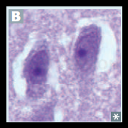
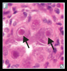
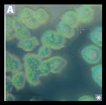
Characteristic “owl eye” inclusions of cells infected with CMV. |
|
|
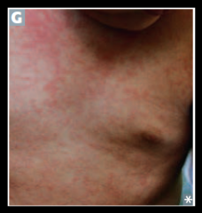
Diffuse macular rash in roseola |
|
|
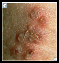
Shingles |
|
|
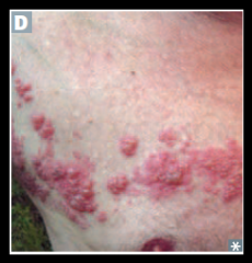
Herpes genitalis |
|
|
Pseudomonas aeruginosa infection. Ecthyma |
|
|
Pseudomonas aeruginosa. Blue-green color due to pyocyanin. |
|
|
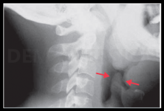
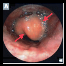
Haemophilus influenzae epiglottitis. Thickening of the epiglottis (“thumbprint sign”) on lateral neck radiograph. |
|
|
Haemophilus influenzae epiglotittis |